Nanoparticles for resisting H1N1 subtype influenza A virus based on self-assembled ferritin as well as preparation method and application of nanoparticles

An influenza virus and nanoparticle technology, applied in the field of genetic engineering, can solve the problem that antibodies cannot provide cross-protection, achieve high sensitivity and curative effect, improve accuracy and repeatability, and shorten the treatment process

Pending Publication Date: 2022-07-29
QINGDAO AGRI UNIV
0 Cites 0 Cited by

AI-Extracted Technical Summary

Problems solved by technology

Influenza A viruses have many subtypes, and most antibodies ...

Abstract

The invention discloses nanoparticles for resisting H1N1 subtype influenza A virus based on self-assembled ferritin as well as a preparation method and application of the nanoparticles, and relates to the technical field of genetic engineering. The nanoparticle is formed by connecting a signal peptide-ST-FE fusion protein and a signal peptide-SC-HA-VHH fusion protein in vitro, the signal peptide-ST-FE fusion protein is obtained by connecting a signal peptide-SpyTag as shown in SEQ ID NO: 3 to the N end of a ferritin monomer subunit; the signal peptide-SC-HA-VHH fusion protein is obtained by connecting the end N of a nano antibody of an anti-H1N1 subtype influenza A virus hemagglutinin protein with a signal peptide-SpyCatcher as shown in SEQ ID NO: 4. The invention further discloses a preparation method of the signal peptide-SC-HA-VHH fusion protein. The nanoparticles for resisting the influenza A (H1N1) subtype virus, disclosed by the invention, have great significance in monitoring, preventing and treating the influenza A (H1N1) subtype virus.

Application Domain

NanomedicineAntivirals +3

Technology Topic

Image

  • Nanoparticles for resisting H1N1 subtype influenza A virus based on self-assembled ferritin as well as preparation method and application of nanoparticles
  • Nanoparticles for resisting H1N1 subtype influenza A virus based on self-assembled ferritin as well as preparation method and application of nanoparticles
  • Nanoparticles for resisting H1N1 subtype influenza A virus based on self-assembled ferritin as well as preparation method and application of nanoparticles

Examples

  • Experimental program(11)

Example Embodiment

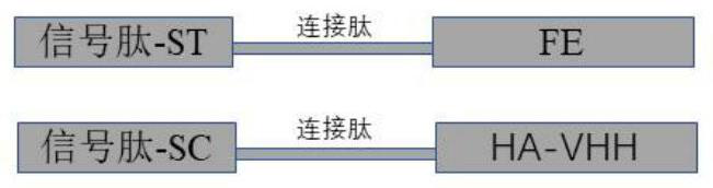
[0048] Example 1 Construction of plasmid signal peptide-SpyTag-ferritin monomer subunit (signal peptide-ST-FE) and signal peptide-SpyCatcher-anti-H1N1 subtype influenza virus hemagglutinin protein Nanobody (signal peptide-SC-HA -VHH)
[0049] First, the gene signal peptide-ST-FE and the signal peptide-SC-HA-VHH were synthesized. The gene tandem structure is as follows figure 1 , and optimized by the codons of the CHO eukaryotic expression system, synthesized by GenScript Biotechnology Co., Ltd. The amino acid sequence of the signal peptide-ST-FE (SEQ ID NO: 1) is as follows:
[0050] MGWSCIILFLVATATGVHSAHIVMVDAYKPTKGGGSGGGSGGGSRMLKALNDQLNRELYSAYLYFAMAAYFEDLGLEGFANWMKAQAEEEIGHALRFYNYIYDRNGRVELDEIPKPPKEWESPLKAFEAAYEHEKFISKSIYELAALAEEEKDYSTRAFLEWFINEQVEEEASVKKILDKLKFAKDSPQILFMLDKELSARAPKLPGLLMQGGE;
[0051] The amino acid sequence (SEQ ID NO: 2) of the signal peptide-SC-HA-VHH is as follows:
[0052] MGWSCIILFLVATATGVHSSYYHHHHHHDYDIPTTENLYFQGSATHIKFSKRDEDGKELAGATMELRDSSGKTISTWISDGQVKDFYLYPGKYTFVETAAPDGYEVATAITFTVNEQGQVTVNGKATKGDAHIGGGSGGGSGGGSQVQLVESGGGLVQSGGSLRLSCAASGSMSRIITMGWYRQAPGMERELVAVIGNNDNTVYGDSVQGRFTVSRDNAKNTAYLQMNSLNAEDTAMYYCKISTLTPPHEYWGQGTQVTVSSHHHHHH;
[0053] The amino acid sequence (SEQ ID NO: 3) of the signal peptide-SpyTag is as follows:
[0054] MGWSCIILFLVATATGVHSAHIVMVDAYKPTK;
[0055] The amino acid sequence of the signal peptide-SpyCatcher (SEQ ID NO: 4) is as follows:
[0056] MGWSCIILFLVATATGVHSSYYHHHHHHDYDIPTTENLYFQGSATHIKFSKRDEDGKELAGATMELRDSSGKTISTWISDGQVKDFYLYPGKYTFVETAAPDGYEVATAITFTVNEQGQVTVNGKATKGDAHI;
[0057] The amino acid sequence of the linker peptide (SEQ ID NO: 5) is as follows:
[0058] GGGSGGGSGGGS;
[0059] The amino acid sequence of the signal peptide (SEQ ID NO: 6) is as follows:
[0060] MGWSCIILFLVATATGVHS.
[0061] After the signal peptide-ST-FE and signal peptide-SC-HA-VHH genes are synthesized, the synthetic gene is used as a template to amplify the target fragment by PCR technology. The amplification primers are:
[0062] Signal Peptide-ST-FE:
[0063] Upstream primer sequence: AATCTCTAGAATGGGCTGGAGCTGCAT (SEQ ID NO: 7);
[0064] Downstream primer sequence: AATCAAGCTTTTACTCGCCGCCCTGCAT (SEQ ID NO: 8);
[0065] Signal Peptide-SC-HA-VHH
[0066] Upstream primer sequence: AATCTCTAGAATGGGCTGGAGCTGCAT;
[0067] Downstream primer sequence: AATCAAGCTTTTAGTGGTGGTGGTGGTG (SEQ ID NO: 9).
[0068] The nucleotide sequences of the signal peptide-ST-FE and signal peptide-SC-HA-VHH genes are as follows:
[0069] Signal Peptide-ST-FE:
[0070] GAATTCATGGGTTGGAGTTGCATCATCCTATTTCTAGTGGCCACCGCTACCGGCGTGCACTCTGCCCACATCGTGATGGTGGACGCCTACAAGCCCACAAAGGGCGGAGGCAGCGGCGGCGGCTCTGGCGGAGGATCTCGGATGCTGAAGGCCCTGAACGACCAGCTGAATCGGGAGCTGTACTCCGCCTACCTGTACTTTGCCATGGCCGCTTACTTCGAGGACCTGGGCCTGGAGGGCTTCGCCAACTGGATGAAAGCTCAGGCCGAGGAAGAGATCGGCCACGCCTTGAGATTCTACAACTACATCTACGACAGAAACGGCAGAGTGGAACTGGATGAGATTCCTAAGCCTCCAAAAGAGTGGGAGAGCCCCCTGAAGGCTTTCGAGGCTGCTTACGAGCATGAGAAGTTCATCTCCAAGTCCATCTACGAGCTGGCTGCTCTGGCAGAGGAAGAAAAGGATTATTCCACCAGAGCCTTCCTGGAATGGTTCATCAACGAGCAAGTCGAAGAAGAGGCCTCCGTGAAGAAGATCCTGGACAAGCTGAAGTTTGCCAAGGACTCCCCTCAGATCCTGTTCATGCTCGATAAAGAACTGTCTGCTCGGGCCCCTAAGCTGCCTGGCCTGCTGATGCAGGGCGGCGAGTGAGGATCC(SEQ ID NO:10);
[0071] Signal Peptide-SC-HA-VHH:
[0072]GAATTCATGGGTTGGAGTTGCATCATCCTATTTCTAGTGGCCACCGCTACCGGCGTGCACTCCTCCTACTACCACCACCACCACCACCACGACTACGACATTCCCACCACCGAGAACCTGTACTTCCAGGGCTCCGCCACACACATCAAGTTCTCCAAGAGAGACGAGGATGGCAAAGAGCTGGCTGGCGCTACAATGGAACTGAGAGATAGCTCTGGCAAAACAATCTCTACCTGGATCAGCGACGGCCAAGTGAAGGACTTCTACCTCTATCCTGGCAAGTACACCTTCGTGGAAACAGCTGCTCCTGATGGCTACGAGGTGGCTACCGCCATCACCTTTACCGTGAACGAGCAGGGCCAGGTCACCGTGAACGGCAAGGCCACCAAGGGCGATGCCCACATCGGCGGAGGATCTGGCGGAGGCTCCGGCGGAGGCTCTCAGGTGCAGCTGGTGGAATCTGGAGGTGGCCTGGTGCAGTCCGGCGGCAGCCTGCGGCTGTCCTGTGCCGCTTCTGGCTCCATGAGCCGGATCATCACCATGGGCTGGTACAGACAGGCCCCAGGCATGGAACGCGAGCTGGTCGCCGTGATCGGCAACAACGACAATACCGTTTACGGCGACTCCGTGCAAGGCAGATTCACCGTGTCTCGGGACAATGCCAAGAACACCGCTTATCTGCAGATGAACTCCCTGAACGCCGAGGACACCGCCATGTACTACTGCAAGATCTCCACCCTGACACCTCCTCACGAGTACTGGGGCCAGGGCACCCAGGTGACCGTGTCCTCTCACCATCACCACCATCATTGAGGATCC(SEQ IDNO:11)。
[0073] The target gene amplification system is shown in Table 1:
[0074] Table 1
[0075]
[0076] The PCR reaction program of the target gene is shown in Table 2:
[0077] Table 2
[0078]
[0079] The amplified products were electrophoresed on a 1% agarose gel, and the results were as follows figure 2 shown, is the same as the expected fragment length.
[0080] The PCR products were recovered, and the two target fragments recovered from the PCR gel were digested with the eukaryotic expression plasmid P3 plasmid using restriction enzymes Xba I and Hind III respectively; T4 DNALigase was used to connect the above two restriction fragments to construct Two target gene vector plasmids were obtained, namely plasmid signal peptide-ST-FE (p3-ST-FE) and signal peptide-SC-HA-VHH (p3-SC-HA-VHH), which were stored at -20°C.

Example Embodiment

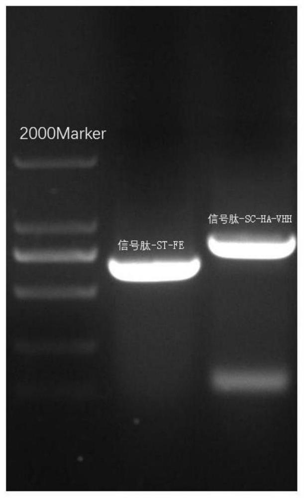
[0081] Example 2 Linearization of Plasmid Signal Peptide-ST-FE and Signal Peptide-SC-HA-VHH
[0082] Take out the plasmid signal peptide-ST-FE and signal peptide-SC-HA-VHH stored at -20°C, and measure their concentrations by NanoDrop one to be 1200ng/μL and 890ng/μL, respectively, and use the restriction endonuclease PvuⅠ for single enzyme Cut, concrete reaction system is shown in following table 3, table 4:
[0083] Table 3 Plasmid signal peptide-ST-FE single digestion reaction system
[0084]
[0085] Table 4 Plasmid signal peptide-SC-HA-VHH single enzyme digestion reaction system
[0086]
[0087] Add each component according to the single enzyme digestion reaction system table, and place it in a 37°C water bath for 2h digestion.
[0088] During digestion, prepare a 1% agarose gel. First, prepare the gel plate, select and insert the comb according to the total enzyme digestion system, then weigh 0.5g of agarose into a 100mL beaker, weigh 50mL of 1×TAE with a graduated cylinder, and add it to it, mix well and place it in a microwave oven for heating Take it out for 30s to observe the dissolution of the agarose, repeat heating to make it fully dissolved, then add 2 μL GoldView I nucleic acid dye to it, shake it gently (at this time, force evenly to avoid bubbles), pour the gel, and let it stand at room temperature for 40min to make the gel fully solidified. Then, the single-enzyme-digested product was mixed with an appropriate amount of 6×Loading Buffer and added to the gel well. The non-single-enzyme-digested knockout plasmid and DL 10000 DNA Marker were used as controls. The electrophoresis conditions were constant voltage 110V, 600mA, 30min. After electrophoresis, the gel block was gently taken out and placed in a blue-light gel cutter for gel cutting. According to the instructions of the gel recovery kit (SanPrepColumn DNA Gel Extraction Kit), the purified single-enzyme digested vector was recovered. The purified product was recovered and the concentration and OD were determined using the NanoDrop one 260/280 , stored at -20°C. 1% agarose gel as image 3 As shown, linearized plasmids run slower than circular plasmids, as expected.

Example Embodiment

[0089] Example 3 Electrotransfection of linear signal peptide-ST-FE and signal peptide-SC-HA-VHH GS gene deletion CHO cells
[0090] (1) Preparation before electroporation of CHO suspension cells lacking GS gene. 24h before electroporation, cells were at a viable cell density of 5×10 5 The cells/mL were inoculated into a 125 mL conical flask for shaking culture, the culture volume was 30 mL, and the rotational speed of the carbon dioxide shaking incubator was set to 110 rpm.
[0091] (2) Collection of CHO suspension cells lacking GS gene. On the day of electroporation, samples were taken to calculate the viable cell density and viability (98%) of suspension culture, and 1×10 cells were collected by calculation. 7 The cell suspension volume required for each viable cell was placed in a sterile centrifuge tube, centrifuged at 1000 rpm for 5 min, and then resuspended in 400 μL of 302 medium (containing glutamine).
[0092] (3) Incubation of cells with plasmids. Add 40 μg of linearized knockout plasmid (linear plasmid p3-ST-FE or linear plasmid p3-SC-HA-VHH) to the above-treated cell suspension, mix by gently pipetting, and incubate at room temperature for 10 min.
[0093] (4) Electric shock. The parameters of the electroporator are set in advance, and the electric shock conditions are: voltage 280V, electric shock time 20ms, and electric shock once. After 10 min incubation at room temperature, the above cell plasmid mixture was quickly added to a pre-cooled 4 mm electric shock cup, and electric shock was performed once.
[0094] (5) Cell treatment after electric shock. After the electric shock, the cells after the electric shock were quickly transferred to 20 mL of pre-warmed 302 medium (containing glutamine), and the T75 cell culture flask was left to culture for 24 hours.
[0095] (6) 24h after electric shock, the cells were sampled to calculate the viability and record. The cell viability was 48% after 24h electroporation with linear plasmid p3-ST-FE, and 52% after 24h electroporation with linear plasmid p3-SC-HA-VHH.
[0096] (7) Continue culturing to a cell viability rate of more than 95% using a medium without glutamine.

PUM

no PUM

Description & Claims & Application Information

We can also present the details of the Description, Claims and Application information to help users get a comprehensive understanding of the technical details of the patent, such as background art, summary of invention, brief description of drawings, description of embodiments, and other original content. On the other hand, users can also determine the specific scope of protection of the technology through the list of claims; as well as understand the changes in the life cycle of the technology with the presentation of the patent timeline. Login to view more.

Similar technology patents

Method of quickly detecting content of cell infective virions in circovirus

Owner:SICHUAN ENTRY EXIT INSPECTION & QUARANTINE BUREAU INSPECTION & QUARANTINE TECHN CENT +1

Whole blood separation membrane for immunofluorescence chromatography detection as well as preparation method and application of whole blood separation membrane

PendingCN114280312ASimplify sample handlingImprove accuracy and repeatabilityBiological testingPolyclonal antibodiesImmunofluorescence
Owner:HEBEI TEWENTE BIOTECH DEV CO LTD

An electrospray ionization device and its application

InactiveCN105719938BImprove biocompatibilityImprove accuracy and repeatabilityMaterial analysis by electric/magnetic meansIon sources/gunsIonizationCell based
Owner:CHINA UNIV OF GEOSCIENCES (BEIJING)

High-precision vehicle steering tracking test method and system

ActiveCN114397120AReduce driving difficulty and trial riskImprove accuracy and repeatabilityVehicle steering/rolling behaviourReal time displayClassical mechanics
Owner:XIANGYANG DAAN AUTOMOBILE TEST CENT

Classification and recommendation of technical efficacy words

  • Improve accuracy and repeatability

Mixer with high output power accuracy and low local oscillator leakage

ActiveUS20090075689A1Improve accuracy and repeatabilityIdentical layoutAutomatic scanning with simultaneous frequency displayResonant long antennasFrequency mixerLocal oscillator
Owner:QUALCOMM INC

System for and Method of Fast Pulse Gas Delivery

ActiveUS20140190571A1Reduce repeatability and accuracyImprove accuracy and repeatabilityOperating means/releasing devices for valvesFlow control using electric meansTraffic volumeProgram instruction
Owner:MKS INSTR INC

Analytical Instrument Having Internal Reference Channel

InactiveUS20090323073A1Improve accuracy and repeatabilityScattering properties measurementsDetector arrayInstrumentation
Owner:REICHERT

Method for controlling growing multiple layer film for making multiple-level micro-reflector

Owner:CHANGCHUN INST OF OPTICS FINE MECHANICS & PHYSICS CHINESE ACAD OF SCI
Why Eureka
Try Eureka