Amanita amanita peptide detection method for non-disease diagnosis purpose
A detection method, the technology of amanitin, which is applied in the detection field, can solve the problems of short detection window period and low detection rate of amanitin, so as to extend the detection window period, meet the sensitivity and detection limit, and be easy to promote The effect of using
- Summary
- Abstract
- Description
- Claims
- Application Information
AI Technical Summary
Problems solved by technology
Method used
Image
Examples
Embodiment 1
[0048] The detection method of protein-bound α-amanita peptide in plasma of exposed mice includes the following operations:
[0049] (1) Preparation of samples to be tested
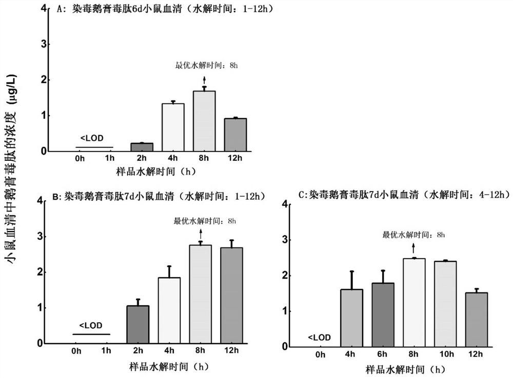
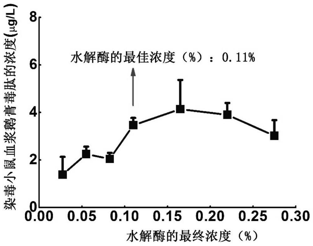
[0050] Take 200 μl of the plasma sample (sample to be tested) of the infected mice in a 1.5 ml centrifuge tube, add 20 μl of 1wt% trypsin solution (prepared by dissolving the solid trypsin in phosphate buffer solution) to make the final concentration of trypsin 0.11 wt%, mixed by vortex, and hydrolyzed in a water bath at 37°C for 8h. Take the solid-phase extraction column HLB, activate it with 1 mL of methanol and 1 mL of water in turn, let it stand for equilibrium, take the hydrolyzed plasma sample (220 μl in volume) and load it, and then use 1 mL of 5% methanol aqueous solution to wash, rinse After washing, 2 mL methanol was used for stepwise elution, and the eluate was collected and evaporated to dryness by vacuum centrifugation. After evaporating the solvent to dryness, add 10 μL of internal standar...
experiment example 1
[0083] This experimental example is used to verify the effect of protease hydrolysis on the detection of free α-amanita peptide in the sample:
[0084] Add α-amanita to the blank plasma of mice to prepare a spiked plasma (sample) containing 10 μg / L α-amanita peptide, then take 200 μl of the spiked plasma and add 20 μl of 0.25% trypsin solution , after mixing, hydrolyzed in water bath at 37°C for 0-12h. Take the plasma samples at different hydrolysis time points, prepare the samples to be tested according to the solid phase extraction method in the operation (1) of Example 1, and detect according to the UPLC-MS / MS method of the operation (3) in Example 1. Two samples were tested in parallel at the hydrolysis time point, and the test results are shown in Table 6. It can be seen from Table 6 that the addition of protease and the hydrolysis time within 12h have little effect on the detection results of free α-amanita peptide content in plasma.
[0085] Table 6 Effect of protease...
experiment example 2
[0088] This experimental example is used to verify the presence of protein-bound α-amanita peptide in plasma samples:
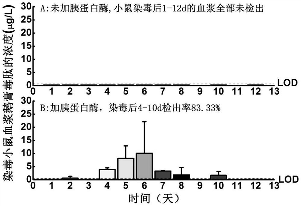
[0089]ICR male mice were injected with α-amanita toxin by intraperitoneal injection at doses of 0.33mg / kg and 0.66mg / kg, respectively, and the mice were collected at different time points within 0.5h-14d after exposure. Plasma, the method of embodiment 1 (protease hydrolysis) and comparative example 1 (unhydrolyzed) was used to detect the α-amanita peptide content in the mouse plasma collected at each time point, and the detection rate was counted. The results are shown in Table 7 shown (LOD = 0.3 μg / L).
[0090] Table 7 Detection results of different detection methods on α-amanita peptide in the plasma of the same infected mice
[0091]
[0092] As can be seen from Table 7, after the mice were exposed to α-amanita for 2d, when the method of Comparative Example 1 (not hydrolyzed) was used for detection, α-amanita could not be detected in the serum of the ...
PUM
 Abstract
Description
Claims
Application Information
 - R&D
- Intellectual Property
- Life Sciences
- Materials
- Tech Scout
- Unparalleled Data Quality
- Higher Quality Content
- 60% Fewer Hallucinations
Browse by: Latest US Patents, China's latest patents, Technical Efficacy Thesaurus, Application Domain, Technology Topic, Popular Technical Reports.
© 2025 PatSnap. All rights reserved.Legal|Privacy policy|Modern Slavery Act Transparency Statement|Sitemap|About US| Contact US: help@patsnap.com


