Preparation method of collagen/chitosan porous scaffold for tissue engineering

A tissue engineering and porous scaffold technology is applied in the field of preparation of collagen/chitosan porous scaffolds for tissue engineering. and degradability, promoting in vitro construction or in vivo regeneration

Inactive Publication Date: 2003-02-05
ZHEJIANG UNIV
View PDF0 Cites 20 Cited by
  • Summary
  • Abstract
  • Description
  • Claims
  • Application Information

AI Technical Summary

Problems solved by technology

Although collagen materials have unparalleled biocompatibility, the mechanical strength of scaffolds constructed of pure collagen is low, and the degradation rate is too fast, which cannot meet the requirements of tissue engineering scaffolds. Therefore, it is necessary to add other components to improve the collagen scaffolds. performance and cross-linked by appropriate methods to obtain a scaffold structure similar to extracellular matrix with a degradation rate that matches tissue regeneration

Method used


Image

  • Preparation method of collagen/chitosan porous scaffold for tissue engineering
  • Preparation method of collagen/chitosan porous scaffold for tissue engineering
  • Preparation method of collagen/chitosan porous scaffold for tissue engineering

Examples


Embodiment 1

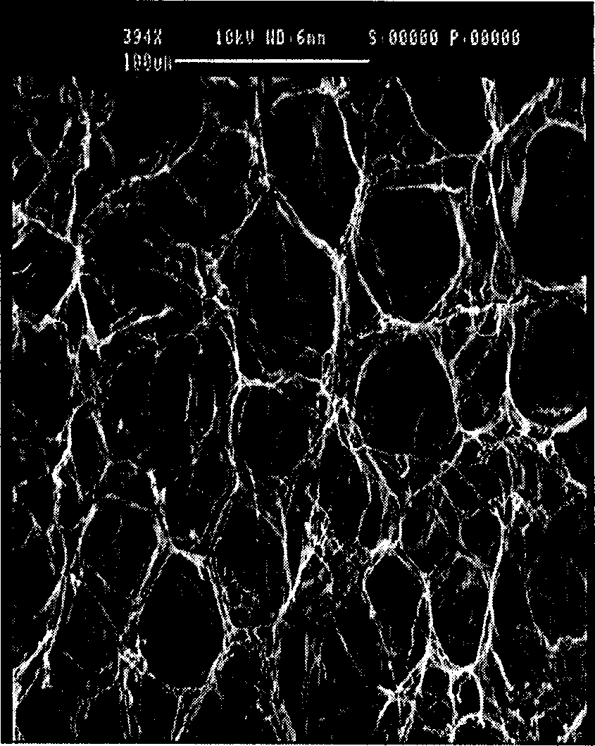
[0027] Embodiment 1: The effect of chitosan content on the microstructure of bovine tendon collagen / chitosan porous scaffold

[0028] The collagen of bovine tendon was extracted from bovine tendon by enzymatic hydrolysis and acid extraction. Prepare respectively the collagen solution that concentration is 0.5% and the chitosan solution that concentration is 0.5% with acetic acid solution, then chitosan solution and collagen solution are uniformly blended and obtain chitosan content to be (by weight) 10%, 20% respectively. %, 30%, 50% collagen / chitosan mixed solution. Freeze at -20°C for 1 hour, and after freeze-drying, porous scaffolds with different pore sizes and shapes can be prepared, see Figure 1a , Figure 1b . Figure 1a , Figure 1b These are scanning electron microscope (SEM) pictures of collagen / chitosan scaffolds with chitosan content of 10% and 50%, respectively.

Embodiment 2

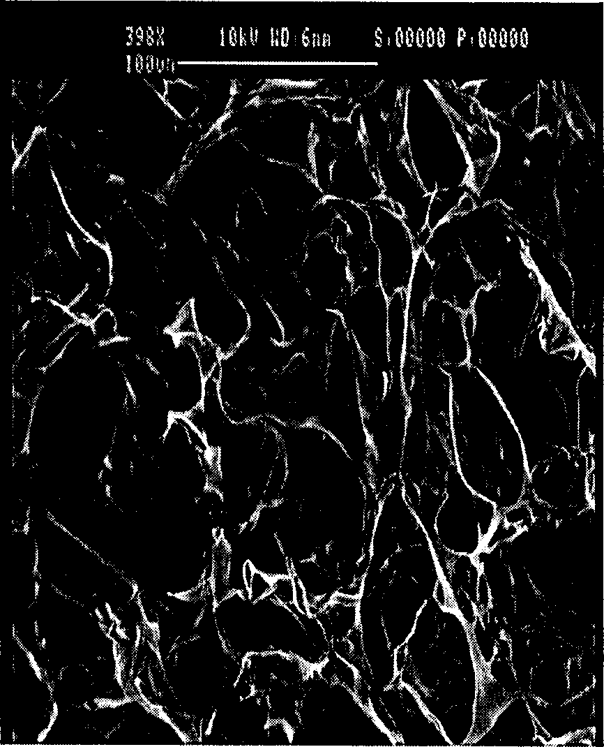
[0029] Embodiment 2: the impact of freezing temperature on the microstructure of bovine tendon collagen / chitosan porous scaffold

[0030] The collagen of bovine tendon was extracted from bovine tendon by enzymatic hydrolysis and acid extraction. With the acetic acid solution of 0.5M, preparation concentration is respectively the collagen solution of 0.5% and the chitosan solution of 0.5%, then the chitosan solution and the collagen solution are uniformly blended to obtain chitosan content and be (by weight) 10% collagen / Chitosan mixed solution. Then freeze at -50°C and -20°C for 1 hour respectively, and porous scaffolds with different pore sizes can be prepared after freeze-drying, see Figure 2a , Figure 2b . Figure 2a , Figure 2b SEM photos of porous scaffolds prepared by freezing at -50°C and -20°C, respectively.

Embodiment 3

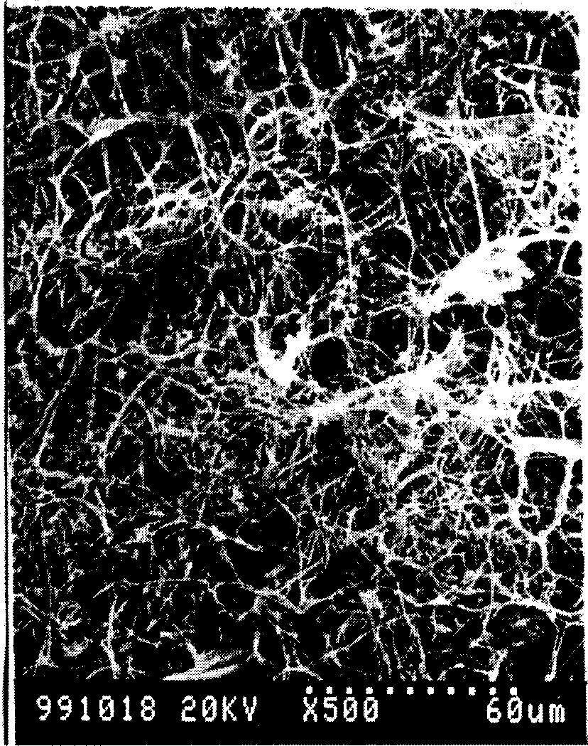
[0031] Embodiment 3: the crosslinking of bovine tendon collagen / chitosan porous scaffold

[0032] The collagen of bovine tendon was extracted from bovine tendon by enzymatic hydrolysis and acid extraction. A 0.5% acetic acid solution was used to prepare a 0.5% collagen solution and a 0.5% chitosan solution, and then the chitosan solution and the collagen solution were uniformly mixed to obtain a chitosan content of 10% (by weight). collagen / chitosan mixed solution. Using the freeze-lyophilization method, freeze at -20°C for 1 hour, and then freeze-dry in a freeze dryer for 24 hours. The freeze-dried collagen / chitosan scaffold was vacuum dry-heated and cross-linked in a vacuum oven at 105° C. for 24 hours to obtain a dry-heat-crosslinked bovine tendon collagen / chitosan porous scaffold. The bovine tendon collagen / chitosan porous scaffold obtained through vacuum dry heat crosslinking was soaked in 100ml of 0.5M acetic acid solution for 1 hour, and then crosslinked with 0.05% to...


PUM


Abstract

The present invention relates to a porous scaffold with controllable microstructure, proper degradation rate and good biological compatibility prepared by using natural biological materials of collagen and chitosan as raw material and adopting the processes of freeze-freeze drying, further vacuum drying and heating and cross-linking of aldehyde compound or carbodiimide compound. The biological compatibility of said invented collagen/chitosan scaffold is good, its degradation rate can be controlled, and its raw material source is extensive and its cost is low, and its reproducibility is good. Said scaffold can be extensively used in the field of tissue engineering technology.

Description

technical field [0001] The invention relates to a preparation method of a collagen / chitosan porous support for tissue engineering, in particular to a preparation method of a histocompatible three-dimensional porous support for guiding tissue and organ regeneration. Background technique [0002] Tissue engineering is an interdisciplinary research topic involving medicine, chemistry, biology, materials science, etc., and the construction of three-dimensional scaffolds is one of the keys. Only by constructing a scaffold with a specific microstructure, excellent mechanical properties, suitable degradation properties, and good biocompatibility can it effectively promote cell adhesion and growth, and then guide the regeneration of tissues and organs. [0003] Collagen is the main component of connective tissue in mammals, constituting about 30% of the protein in the human body and 72% of the dry weight in the skin. There are 19 types of collagen, the mos...

Claims


Application Information

Patent Timeline
Patent Type & Authority Applications(China)
IPC IPC(8): A61L31/04
Inventor 高长有马列王登勇毛峥伟沈家骢韩春茂
Owner ZHEJIANG UNIV
Why Eureka
Try Eureka