Method for kidney disease detection by protein profiling

A protein map and kidney disease technology, applied in the field of kidney disease complications, can solve problems such as inability to detect quickly, disappointing effectiveness, and unrepeatable technology

Inactive Publication Date: 2004-08-11
MONASH UNIV
View PDF2 Cites 12 Cited by
  • Summary
  • Abstract
  • Description
  • Claims
  • Application Information

AI Technical Summary

Problems solved by technology

[0010] However, previous attempts to use urine protein profiling for clinical diagnostic purposes have been largely disappointing in their effectiveness, in part because these techniques are not reproducible, sensitive, and rapid

Method used


Image

  • Method for kidney disease detection by protein profiling
  • Method for kidney disease detection by protein profiling
  • Method for kidney disease detection by protein profiling

Examples


Embodiment 1

[0139] Example 1: Size Exclusion Chromatography of Human Serum Albumin (HSA)

[0140] The distribution of albumin in urine was analyzed using urine samples provided by normal healthy volunteers.

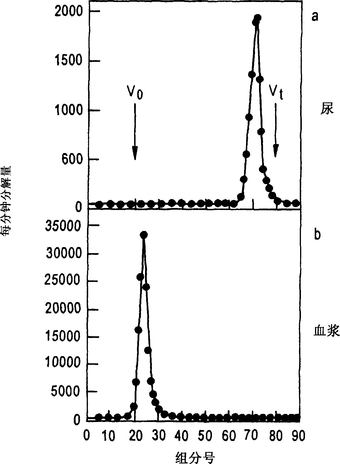
[0141] Will 3 H[HAS] (human serum albumin) was injected into healthy volunteers, urine samples and plasma were collected, and they were analyzed by size exclusion chromatography with G-100 column. The column was eluted with PBS (pH=7.4) at 4°C at a rate of 20 mL / hr. The external water volume (Vo) of the column was determined with blue dextran T2000, while the total volume was determined with tritiated water.

[0142] Tritium radioactivity in 1 mL of aqueous samples was determined with 3 mL of scintillator and detected on a Wallac 1410 liquid scintillation counter (Wallal Turku, Finland).

[0143] Figure 2 shows the distribution of albumin in urine and plasma.

Embodiment 2

[0144] Example 2: Albumin secretion in normal healthy volunteers and diabetic patients

[0145] Will use in embodiment 1 3 H[HAS] was injected into normal healthy volunteers and diabetic patients. Collect urine sample, determine with the method of embodiment 1 3 H[HAS].

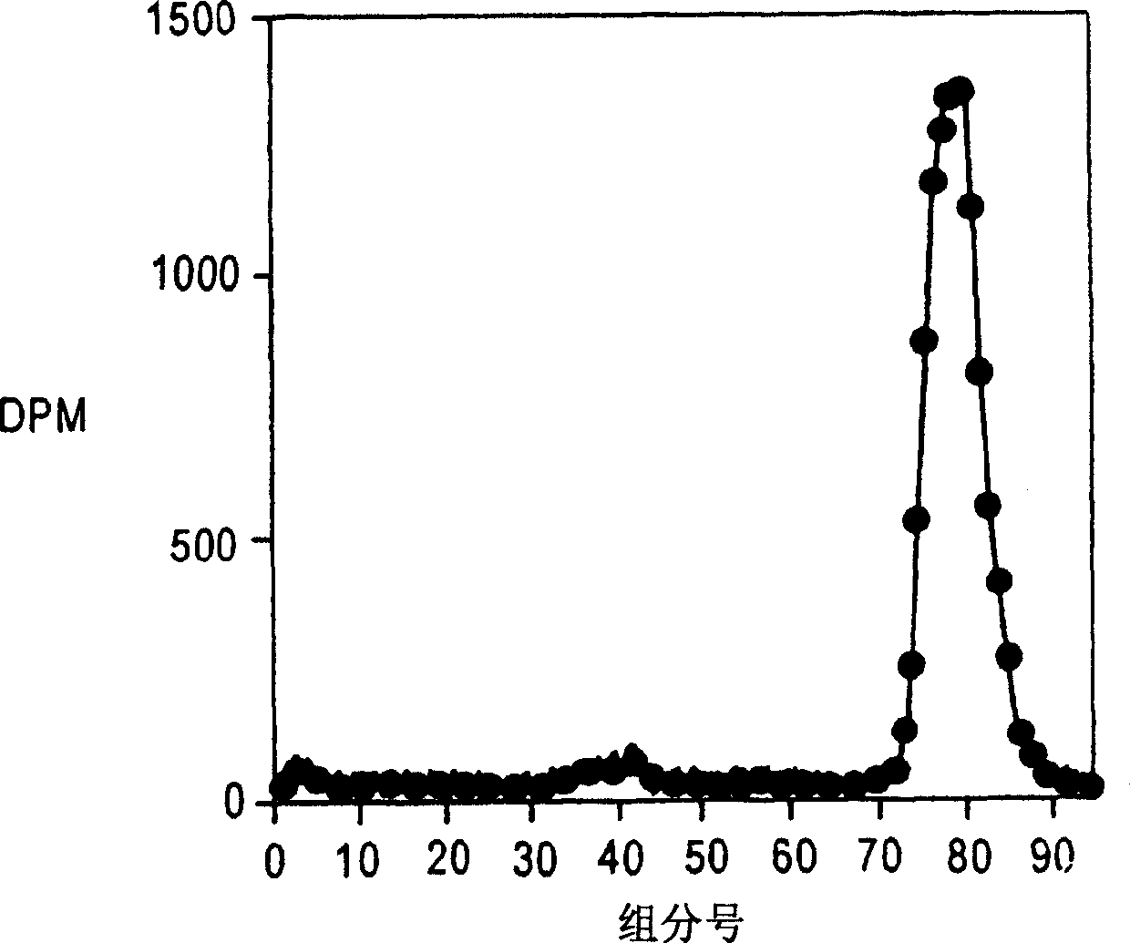
[0146] Normal healthy volunteers ( FIG. 3 ) were shown to secrete fragmented albumin on size exclusion chromatography performed as in Example 1 .

[0147] Diabetic patients (Figure 4) showed the presence of essentially full-length and fragmented albumin on size exclusion chromatography. However, albumin secretion rates detected with these methods were of the order of 5 μg / min (control) and 1457 μg / min (diabetic patients).

Embodiment 3

[0148] Example 3: Determination of intact albumin and intact / modified albumin on HPLC

[0149] Urine samples were collected from normal healthy volunteers, normoalbuminuric diabetics and macroalbuminuric patients. Collect midstream urine into a 50 μL urine sample container. Urine samples were frozen until further use. Urine samples were centrifuged at 5000g prior to HPLC analysis.

[0150] Urine samples were analyzed by HPLC with a hydrophobic column Zorbax 300SB-CB (4.6mm×150mm). Utilize a 50 µL sample loop.

[0151] The sample was eluted from the column using the following conditions

[0152] Solvent A Water 1% trifluoroacetic acid

[0153] Solvent B 60% acetonitrile, 0.09% TFA

[0154]Solvent A2 99.96 > 00.00: 49.58 minutes

[0155] Pressure 9.014M Pascal (~1100psi).

[0156] Solvent B2 0.04>100.0: 49.58 minutes

[0157] Pressure 7.154M Pascal

[0158] The wavelength used is 214 nm.


PUM


Abstract

The invention provides a method of generating and analysing a urinary protein fragmentation profile, in terms of the size, and sequence of particular fragments derived from intact filtered proteins together with the position where enzymes scission occurs along the protein polypeptide chain is characteristic of the diseased state of the kidney. With the recognition that filtered proteins are degraded during renal passage, the methods described in this application will be able to detect protein fragments derived from proteins generated by non-renal disease. The urinary analysis of these filtered proteins would currently not detect the intact form of these proteins. Therefore, a method as described to detect and analyse fragments resulting from degradation during renal passage that will be able to detect the seriousness of the disease.

Description

[0001] Related application reference [0002] This application claims priority to US Provisional Application 60 / 301,251, filed June 28,2001. field of invention [0003] The present invention relates to an improved method of detecting early stage renal disease and / or renal complications of the disease, especially diabetic complications. Background of the invention [0004] Excessive protein, such as albumin, in the urine indicates kidney disease. Diabetic nephropathy is one such disease. [0005] Applicants have discovered that proteins, including albumin, are normally secreted as a mixture of native proteins and fragments that are specifically produced during renal passage (Osicka T.M et al., Nephrology, 2:199-212( 1996)). During renal passage, proteins are severely degraded by the posterior glomerulus (basement membrane) including tubular cells. Lysosomes in tubular cells are responsible for degrading proteins secreted during renal passage. Figure 1 shows the progressi...

Claims


Application Information

Patent Timeline
Patent Type & Authority Applications(China)
IPC IPC(8): C12Q1/37G01N27/447G01N27/62G01N27/64G01N30/26G01N30/34G01N30/74G01N30/88G01N33/68
CPCG01N33/6803G01N33/6827Y10T436/24C12Q1/37
Inventor 韦恩·D·康珀
Owner MONASH UNIV
Why Eureka
Try Eureka