
Tumor marker, monoclonal antibodies and methods of use thereof

a monoclonal antibody and tumor technology, applied in the field of tumor markers and monoclonal antibodies, can solve the problems of insufficient specificity and sensitivity of most biomarkers commonly used in clinical practice, insufficient specificity and sensitivity to unambiguously distinguish tumors, and insufficient specificity of ca 125 measurement to be used to screen all women

Inactive Publication Date: 2019-07-23
EXTERNAUTICS
View PDF1 Cites 0 Cited by
  • Summary
  • Abstract
  • Description
  • Claims
  • Application Information

AI Technical Summary

Benefits of technology

[0029]Scientific literature has reported that FAT1 has a predominant tumor-suppressive effect. The human FAT1 gene is homozygously deleted in 23% of oral cancer cell lines and in 80% of primary oral cancer cases and FAT1 mRNA expression is repressed in oral cancer cell lines due to homozygous deletion and / or promoter CpG hypermethylation (14). Loss of heterozygosity (LOH) of the FAT1 gene occurs in 42% of low-grade diffuse astrocytoma and 63% of glioblastoma multiforme (15). FAT1 mRNA level in ductal carcinoma in situ is significantly higher than that in invasive breast cancer and FAT1 knockdown promotes progression from ductal carcinoma in situ to invasive breast cancer, indicating that lower FAT1 expression is associated with aggressive breast cancers (16).
[0030]Available in vitro and in vivo studies reported that FAT1 depletion leads to markedly accelerated cell growth and proliferation, while expression of FAT1 robustly suppresses tumor growth. These growth-suppressive effects are abrogated when mutations observed in tumors are present (17). Recent data now implicates FAT1 mutation as a driver of Wnt activation in many cancers, through the involvement the b-catenin. As other cadherins, FAT1 can bind to b-catenin and limits its translocation to the nucleus. Mutations in FAT1 intracytoplasmic domain result in a loss of this ability to regulate b-catenin. Consequently, loss of FAT1 in cells activates the Wnt signaling pathway, unleashing b-catenin-dependent transcriptional activity and upregulating pro-growth wnt transcriptional targets. Consistent with this, primary cancer samples with FAT1 alterations are characterized by significant enhancement of Wnt signaling. The growth-suppressive functions of FAT1 are mediated by its intracytoplasmic, b-catenin binding domain, but the extracellular domain also mediates cell adhesion, which may be a secondary mechanism by which FAT1 loss promotes tumor growth.
[0037]Antibodies generated against FAT1 are able to specifically recognize the protein on the surface of different cancer cell lines. In particular, experiments carried out with two specific anti-FAT1 antibodies showed that the latter, upon surface binding, are efficiently internalized by cancer cells, indicating that they are suitable for the generation of antibody-drug conjugate (ADC). In addition one of these antibodies, when administered to mice bearing tumor, is able to reduce cancer growth.
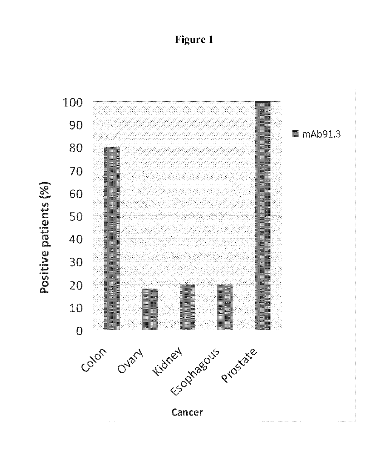
[0038]Finally, antibodies generated against FAT1 are also able to detect the protein in exosomes from cancer cells, indicating that the marker can be detected in biological fluids of oncologic patients. The exosomes carrying FAT1 can be exploited as tools for the development of vaccines to prevent or treat cancers affecting colon, ovary, esophagus, kidney, and prostate.
[0045]A further aspect of this invention is a method of screening a tissue sample for malignancy, which comprises determining the presence in said sample of the above-mentioned tumor marker. This method includes detecting either the marker protein, e.g. by means of labeled monoclonal or polyclonal antibodies that specifically bind to the target protein, or the respective mRNA, e.g. by means of polymerase chain reaction techniques such as RT-PCR. The methods for detecting proteins in a tissue sample are known to one skilled in the art and include immunoradiometric, immunoenzymatic or immunohistochemical techniques, such as radioimmunoassays, immunofluorescent assays or enzyme-linked immunoassays. Other known protein analysis techniques, such as polyacrylamide gel electrophoresis (PAGE), Western blot or Dot blot are equally suitable. Preferably, the detection of the protein marker is carried out with the immune-histochemistry technology, particularly by means of High Through-Put methods that allow the analyses of the antibody immune-reactivity simultaneously on different tissue samples immobilized on a microscope slide. Briefly, each Tissue Micro Array (TMA) slide includes tissue samples suspected of malignancy taken from different patients, and an equal number of normal tissue samples from the same patients as controls. The direct comparison of samples by qualitative or quantitative measurement, e.g. by enzymatic or colorimetric reactions, allows the identification of tumors.
[0074]Therefore, in a further aspect the invention provides the use of antibodies to FAT1 protein for the preparation of a therapeutic agent for the treatment of proliferative diseases of colon, ovary, esophagus, kidney and prostate tissues. For use in therapy, the antibodies can be formulated with suitable carriers and excipients, optionally with the addition of adjuvants to enhance their effects.

Problems solved by technology

Currently, although an abnormal tumor marker level may suggest cancer, this alone is usually not enough to accurately diagnose cancer and their measurement in body fluids is frequently combined with other tests, such as a biopsy and radioscopic examination.
Most biomarkers commonly used in clinical practice do not reach a sufficiently high level of specificity and sensitivity to unambiguously distinguish a tumor from a normal state.
For example, prostate-specific antigen (PSA) levels are often used to screen men for prostate cancer, but this is controversial since elevated PSA levels can be caused by both prostate cancer or benign conditions, and most men with elevated PSA levels turn out not to have prostate cancer.
So far, CA 125 measurement is not sensitive or specific enough to be used to screen all women for ovarian cancer.
However so far different reasons are delaying their use in the common clinical practice, including the higher analysis complexity and their expensiveness.
However, this scientific progress in the molecular oncology field has not been paralleled by a comparable progress in cancer diagnosis and therapy.
However, these treatments are effective only at initial phases of the disease and in particular for solid tumors of epithelial origin, as is the case of colon, breast, ovary, prostate and others, while they are not effective for distant recurrence of the disease.
However, so far many cancer therapies had limited efficacy due to severity of side effects and overall toxicity.
However, at present the number of therapeutic antibodies available on the market or under clinical studies is very limited and restricted to specific cancer classes.
In summary, available screening tests for tumor diagnosis are uncomfortable or invasive and this sometimes limits their applications.
Moreover tumor markers available today have a limited utility in clinics due to either their incapability to detect all tumor subtypes of the defined cancers types and / or to distinguish unambiguously tumor vs. normal tissues.
Similarly, licensed monoclonal antibodies combined with standard chemotherapies are not effective against the majority of cases.
Although often successful, these approaches have several limitations and often, do not provide firm indications on the association of protein markers with tumor.
A first limitation is that, since frequently mRNA levels not always correlate with corresponding protein abundance (approx.
A second limitation is that neither transcription profiles nor analysis of total protein content discriminate post-translation modifications, which often occur during oncogenesis.
As a consequence, large scale studies generally result in long lists of differentially expressed genes that would require complex experimental paths in order to validate the potential markers.
However, large-scale genomic / proteomic studies reporting novel tumor markers frequently lack of confirmation data on the reported potential novel markers and thus do not provide solid demonstration on the association of the described protein markers with tumor.

Method used


Image

  • Tumor marker, monoclonal antibodies and methods of use thereof
  • Tumor marker, monoclonal antibodies and methods of use thereof
  • Tumor marker, monoclonal antibodies and methods of use thereof

Examples


example 1

and Confirmation of FAT1 Over-Expression in Cancer by Immune-Histochemistry

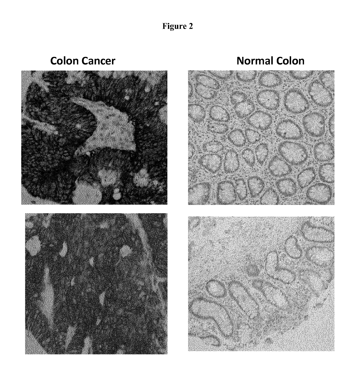
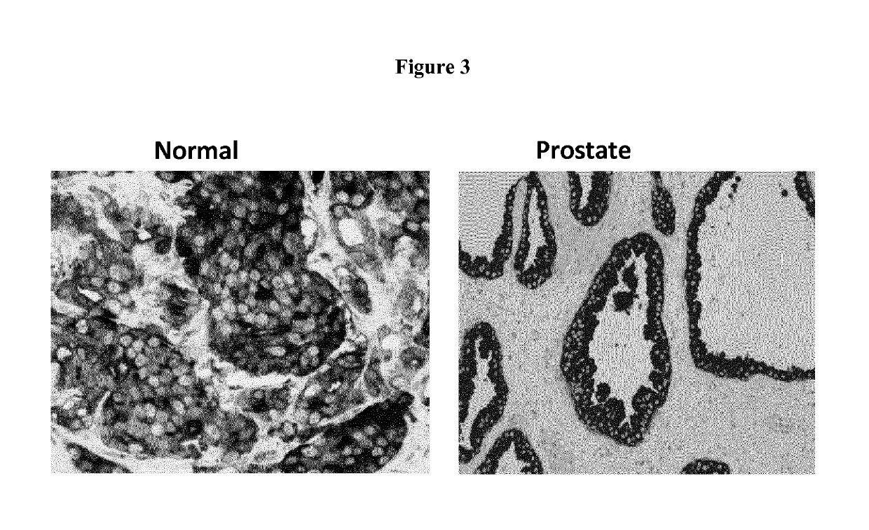
[0106]A proprietary collection of polyclonal and monoclonal antibodies raised against human recombinant proteins was used to identify proteins over-expressed in cancer by immune-histochemistry (IHC). The antibody library was used screen clinical samples by Tissue Micro Array (TMA), a miniaturized immuno-histochemistry technology suitable for HTP analysis that allows to analyse the antibody immuno-reactivity simultaneously on different tissue samples immobilized on a microscope slide. Since the TMAs include both tumor and healthy tissues, the specificity of the antibodies for the tumors can be immediately appreciated. The use of this technology, differently from approaches based on transcription profile, has the important advantage of giving a first hand evaluation on the potential of the markers in clinics. Conversely, since mRNA levels not always correlate with protein levels (approx. 50% correlation), studi...

example 2

n and Localization of FAT1 Protein in Cancer Cells

[0114]The expression and localization of FAT 1 protein in cancer cells was investigated using an anti-FAT1 monoclonal antibody to confirm that FAT1 is expressed by cancer cell lines derived from the human cancers found positive in the IHC screening. Moreover, FAT1 surface localization surface was verified to confirm that FAT1 could be exploited as therapeutic target of anti-cancer therapies. FAT-1 affinity ligands, such as small molecules or antibodies, able to recognize the protein on the cell surface can be developed as novel therapeutic antigens. Finally the association of FAT1 with cell-derived exosomes was investigated to assess whether FAT1 is released by cancer cells and could be detected in patients' biological fluids. This property would allow developing non-invasive diagnostic assays based on FAT1 detection. Moreover, exosomes could be exploited in vaccines based on the elicitation of antibody and T cell response against FA...

example 3

ion of the Specificity of the Anti-FAT1 Antibody by Gene Silencing

[0126]The specifity of the anti-FAT1 monoclonal antibody mAb91.3 showing selective cancer reactivity in IHC was further verified by specific FAT1 knock-down in FAT1 positive tumor cell lines by the siRNA technology and the knock-down of FAT1 expression was monitored at transcriptional and protein level.

[0127]Method

[0128]FAT1 expression was silenced in the HCT15 colon cell lines with two FAT1-specific siRNAs (10 nM) (whose target sequences are reported in the Table) using the HiPerfect transfection reagent (QIAGEN) following the manufacturer's protocol. As control, cells treated with equal concentrations of irrelevant siRNA (scrambled siRNA) were analysed in parallel. At different time points (ranging from 24 to 72 hours) post transfection, the reduction of gene transcription was assessed by quantitative RT-PCR (Q-RT-PCR) on total RNA, by evaluating the relative marker transcript level, using the beta-actin, GAPDH or M...


PUM

PropertyMeasurementUnit
weightaaaaaaaaaa
weightaaaaaaaaaa
diameteraaaaaaaaaa

Abstract

Newly identified proteins as markers for the detection of colon, ovary, kidney, esophagus and prostate tumors, or as therapeutic targets for their treatment; affinity ligands and particularly antibodies capable of selectively interacting with the tumor markers and methods for tumor diagnosis and therapy using such antibodies.

Description

[0001]This application is a U.S. national stage of PCT / EP2014 / 062419 filed on 13 Jun. 2014, which claims priority to and the benefit of European Application No. EP13172028.6 filed on 14 Jun. 2013, the contents of which are incorporated herein by reference in their entireties.[0002]The present invention relates to the use of the FAT Tumor Suppressor Homolog 1 (FAT1) protein or encoding polynucleotides as markers for the detection of tumors, or as targets for their treatment, particularly of tumors affecting colon, ovary, prostate, esophagus and kidneys. Also provided are affinity ligands capable of selectively interacting with the newly identified markers, as well as methods for tumor diagnosis and therapy using such ligands.BACKGROUND OF THE INVENTION[0003]Tumor Markers (or Biomarkers)[0004]Tumor markers are substances that can be produced by tumor cells or by other cells of the body in response to cancer. In particular, a protein biomarker is either a single protein or a panel of d...

Claims


Application Information

Patent Timeline
Patent Type & Authority Patents(United States)
IPC IPC(8): C07K16/30A61K39/00G01N33/574A61K47/68
CPCG01N33/57484A61K47/6843C07K16/30C07K16/3046C07K16/3069G01N33/57419A61K39/0011C07K2317/77A61K2039/55505C07K2317/21C07K2317/24C07K2317/34C07K2317/54C07K2317/55C07K2317/565C07K2317/622C07K2317/626
Inventor GRIFANTINI, RENATAPILERI, PIEROCAMPAGNOLI, SUSANNAGRANDI, ALBERTOPARRI, MATTEOBOQUAN, JIN
Owner EXTERNAUTICS
Learn More