Methods for quantitating DNA using digital multiple displacement amplification

a technology of multiple displacement and quantitation method, applied in the field of methods for quantitating nucleic acids, can solve the problems of insufficient basis on which inability to distinguish composite genomes assembled from metagenomic data, and inability to accurately model the response of single-gene studies, etc., to reduce the impact of contaminates and reagent costs, improve the reliability and throughput of single-cell wga, and eliminate background amplification

Active Publication Date: 2019-11-26
THE BROAD INST INC +1
View PDF6 Cites 2 Cited by
  • Summary
  • Abstract
  • Description
  • Claims
  • Application Information

AI Technical Summary

Benefits of technology

[0012]Technological advances improve the reliability and throughput of single-cell WGA and sequencing. The contamination of reagents with bacterial DNA that plagued early efforts in single-cell microbial genomics is now well-understood, and effective measures to quantify, control, and eliminate such contamination are disclosed herein. The elimination of background amplification not only yields clean datasets, but also allows the facile application of screens for successful WGA reactions. The move to lower-volume WGA reaction platforms further reduces the impact of contaminates and WGA reagent costs, opening the way to massive increases in reaction throughput.
[0013]The key drivers of new technology for single-cell genomics are advances in throughput, integration of isolation and WGA with selection and sequencing procedures, improvements in more uniform representation of template sequences in the WGA product, fewer artifact sequences such as chimeras, expanded sample / cell type compatibility, and contextualization of single-cell genome data by in-line collection of other information about the processed cells (eg phenotyping, imaging, RNA, protein, metabolite analyses). The ability to obtain sequence data from individual cells from a known biological setting or with a known history (e.g. interaction with other cells prior to the analysis) is especially informative. Because sample preparation costs already dominate single-cell microbial genomics workflows today, the most impactful advances will scale to large numbers of cells at reasonable cost.

Problems solved by technology

Single-gene studies, metagenomic assemblies, and the genome sequences of a limited number of cultured isolates are not a sufficient basis on which to accurately model the responses of natural microbial networks or engineer the function of artificial communities.
For example, gene catalogs and composite genomes assembled from metagenomic data do not presently distinguish between genes that are tightly coupled within the context of the same organism and genes that are coupled across different organisms.
This is a critical limitation, because only gene products encoded by the same organism can freely come into contact with one another to form complexes, drive signaling pathways, or carry out multi-step enzymatic transformations of diffusible substrates at maximum efficiency.
Such high fold-amplification from sub-nanogram samples (Dean, et al., 2002, Proceedings of the National Academy of Sciences 99: 5261) and individual bacteria (Raghunathan, et al., 2005, Applied and Environmental Microbiology 71: 3342-3347) with good representation of the genome were first achieved by the multiple displacement amplification (MDA) whole-genome amplification (WGA) chemistry, but produced material with undesirable characteristics such as uneven representation and dislocated sequences.
Given the concentration of contaminating fragments present in commercial WGA reagents (varying from 5 to 50 fragments per reaction microliter in the enzyme alone), volume reduction by itself does not necessarily eliminate reagent contamination (Blainey & Quake, 2011, Nucleic Acids Res 39: e19).
QPCR is insufficiently sensitive since only contaminant molecules with an intact sequence locus matching specific PCR primers can be detected.

Method used


Image

  • Methods for quantitating DNA using digital multiple displacement amplification
  • Methods for quantitating DNA using digital multiple displacement amplification
  • Methods for quantitating DNA using digital multiple displacement amplification

Examples


example 1

dic Droplets

[0095]Fabricating the Microfluidic Devices.

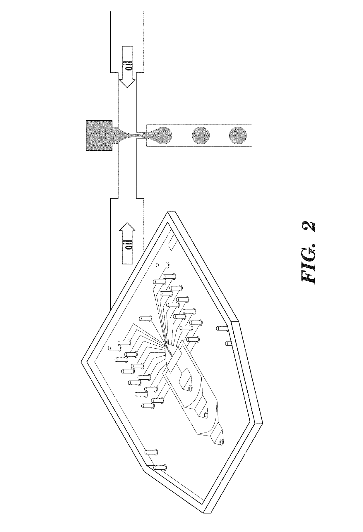
[0096]Microfluidic devices were constructed using standard PDMS soft lithography (McDonald J C, et al. (2000) Fabrication of microfluidic systems in poly(dimethylsilox-ane). Electrophoresis 21:27-40) with 18 micron SU8-on-Si wafer molds formed using transparency masks from FineLine Imaging. After curing, the PDMS device is adhered to glass for 2 hours at 80° C.

[0097]Multiple Displacement Reaction.

[0098]The MDA reactions were prepared according to protocol (Qiagen Repli-G Midi Kit) with Lambda DNA added to 5e-5, 5e-4, and 5e-3 ng / uL in the standard samples. Each reaction was supplemented with 0.3% Tween-20 and Evagreen prior to drop formation. Drop and bulk samples incubated at 30° C. for 16 h, and 65° C. for 20 m. Half of the post-incubation drop samples were converted to bulk with half volume 1H,1H,2H,2H-Perfluoro-1-octanol (Sigma Aldrich) and half of the post-incubation bulk samples were made into drops.

[0099]Forming Monodispe...

example 2

Matrices

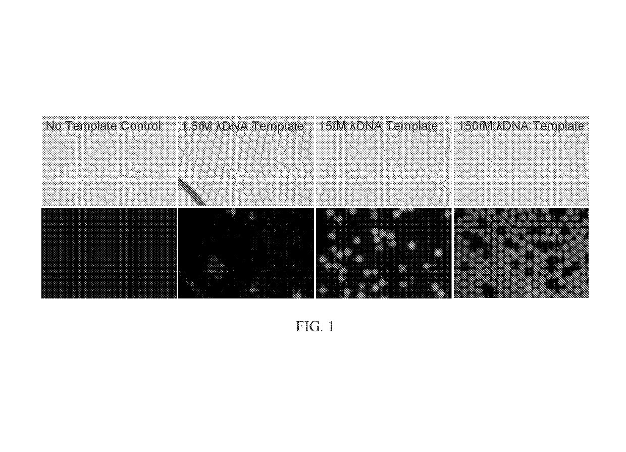
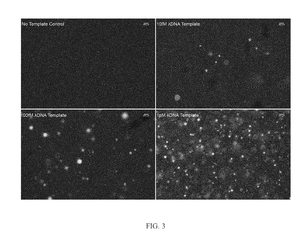
[0108]FIGS. 3-6 depict MDA reactions in a gel matrix with lambda phage genome as a template, which is representative of single genome amplification as well as single cell analysis.

[0109]FIG. 3 depicts digital MDA products at 30° C. after 16 hours stained with 500 nM SYTOX Orange dye in 7% 4-Arm PEG Acrylate-Dithiol hydrogels. The initial Lambda Phage DNA template concentrations were 10 fM, 100 fM, and 1 pM. (or convert to mass / volume, 1.3e-5 ng / μL, 1.3e-4 ng / μL, and 1,3e-3 ng / μL).

[0110]FIG. 4 depicts a multiple displacement amplification in gel. The figure depicts the number of DNA amplification colonies observed vs. initial lambda phage DNA concentration in 4-arm PEG acrylate gel after 16 hours of multiple displacement amplification. DNA was stained with 500 nM SYTOX Orange.

[0111]FIG. 5 depicts 216 bp PCR products using Vent(exo-) polymerase (a polymerase with strong strand displacement) after 40 thermal cycles stained with 500 nM SYTOX Orange dye in 7% 4-Arm PEG Acrylate-D...

example 3

and Methods

[0113]4-Ami PEG Hydrogel. Hydrogel components, including 4-arm PEG acrylate (MW 10,000) and HS-PEG-SH (MW 3,400), were obtained from Layson Bio. For every 25 μl of 10% weight percentage cross-linked hydrogel, 1.6 mg of 4-arm PEG acrylate and 1.1 mg of HS-PEG-SH were dissolved in PBS buffer (Invitrogen). It was briefly vortexed and centrifuged to ensure mixing and it was allowed to sit for 10 minutes while the hydrogel components cross-linked through the reaction between the thiol and acrylate groups

[0114]Polymerase Chain Reaction (PCR). The primers used for PCR on purified λ DNA (48 kbp, NEB) were purchased from IDT. Primer F 5′: CGGCAAACGGGAATGAAACGCC. Primer R 5′: Alyson. A 25 μl hydrogel PCR reaction consisted of 2 U of VentR(exo-) polymerase (NEB), 1× ThermoPol Reaction Buffer (NEB), 0.4 mM dNTP (NEB), 1 μM Primers, 5% DMSO (Sigma), 0.5 mg / ml BSA(NEB), 1.6 mg 4-arm PEG acrylate in PBS, 1.1 mg HS-PEG-SH in PBS, and λDNA template (NEB) of various concentrations. The 25 ...


PUM

PropertyMeasurementUnit
waiting timeaaaaaaaaaa
concentrationsaaaaaaaaaa
volumeaaaaaaaaaa

Abstract

The present invention relates to methods of quantifying nucleic acids involving (a) contacting a sample to be tested with whole-genome amplification (WGA) reaction components and a dye molecule to form a reaction sample, wherein the dye molecule detects the nucleic acid; (b) partitioning the reaction sample wherein each partitioned reaction sample corresponds to a single reaction; (c) allowing the reaction to occur in the partitioned reaction sample and (d) determining the number of partitioned reaction samples having the dye molecule, wherein the dye molecule indicates the presence of the nucleic acid; thereby quantifying the nucleic acid in the sample to be tested.

Description

RELATED APPLICATIONS AND INCORPORATION BY REFERENCE[0001]This application is a continuation-in-part of international patent application Serial No. PCT / US2014 / 028941, filed Mar. 14, 2014 and published as PCT Publication No. WO 2014 / 153071 on Sep. 23, 2014 and which claims benefit of and priority to U.S. provisional patent application Ser. No. 61 / 782,826 filed Mar. 14, 2013.FEDERAL FUNDING LEGEND[0002]This invention was made with government support under Grant No. R01HG004863 awarded by the National Institutes of Health. The government has certain rights in the invention.[0003]The foregoing application, and all documents cited therein or during their prosecution (“appln cited documents”) and documents cited or referenced herein (“herein cited documents”), and all documents cited or referenced in herein cited documents, together with any manufacturer's instructions, descriptions, product specifications, and product sheets for any products mentioned herein or in any document incorporate...

Claims


Application Information

Patent Timeline
Patent Type & Authority Patents(United States)
IPC IPC(8): C12P19/34C12Q1/6851
CPCC12Q1/6853C12Q1/6851C12Q2531/119C12Q2565/629
Inventor BLAINEY, PAULXU, LIYI
Owner THE BROAD INST INC
Why Eureka
Try Eureka