Methods and tools for detecting interactions in eukaryotic cells using microtubule structures and dynamics

a microtubule and eukaryotic cell technology, applied in the field of methods and tools for detecting interactions in eukaryotic cells using microtubule structures and dynamics, can solve the problems of affecting the accuracy of eukaryotic cells, so as to achieve high throughput methods, easy detection, and high sensitivity protein partners

Active Publication Date: 2020-03-17
INST NAT DE LA SANTE & DE LA RECHERCHE MEDICALE (INSERM) +1
View PDF8 Cites 0 Cited by
  • Summary
  • Abstract
  • Description
  • Claims
  • Application Information

AI Technical Summary

Benefits of technology

[0013]Surprisingly, it has been found that the prey, when interacting with the bait, becomes also localized along microtubules which makes it thus easily detectable. The invention presents the following advantages: (i) a high sensitivity protein partners detection system which can be automatized and which is thus suitable to high throughput methods. (ii) the detection of bait / prey interaction is performed in a living cell environment which preserves any post-translationnal modifications of the partners.

Problems solved by technology

The function of a number of proteins remains elusive, which slows down understanding of fundamental aspects of cell mechanisms and impedes advance in diagnostic and treatment of many diseases and in personalized medicine.
One of the main obstacles to better understand a given protein's functions is that proteins generally interact with many other partners (proteins, nucleic acids, sugars, lipids .
. . ) and many of these interactions are weak and transient, albeit of critical importance.
Each of these methods suffers from numerous drawbacks.
Two-hybrid systems can detect protein interactions in living cells but require two or more chimeric proteins and are unable to monitor rapid biochemical events taking place in the cells (Lievens et al., Mammalian two-hybrids come of age.
In addition, weak and transient interactions are not detected.
The detection of weak interactions is also challenging due to the low signal to noise ratio (Lam et al., Improving FRET dynamic range with bright green and red fluorescent proteins.

Method used


Image

  • Methods and tools for detecting interactions in eukaryotic cells using microtubule structures and dynamics
  • Methods and tools for detecting interactions in eukaryotic cells using microtubule structures and dynamics
  • Methods and tools for detecting interactions in eukaryotic cells using microtubule structures and dynamics

Examples


example 1

rotocol for High-Throughput Analysis and Detection System

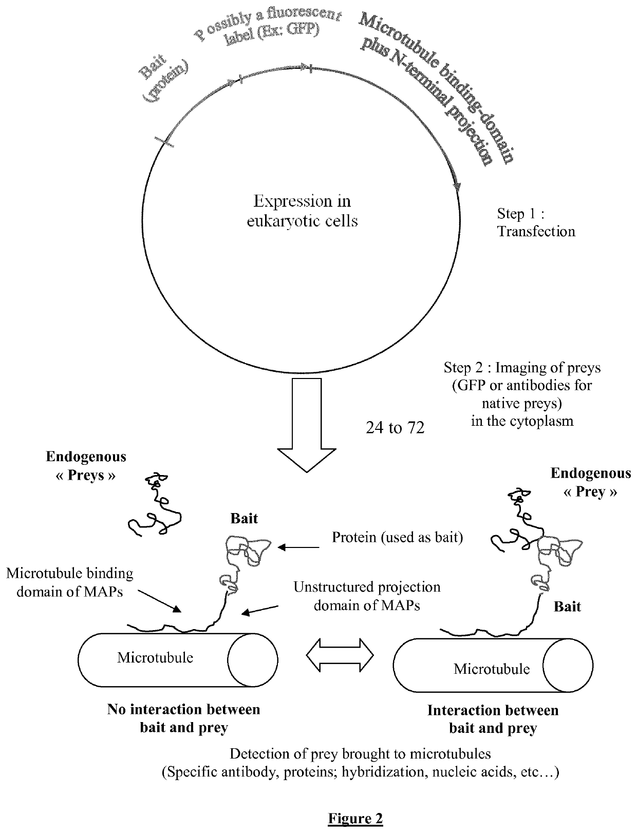
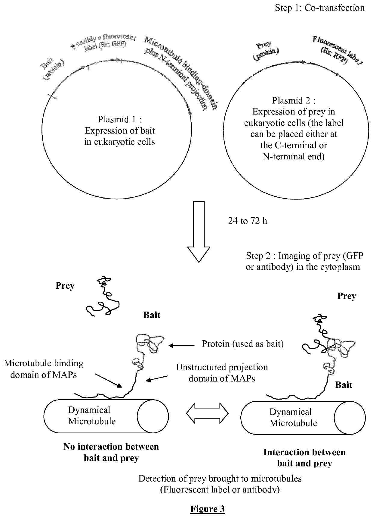
[0250]1) The cDNA sequences encoding for a known protein or a polypeptide chain called “bait” are inserted in a plasmid which directs, within the cell the synthesis of this bait fused to a microtubule binding protein (tau, MAP2) or domain linked to a projection domain to favor bait's accessibility to prey. The bait can additionally be fused to fluorescent label like a fluorescent protein (GFP, RFP, . . . ).

[0251]2) cDNA sequences encoding the “prey”, a polypeptide or protein, are inserted in a plasmid to direct its expression within cell. The prey is possibly fused to a fluorescent label such as a fluorescent protein (i.e. GFP or XFP) for its detection in living cells via fluorescence microscopy.

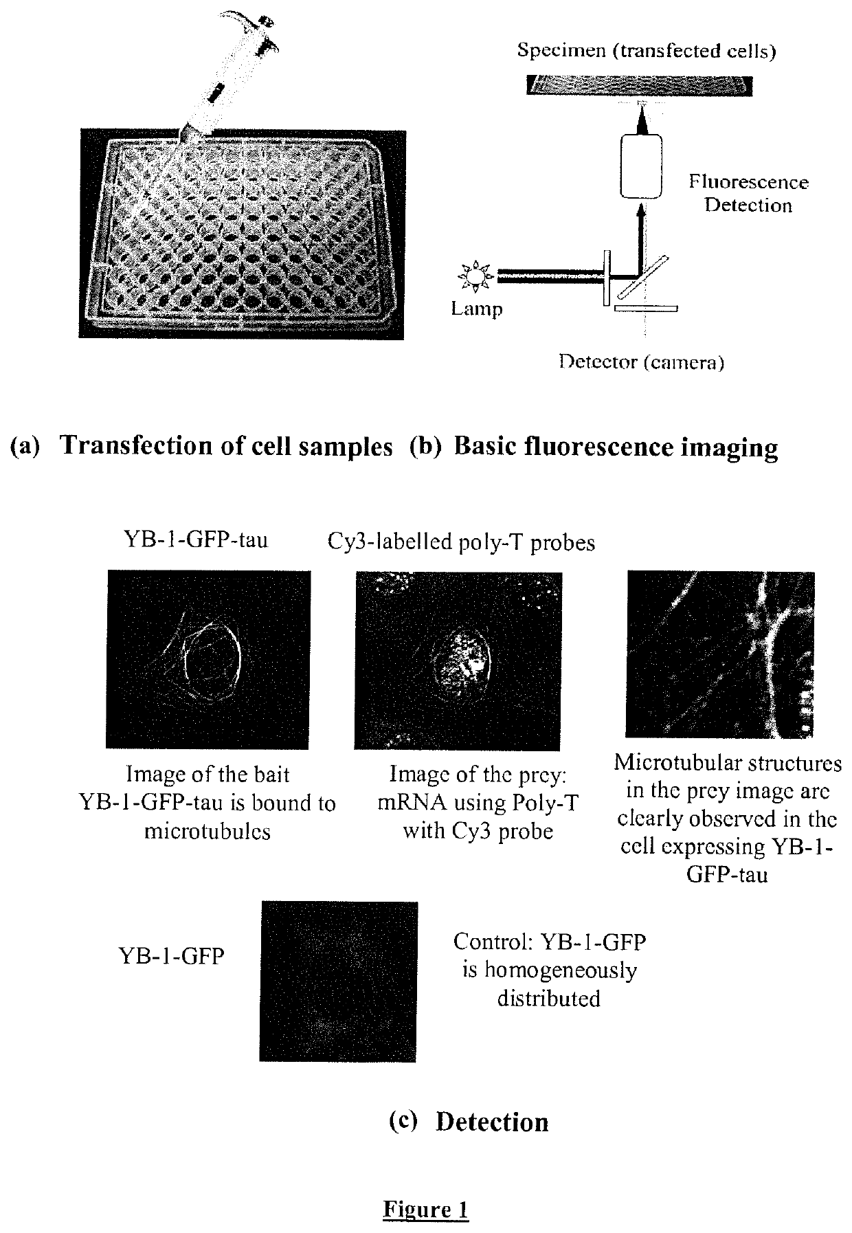
[0252]3) Array of cell samples in 6 to 96-well plates are co-transfected with different preys (see FIG. 1) to identify various interacting partners for given bait at high throughput. An automatic detection system allows the recognition...

example 2

of YB-1 Self-Attraction and YB-1 Binding to cy3-Labeled-Poly-T-Probes in Fixed and Living NRK Cells

[0261]YB-1 (Accession number: NP_004550.2) is known to be one of the core mRNA-binding proteins (Nekrasov et al., The mRNA-binding protein YB-1 (p50) prevents association of the eukaryotic initiation factor eIF4G with mRNA and inhibits protein synthesis at the initiation stage. J Biol Chem 278, 13936-13943; 2003). We then investigated whether or not mRNA molecules can be brought to microtubules using YB-1-tau as bait (FIG. 1C).

[0262]In FIG. 1C we show NRK cells transfected with the human YB-1 protein, an mRNA binding protein, fused to tau and GFP. The bait, YB-1-GFP-tau, was then indeed detected on microtubules in the GFP fluorescence image, in contrast to YB-1-GFP or endogenous YB-1 which remained dispersed in the cytoplasm.

[0263]mRNA molecules were clearly detected on microtubules using in situ hybridization (cy3-labeled poly-T probes) on transfected and fixed cells but not on those ...

example 3

of Binding of eIF2A to eIF2B After eIF2A Phosphorylation in Living Cells

[0268]In FIG. 6A, in HeLa cells transfected with eIF2B-tau, eIF2B-tau is bound to microtubules as clearly observed using anti-eiF2B labeling.

[0269]One of the early responses in mammalian cells exposed to environmental stress (hyperthermia, osmotic stress, hypoxia, oxidative stress, virus) is to block the initiation of translation. Such initiation block is mediated by the phosphorylation of the initiation factor eIF2A via various kinases like PKR and PERK. This phosphorylation occurs at serine 51 and leads to the strong binding of eIF2A to eIF2B. The formation of this complex prevents the regeneration of eIF2A-GDP (GDP / GTP regeneration) by eIF2B (Krishnamoorthy et al.; Tight binding of the phosphorylated alpha subunit of initiation factor 2 (eIF2alpha) to the regulatory subunits of guanine nucleotide exchange factor eIF2B is required for inhibition of translation initiation. Mol Cell Biol 21, 5018-5030; 2001).

[02...


PUM

PropertyMeasurementUnit
lengthaaaaaaaaaa
lengthaaaaaaaaaa
inner diameteraaaaaaaaaa

Abstract

A method for detecting an interaction between one or more protein bait and one or more candidate prey in a eukaryotic cell, comprising the steps of: a) providing an eukaryotic cell expressing (i) one or more protein bait, and (ii) one or more candidate prey, wherein said protein bait comprises a bait moiety and a polymerized-tubulin binding moiety. b) determining the occurence of an interaction between said one or more protein bait and said one or more candidate prey in the eukaryotic cell, wherein said protein bait is bound to polymerized tubulin in the eukaryotic cell, thereby localizing said one or more candidate prey along said polymerized tubulin, thereby detecting said interaction.

Description

FIELD OF THE INVENTION[0001]The invention relates to methods, uses and tools for studying ligand interactions in an eukaryotic cell.BACKGROUND OF THE INVENTION[0002]The function of a number of proteins remains elusive, which slows down understanding of fundamental aspects of cell mechanisms and impedes advance in diagnostic and treatment of many diseases and in personalized medicine. One of the main obstacles to better understand a given protein's functions is that proteins generally interact with many other partners (proteins, nucleic acids, sugars, lipids . . . ) and many of these interactions are weak and transient, albeit of critical importance.[0003]A series of methods have been developed to provide a better view on protein interaction (Lowder et al., Visualizing protein partnerships in living cells and organisms. Curr Opin Chem Biol 15, 781-788, 2011), among them immuno-precipitation assays, two-hybrid systems and variants like substrate complementation systems, Fluorescence E...

Claims


Application Information

Patent Timeline
Patent Type & Authority Patents(United States)
IPC IPC(8): G01N33/68G01N33/58C07K14/47C12N15/10
CPCG01N33/582G01N33/6845C12Q2563/107C12N15/1055C07K14/47C07K2319/30C07K2319/70C07K2319/20C07K2319/60G01N2333/47
Inventor PASTRE, DAVIDCAILLERET, MICHELCURMI, PATRICKBOCA, MIRELA
Owner INST NAT DE LA SANTE & DE LA RECHERCHE MEDICALE (INSERM)
Why Eureka
Try Eureka