

Medical/surgical implant
a technology for surgical implants and implants, applied in the field of implants, can solve the problems of significant soft tissue irritation, negative psychological effect of procedures on patients' well-being, undesirable breast appearance, etc., and achieve the effects of reducing necrosis and resorption of transplanted cells, reducing post-operative healing time, and improving patient well-being
- Summary
- Abstract
- Description
- Claims
- Application Information
AI Technical Summary
Benefits of technology
Problems solved by technology
Method used
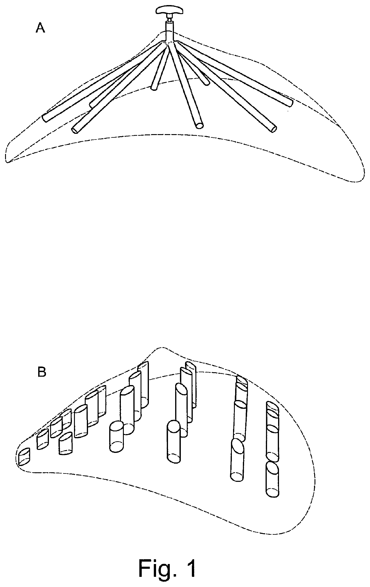
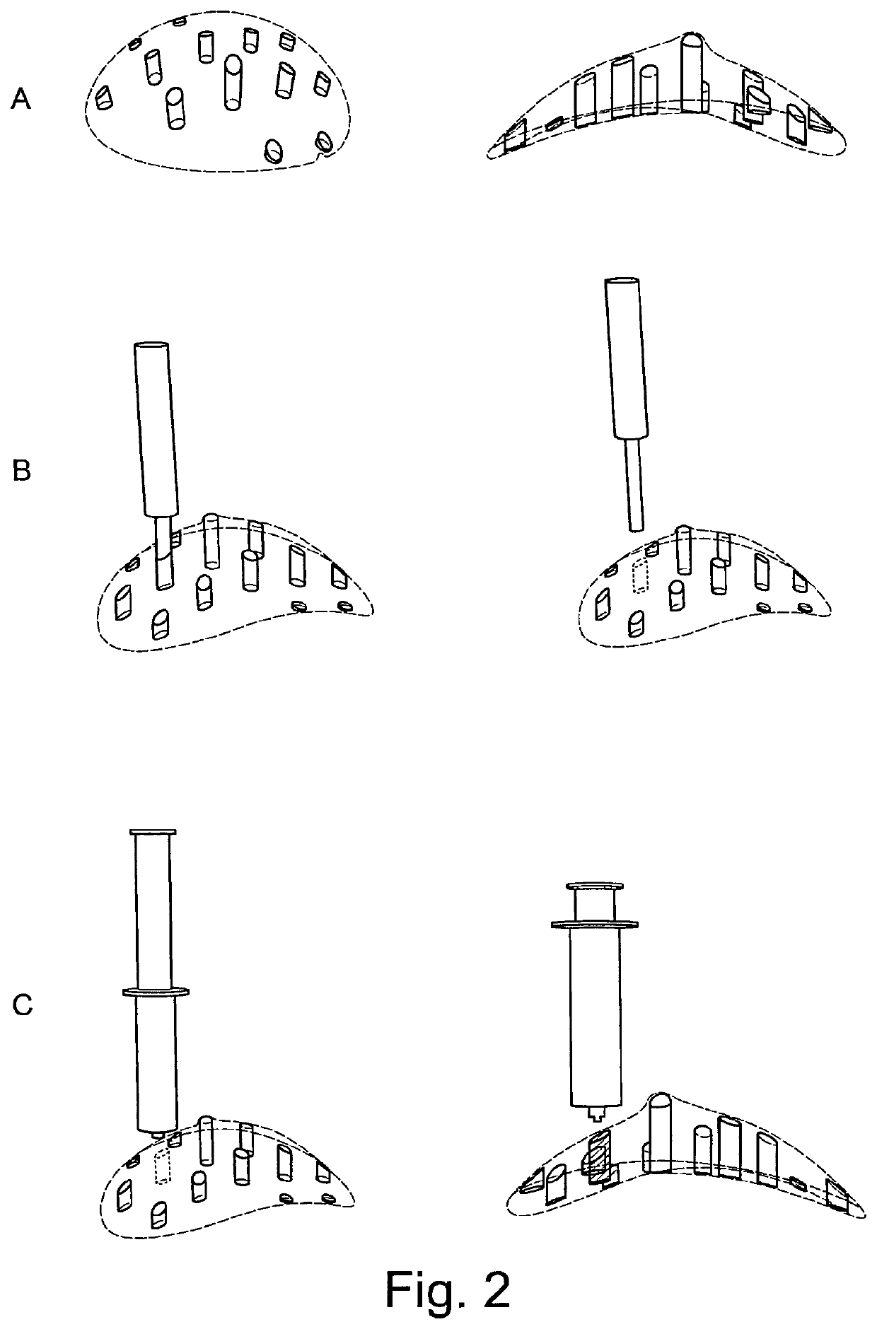
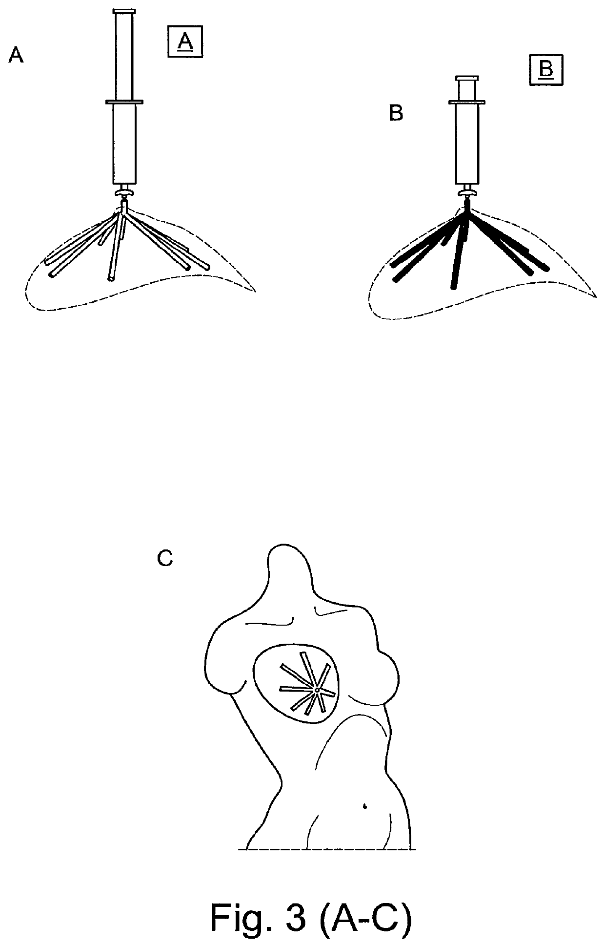
Image

Examples
example 1
[0223]This example combines delayed fat injection with an acellular biodegradable scaffold. In this method of implantation, the scaffold is first implanted with no fat tissue into the implantation site. A fibrin clot is formed immediately after implantation of the scaffold from the hematoma caused by the surgical procedure (Henkel et al., 2013; Salgado et al., 2004). The clot consists of platelets embedded in a mesh of cross-linked fibres, together with a growth-factor rich cocktail of fibronectin, vitronectin and thrombospondin. The fibrin clot and the associated growth-factor cocktail may stimulate a strong angiogenic response and induce highly organised connective tissue to penetrate into the scaffold. After a fixed period of time, fat is isolated from a donor site within the patient's body and injected into the scaffold (see FIG. 12 for a visualisation of this concept). The amount of fat that can be harvested from the patient without encountering donor site morbidity depends on ...
example 2
[0275]Breast shaped scaffolds made of poly(D,L)-lactide polymer and containing voids and space-occupying structures essentially as shown in FIG. 21, scaled up to a scaffold volume of 125 cm3, were prepared. The scaffolds were fabricated with a 3D printer fitted with two extruders (one for printing poly(D,L)-lactide polymer for the scaffold structure, one for printing polylactic acid including a black dye for the space-occupying structures). Such a dual 3D print strategy also allows to prepare complex channel designs, if desired (e.g. a radially convergent design). Since the space-occupying structures are made of solid material, they are not degraded as quickly as the scaffold and thus are capable of preventing tissue / cell invasion within the rather short period of prevascularization in this example. With space-occupying structures made of an undegradable material, prevention of tissue / cell invasion is even better.
[0276]With such scaffolds, a pilot study was undertaken whereby n=6 sc...
PUM
| Property | Measurement | Unit |
|---|---|---|
| time | aaaaa | aaaaa |
| length | aaaaa | aaaaa |
| length | aaaaa | aaaaa |
Abstract
Description
Claims
Application Information