
Antigenic composition for detecting auto-antibody with specific response to exosomal protein EIF3A, and method for diagnosing liver cancer using antigenic composition

an antigen composition and exosomal protein technology, applied in the field of antigens, can solve the problems of low sensitivity and specificity for cancer diagnosis, obstacles to obtaining usefulness, acquisition of autoantibodies to be analyzed,

Active Publication Date: 2020-10-20
KOREA RES INST OF BIOSCI & BIOTECH +1
View PDF10 Cites 0 Cited by
  • Summary
  • Abstract
  • Description
  • Claims
  • Application Information

AI Technical Summary

Benefits of technology

This patent describes a newly-discovered autoantibody that targets a protein called EIF3A, which is associated with liver cancer. The invention also includes a hybridoma cell line that produces the autoantibody, a polypeptide that identifies the specific part of the protein that the autoantibody targets, a diagnostic composition for liver cancer that measures the expression level of the autoantibody, a kit for diagnosing liver cancer, and a method for diagnosing and screening for a therapeutic agent for liver cancer.

Problems solved by technology

However, the above-mentioned antigens have low sensitivity and specificity for cancer diagnosis, and thus they have been used only as an auxiliary means of cancer diagnosis.
It was also an important issue that due to extracellular secretion by post-translational modification or extracellular release of intracellular proteins by necrosis of a tumor site, new epitopes stimulate the immune system to induce antibodies.
However, some problems that arise during processes of identifying cancer-associated autoantibodies and acquiring antigens are obstacles to obtaining useful results.
A first problem is acquisition of autoantibodies to be analyzed.
However, since it is difficult to continuously obtain patients' sera, there is a disadvantage that a sufficient amount thereof cannot be used for characterization.
A second problem is that it is difficult to obtain recombinant proteins sufficiently reflecting the structure of the antibody-specific epitope even though antigen proteins are identified.
However, in the case of antibodies recognizing high order structures, there is a difficulty in acquiring antigens for detection.
Considering that antibodies against in vivo antigens commonly induce structure-recognizing antibodies, there are obvious limitations in the studies on the use of autoantibodies, which have been conducted so far.

Method used


Image

  • Antigenic composition for detecting auto-antibody with specific response to exosomal protein EIF3A, and method for diagnosing liver cancer using antigenic composition
  • Antigenic composition for detecting auto-antibody with specific response to exosomal protein EIF3A, and method for diagnosing liver cancer using antigenic composition
  • Antigenic composition for detecting auto-antibody with specific response to exosomal protein EIF3A, and method for diagnosing liver cancer using antigenic composition

Examples


example 1

on of XC90 Autoantibody Derived from HBx Mouse

[0102]In order to acquire autoantibodies produced during carcinogenesis, an HBx transgenic mouse which develops liver cancer similar to human liver cancer was used. Spleen cells were obtained from HBx transgenic mice, in which development of liver cancer was confirmed, as a B cell group, and fused with a mouse myeloma cell Sp2 / 0 to prepare a B cell hybridoma cell line according to a common B cell hybridoma preparation method. Primary selection of the fused cells was performed using HAT medium (hypoxanthine-aminopterin-thymidine medium), and only clone-forming cells were cultured separately. The cells, in which cancer cell-reactive antibodies were detected in the culture medium of the clone-forming cells, were only selected and maintained.

[0103]The reactivity of cancer model mouse-derived autoantibodies against cancer cells was examined by flow cytometric analysis of cancer cell line after intracellular staining following the fixation and...

example 2

tion and Purification of XC90 Antibody Isotype

[0106]An isotype of XC90 antibody was determined by analysis using a mouse antibody isotyping kit, and as a result, it was IgM type.

[0107]For analysis of XC90 antibody-specific reaction, a monoclonal antibody was purified. Cell culture medium obtained by culturing a large amount of XC90 antibody-producing B cells or an antibody-producing cell was injected into the peritoneal cavity of mice, thereby acquiring ascites fluid. The ascites fluid was applied to mannose-binding protein-agarose (Pierce) or protein L agarose to purify IgM type antibody.

[0108]After SDS-PAGE, the purified antibodies were confirmed by Coomassie staining, and protein quantification was performed by a Bradford method.

example 3

n of XC90 Antibody-Specific Antigen in Various Cancer Cells and Identification of Antigen Protein

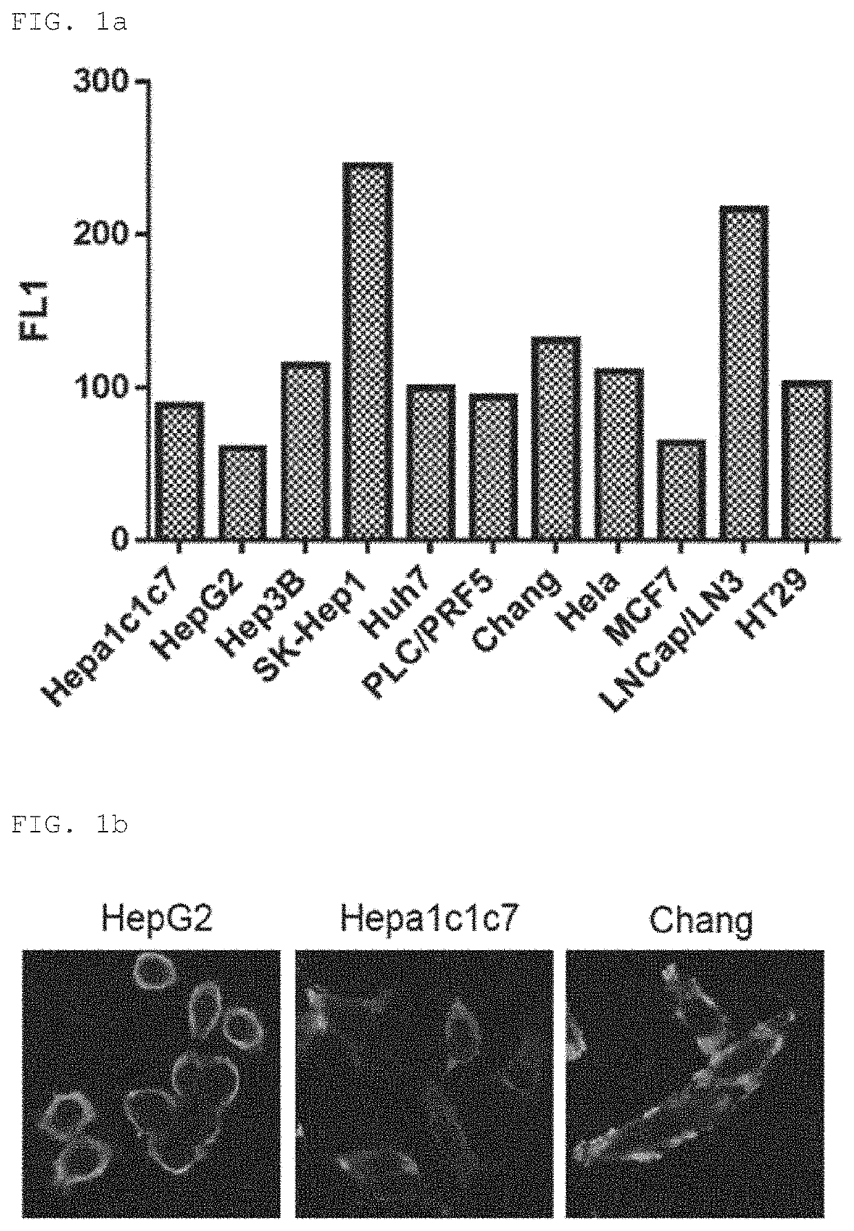
[0109]To examine reactivity of the XC90 monoclonal autoantibody purified in Example 2 for various cancer cell lines, intracellular staining of the various cancer cell lines was performed in the same manner as in Example 1, followed by flow cytometry (FIG. 1A). As shown in FIG. 1A, expression of XC90 antibody-reactive antigen was observed in liver cancer cell lines including HepG2, SK-Hep-1, Huh7, etc., and other cancer cell lines including HeLa, HT29, etc., and in particular, overexpression of the autoantibody was observed in SK-Hepl liver cancer cell line and LNCap / LN3 prostate cancer cell line.

[0110]Further, to examine the intracellular localization of the antibody-reactive antigen expression in cells, confocal microscope was used to observe the cells after intracellular staining, and as shown in FIG. 1B, strong staining was observed in the cytoplasm.

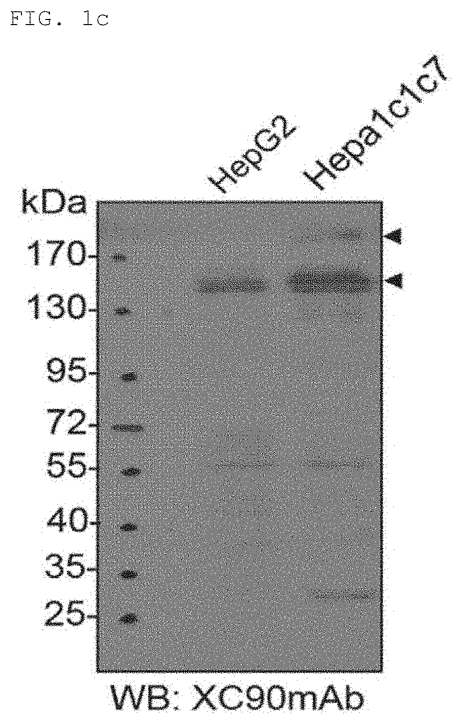
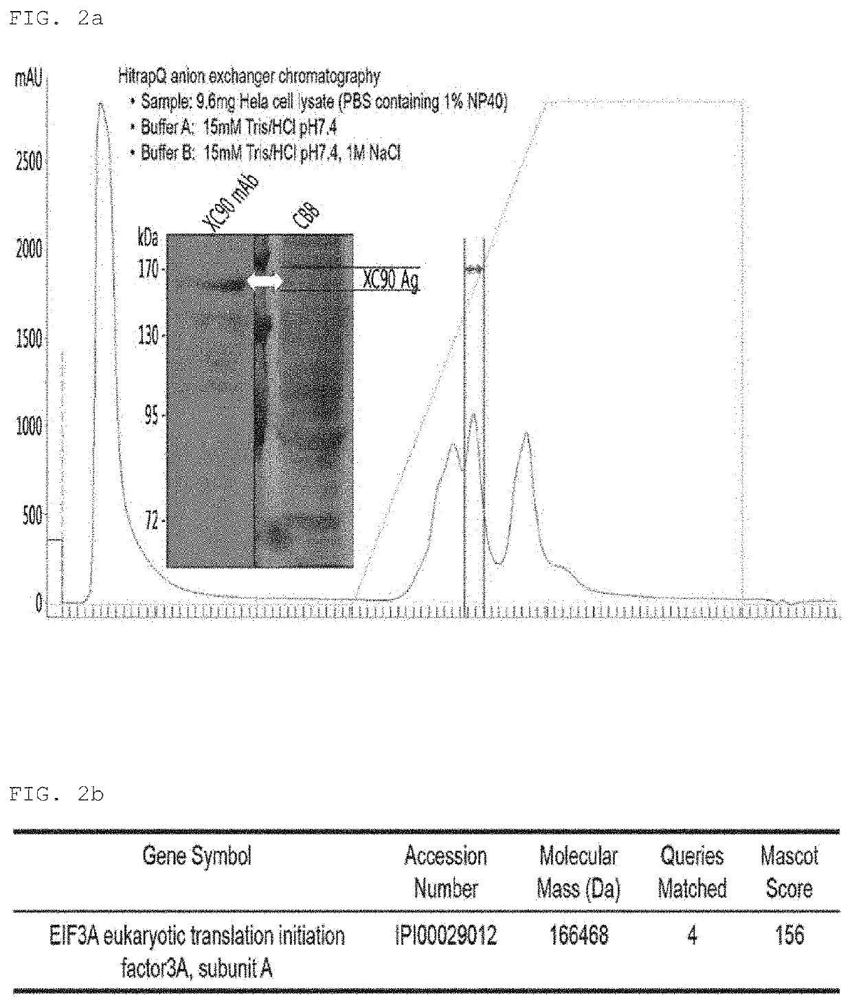
[0111]Expression of the XC90 antibo...


PUM

PropertyMeasurementUnit
concentrationaaaaaaaaaa
molecular weightaaaaaaaaaa

Abstract

Provided are an autoantibody specifically binding to an exosomal protein EIF3A (Eukaryotic translation initiation factor 3 subunit A) or a fragment including an antigen-binding site (paratope) thereof, a hybridoma cell line producing the autoantibody, a polypeptide having an amino acid sequence of an antigenic determinant (epitope) specifically binding to the autoantibody, a composition for diagnosing liver cancer including an agent measuring an expression level of the autoantibody or the fragment including the antigen-binding site thereof, a kit for diagnosing liver cancer including the composition, and a method of providing information for diagnosis of liver cancer by using the composition. Further, provided is a method of screening for a therapeutic agent for liver cancer by using an expression level of the autoantibody. When anti-EIF3A autoantibody of the present invention is used as a diagnostic marker for liver cancer, the incidence of liver cancer may be diagnosed at a high level only by using non-invasive biological samples. Furthermore, liver cancer may be easily diagnosed by using only the amino acid sequence identified in the present invention, leading to the effective development of diagnostic products such as a diagnostic kit for liver cancer.

Description

CROSS-REFERENCE TO RELATED APPLICATIONS[0001]This application is a § 371 national stage of PCT International Application No. PCT / KR2016 / 007415, filed Jul. 8, 2016, claiming priority of Korean Patent Application No. KR 10-2015-0098665, filed Jul. 10, 2015, the contents of each of which are hereby incorporated by reference into the application.REFERENCE TO A SEQUENCE LISTING[0002]This application incorporates by reference nucleotide and / or amino acid sequences which are present in the file named “200206_90283_Substitute_Sequence_Listing_BI.txt”, which is 9.43 kilobytes in size, and which was created Feb. 5, 2020 in the IBM-PC machine format, having an operating system compatibility with MS-Windows, which is contained in the text file filed Feb. 6, 2020 as part of this application.BACKGROUND OF THE INVENTION1. Field of the Invention[0003]The present invention relates to an antigenic composition for detecting an autoantibody specific to an exosomal protein EIF3A (eukaryotic translation ...

Claims


Application Information

Patent Timeline
Patent Type & Authority Patents(United States)
IPC IPC(8): G01N33/574C07K14/47G01N33/564G01N33/58C07K16/30C12N5/16C07K16/18A61P35/00
CPCG01N33/564C07K14/4748C07K16/303C12N5/163G01N33/57438A61P35/00G01N33/58C07K16/18C07K2317/34G01N2800/52C07K2317/14
Inventor CHO, EUN WIEHEO, CHANG KYUHWANG, HAI MINKO, JEONG HEONLIM, KOOK JINLEE, HYE JUNG
Owner KOREA RES INST OF BIOSCI & BIOTECH
Learn More