Method for selective expression of therapeutic genes by hyperthermia

a gene therapy and hyperthermia technology, applied in the field of promoting antitumor activity in mammals, can solve the problems of lack of temporal, limited use of gene therapy methods, and prohibitive us

Inactive Publication Date: 2003-03-06
DUKE UNIV
View PDF5 Cites 23 Cited by
  • Summary
  • Abstract
  • Description
  • Claims
  • Application Information

AI Technical Summary

Benefits of technology

0063] FIG. 18 is a graph depicting percentage specific lysis response of splenocytes from differently treated, B16.F10 tumor-bearing C57 mice against B16.F10, Tramp, or YAC1 cells. (solid line without symbol), (x), (+), splenocytes of untreated control animals against B16.F10 (solid line without symbol), Tramp (X), or YAC1 (+); (.diamond.), (.quadrature.), (.DELTA.), splenocytes of animals injected intratumorally with AdGFP against B16.F10 (.diamond.), Tramp (.quadrature.), or YAC1 (.DELTA.); (.diamond-solid.), (.box-solid.), (.tangle-solidup.), splenocytes of animals injected intratumorally with AdIL12 / B7.1 against B16.F10 (.diamond-solid.), Tramp (.box-solid.), or YAC1 (.tangle-solidup.).
0064] FIG. 19A is a photomicrograph (25.times. magnification) depicting a central region of a B16.F10 melanoma treated by intratumoral injection of the control vector AdGFP. The tumor tissue has been stained with an anti-collagen IV antibody to detect blood vessels, and an extensive network of blood vessels is observed as a dark staining product precipitate.
0065] FIG. 19B is a photomicrograph of a center region of a tumor treated by intratumoral injection of AdIL12 / B7.1. The tumor tissue has been stained with an anti-collagen IV antibody to detect blood vessels. Blood vessels are notably absent (fewer regions of dark staining product precipitate), and tumor necrosis is evidenced by the disrupted tissue morphology.

Problems solved by technology

However, the usefulness of gene therapy methods has been limited due to systemic toxicity of anti-tumor polypeptides encoded by gene therapy constructs (Spriggs & Yates (1992) in Bentler, ed., Tumor Necrosis Factor: The Molecules and Their Emerging Roles in Medicine, pp.
Although each of the afore-mentioned systems display inducibility, a lack of temporal and spatial precision of gene induction, an ineffective magnitude of gene induction, a background level of non-induced gene expression, or an inability to regulate termination of gene expression, can still prohibit their usefulness.

Method used


Image

  • Method for selective expression of therapeutic genes by hyperthermia
  • Method for selective expression of therapeutic genes by hyperthermia
  • Method for selective expression of therapeutic genes by hyperthermia

Examples


example 1

In vitro Expression of Adenoviral Gene Therapy Constructs

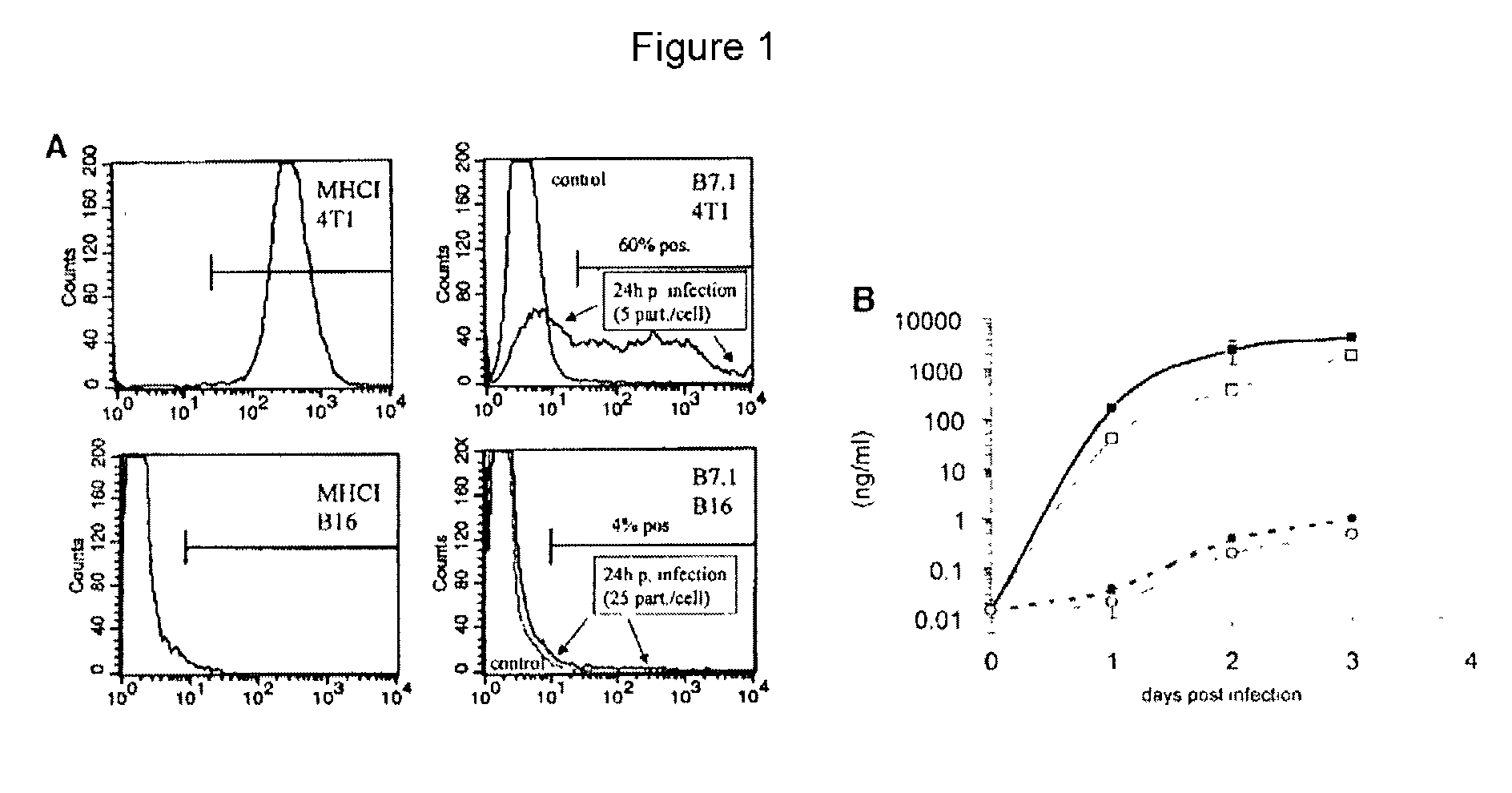
[0294] Eighty percent confluent 4T1 cells were infected with AdIL12 / B7.1 at a MOI of 5 / cell for 5 hours. Twenty-four hours after the beginning of infection, cells were detached with 0.05% trypsin (Gibco / Life Technologies of Rockville, Md.) and resuspended in PBS with 3% normal rat serum. Cells were incubated with monoclonal biotin-labeled anti-mouse B7.1 antibody (Caltag of Burlingame, Calif.) for 30 minutes, washed in PBS, and incubated in streptavidin-phycoerythrin (Pharmingen of San Diego, Calif.) for 30 minutes. Incubations and washes were performed at 4.degree. C. Samples were measured on a FACStar flow cytometer (Becton-Dickinson of Franklin Lakes, N.J.).

[0295] FIG. 1A presents levels of expression of the reporter gene GFP in cell cultures infected with an adenovirus encoding GFP. Levels of GFP were measured using FACS analysis. FIG. 1B presents levels of expression of IL12 in cell cultures infected with an adenoviral co...

example 2

In vivo Expression and Systemic Dissemination of Reporter Gene Adenovirus

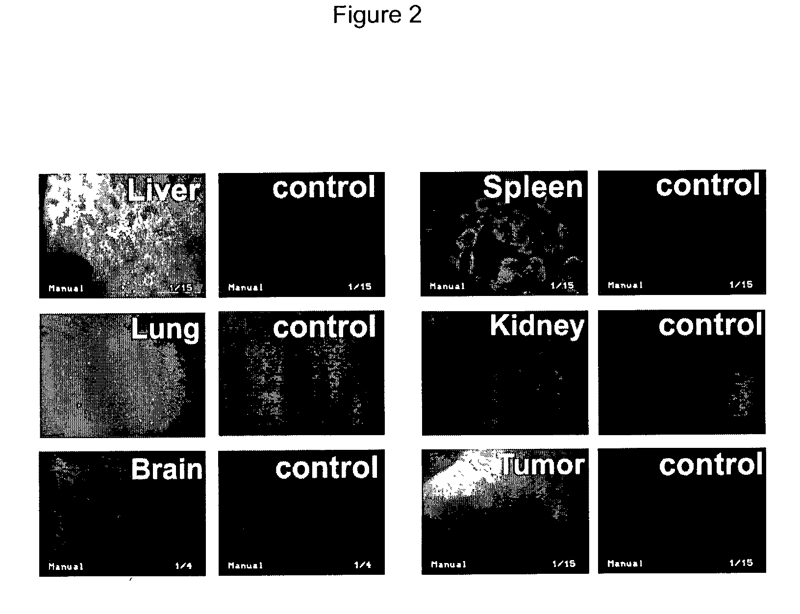
[0296] 3.times.10.sup.8 pfu (plaque forming unit) of AdGFP in 50 .mu.l PBS were intratumorally injected when subcutaneous B16.F10 melanoma tumors reached a diameter of 5-7 mm. The mice were sacrificed 24 hours later. Fluorescence microscopy of harvested tumors and organs (FIG. 2) demonstrated high numbers of GFP expressing cells in the liver and spleen and to a lesser extent in the lung of C57BL / 6 mice bearing B16 melanoma at 24 hours after virus injection. The GFP-expressing area in the tumor was limited to a small volume around the presumed needle track. GFP expression in the liver was almost homogeneous and infected areas were seen throughout the whole spleen. No relevant GFP expression was detected in kidneys or brain of AdGFP injected animals or in any organs of the control animals.

example 3

Intratumoral Expression of Plasmid Gene Therapy Vectors

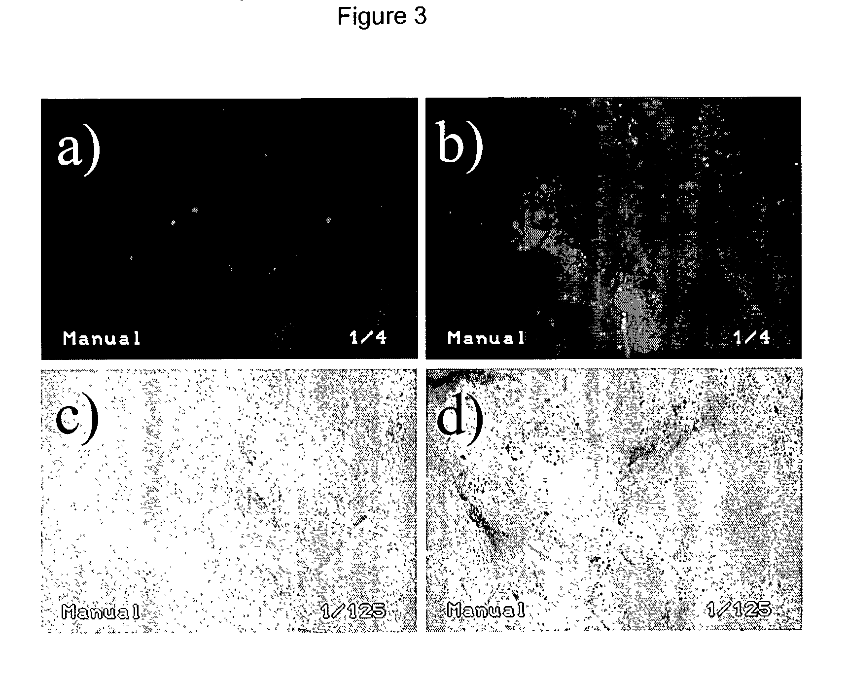
[0297] B16.F10 tumors grown to 5-7 mm in diameter (or 65-179 mm.sup.3) in volume were injected with EGFP or .beta.-gal reporter plasmids and electroporated to promote cell transduction. Expression of reporter genes (EGFP and .beta.-gal) after plasmid injection and electroporation in tumor tissue was assessed in fresh tissue sections (at 250 .mu.m) by fluorescence microscopy and transmission light microscopy (FIG. 3). For both reporter systems, few cells were positive when only naked DNA without consecutive electroporation was injected. The combination with electroporation resulted in consistently efficient transduction of a higher number of cells with both reporter genes. In EGFP experiments, plasmid injection with electroporation allowed 3-8% (as evaluated by fluorescence-activated flow cytometry) of all the cells in the tumor mass to be transduced with the EGFP gene in comparison to <0.1% in tumors injected with EGFP plasmid a...


PUM

PropertyMeasurementUnit
temperatureaaaaaaaaaa

Abstract

A method for controlling in vivo heat-inducible gene expression for provision of therapeutic polypeptide. The method includes the steps of: (a) providing a construct comprising a polynucleotide encoding a therapeutic polypeptide, wherein the polynucleotide is operatively linked to a heat-inducible promoter; (b) administering the construct to a warm-blooded vertebrate at a site at or near the tumor; and (c) applying heat to the site at or near the tumor to express the therapeutic polypeptide, whereby anti-tumor activity is observed.

Description

RELATED APPLICATION INFORMATION[0001] This application is based on and claims priority to U.S. Provisional Patent Application Serial No. 60 / 298,305, filed Jun. 14, 2001, the entire contents of which are herein incorporated by reference.[0003] The present invention generally relates to control of gene expression in tumor cells. Specifically, the present invention provides methods for using a heat inducible promoter operatively linked to a therapeutic gene to preferably induce both immune and anti-angiogenic responses in vivo.1 Table of Abbreviations 4TI cells mouse mammary cancer cells AdCMVIL12 Adenoviral vector encoding IL12 under the control of a CMV promoter (AdmIL12) AdGFP Adenoviral vector encoding GFP under the control of a CMV promoter AdhspEGFP Adenoviral vector encoding EGFP under the control of a hsp70B promoter AdhspIL12 Adenoviral vector encoding IL12 under the control of a hsp70B promoter AdhspTNF.alpha. Adenoviral vector encoding TNF-.alpha. under the control of a hsp7...

Claims


Application Information

Patent Timeline
Patent Type & Authority Applications(United States)
IPC IPC(8): A61K38/19A61K38/20A61K48/00C12N15/861
CPCA61K38/191A61K38/208A61K48/00A61K48/0058C12N15/86C12N2710/10343C12N2830/002C12N2830/85C12N2840/203
Inventor LI, CHUAN-YUANHUANG, QIANDEWHIRST, MARK W.
Owner DUKE UNIV
Why Eureka
Try Eureka