
Contrast Medium for Use in Imaging Methods

a contrast medium and imaging method technology, applied in the direction of magnetic variable regulation, sensors, diagnostics, etc., can solve the problem of not being able to achieve the radiation exposure of disassembly, accumulation, and target of superparamagnetic particles, for example, in the bone, etc., to achieve the effect of avoiding radiation exposure or side effects

Inactive Publication Date: 2003-12-11
EUCRO EURON CONTRACT RES
View PDF5 Cites 22 Cited by
  • Summary
  • Abstract
  • Description
  • Claims
  • Application Information

AI Technical Summary

Benefits of technology

[0012] The present invention has therefore the object to provide an auxiliary for the use in imaging methods that can be enriched in a targeted way in the organ or body part to be examined, wherein the disadvantages or side effects of radiation exposure, as they would occur by radioactive substances or x-rays, are reduced and, if possible, entirely eliminated.
[0014] The term auxiliaries in the context of the present invention means that the superparamagnetic particles according to the invention are used as contrast agents or for sensibilization of the physical parameters to be measured, such as nuclear magnetic resonance. For example, the superparamagnetic particles can be employed in x-ray methods as contrast agents, wherein advantage is taken of the effect that the superparamagnetic particles absorb x-rays more weakly or more strongly than the neighboring body tissues / bones so that the representation of the body structures as an image is possible. In the case of nuclear magnetic resonance methods, the superparamagnetic particles according to the invention can effect a contrast enhancement in that they excite / sensibilize hydrogen bonds in their environment, which leads to a stronger measured signal in nuclear magnetic resonance methods.
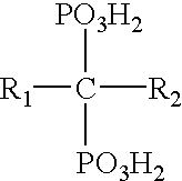
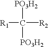
[0053] The contrast agent according to the invention contains superparamagnetic particles as well as a tissue-specific substance, wherein the tissue-specific substance preferably is adsorbed on the superparamagnetic particles. In one possible embodiment, the superparamagnetic particles and the tissue-specific substances can be mixed together as solid materials. However, it was found to be particularly advantageous when the tissue-specific substance is already present when forming the magnetic particles, for example, when the superparamagnetic particles are produced by a size-controlled precipitation in aqueous medium by means of alkaline substances or by reduction of metal cations. As a result of the large particle surfaces produced in situ, an optimal adsorption of the tissue-specific substances on the surface of the particles by means of reactive groups such as OH, SH, hydroxide, amino, carboxyl, ether, sulfo, phosphonic acid groups and so on. It is also possible to apply the tissue-specific substance subsequently onto the precipitated superparamagnetic particles, for example, by suspension of the un-coated (unmodified) superparamagnetic particles in a liquid phase, preferably water, containing the tissue-specific substance or a substance mixture.

Problems solved by technology

While the conventionally employed contrast agents have disadvantages and side effects such as not being tolerated by the patient, the contrast agents used in scintigraphy are in the form of radio nucleotides which exhibit the long-term side effects of radioactive substances.
A targeted accumulation of the superparamagnetic particles, for example, in the bone, is not possible with the agents known in the prior art.

Method used


Image

  • Contrast Medium for Use in Imaging Methods
  • Contrast Medium for Use in Imaging Methods
  • Contrast Medium for Use in Imaging Methods

Examples


Embodiment Construction

Preparative Examples

[0060] 1. In 40 g of deionized water, 6.48 g FeCl.sub.3 were dissolved. Also, 3.97 g FeCl.sub.2 C4H.sub.2O was dissolved in a mixture of 8 ml deionized water and 2 ml 37% hydrochloric acid. The two mixtures were combined shortly before use of the solutions in the precipitation process.

[0061] 2. In a beaker, 400 ml deionized was stirred with 10 g NaOH and 0.2 g 1-methyl-1-hydroxy-1,1-diphosphonic acid (MDP). After cooling, the hydrochloric acid iron solution prepared in 1 was added with intense agitation. By means of a magnetic field, the formed black precipitate was sedimented and the solution above was decanted. Subsequently, water was added several times to the precipitated material and decanted in order to remove foreign ions. Subsequently, 0.5 g MDP and 100 ml water were added. After stirring for an hour at 40 degrees C., the mixture was stirred for 12 hours at room temperature. Portions that were not suspended were separated by centrifugation (5,000-11,000 r...


PUM


Abstract

An imaging auxiliary for medical imaging methods contains stabilizer-free superparamagnetic particles such as metal oxides and metals. Preferred are Î<3>-Fe2O3, Fe3O4, MnFe2O4, NiFe2O4, and CoFe2O4 and their mixtures. The auxiliary can additionally contain biologically and pharmacologically active substances such as proteins, antibodies, peptides, or oligonucleotides. The the superparamagnetic particles have a particle size of 1 nm to 500 nm. A tissue-specific substance in the form of diphosphonic acids and physiologically innocuous salts of diphosphonic acids can be added.

Description

BACKGROUND OF INVENTION[0001] The present invention relates to the use of superparamagnetic particles as auxiliaries in imaging methods as well as a contrast agent or medium for use in medical diagnostics, in particular, in imaging methods.[0002] Imaging methods, for example, x-ray examinations, MR tomography, scintigraphy etc., represent important resources in medical diagnostics for imaging morphological and metabolic processes in the area of the skeletal systems as well as pathological changes in the area of the soft tissue. When employing these methods, the use of so-called contrast media or agents is generally required. They effect in the body parts to be examined a sharp delimitation between healthy tissue and pathological changes.[0003] Contrast agents in MR tomography are in the form of magnetic particles, for example, superparamagnetic iron oxide particles. A review of such contrast agents is provided in Eur. Radiol. (2001) 11, 2319-2331. The field of application of the des...

Claims


Application Information

Patent Timeline
Patent Type & Authority Applications(United States)
IPC IPC(8): A61B5/055G01R33/28A61B6/00A61B6/03A61K49/00A61K49/04A61K49/06
CPCA61K49/1818B82Y5/00A61K49/1842A61P43/00
Inventor GREB, WOLFGANGBLUM, HELMUTROTH, MARCEL
Owner EUCRO EURON CONTRACT RES
Learn More