
Fibrocyte-based vaccine formulations

a technology of fibrocyte and vaccine formulation, which is applied in the direction of immunological disorders, antibody medical ingredients, peptide/protein ingredients, etc., can solve the problem of insufficient first signal to activate t-cells, and achieve the effect of convenient separation

Inactive Publication Date: 2004-02-05
RICE GLENN C +1
View PDF2 Cites 8 Cited by
  • Summary
  • Abstract
  • Description
  • Claims
  • Application Information

AI Technical Summary

Benefits of technology

[0034] Therefore, the inventive fibrocytes fused with tumor cells or tumor cell membranes are effective as tumor vaccines for treating or preventing cancer. Fibrocytes are fused with tumor cells to form a fused fibrocyte / tumor cell and the fused fibrocyte / tumor cells isolated, cultured and utilized as a tumor immunogen. For example, murine MC38 adenocarcinoma cells that stably express the marker DF3 / MUC1 antigen were fused to peripheral blood derived fibrocytes. Cells were maintained in DMEM supplemented with 10% heat-inactivated fetal calf serum (FCS), 2 mM gluitamine, 100U / ml penicillin and 100 .mu.g / ml streptomycin. Fusion efficiencies may be enhanced by pre-culturing tumor cells and fibrocytes prior to fusion as well as fusing at relatively low cell densities (<10.sup.6 / ml). However, fusion at low cell densities is not a requirement for fusion. Agents that act to enhance fiborycte:tumor cell adherence also enhance fusion efficiencies. These agents include, for example, IL-1.alpha. and IL-1.beta.. In addition, a fibrocyte may be modified prior to fusion with a tumor cell to increase the immuno-potency of the resultant hybrid. This modification can take several different means, including gene or protein transfer. Examples of proteins that can be expressed or inhibited in a fibrocyte include cytokines, chemokines, adhesion receptors, or co-stimulatory molecules.
[0042] The fused fibrocyte / tumor fusion cells may be used either in a prophylactic or treatment procedure generally with autologous but also allogeneic procedures. The use of fusion with fibrocytes provides substantial advantages for practicing tumor cell engineering and enhancing tumor cell immunogenicity. There is no need for decoding the precise tumor antigens that may be expressed in the particular tumor. Cell fusion is applicable to diverse tumor types, including solid and blood tumors. It is a simple process and can utilize fusion methods involving chemical fusion, or methods of generating cell fusion that involve electric fields or high pressure. The tumor cell contributes relevant tumor-specific antigens to the hybrid cell or membrane preparation and the fibrocyte-like cell contributes cell surface co-stimulators, soluble cytokines, MHC molecules, adhesion molecules, chemokines and other undefined molecular factors to the hybrid cell. This results in a highly antigenic and immunogenic phenotype for the hybrid cell or hybrid cell membrane preparations. In turn, the hybrid cell can be used as an effective cellular vaccine.
[0045] This invention also provides a fibrocyte-vaccine formulation wherein the antigenic component is a pulsed antigen protein or peptide, or a gene encoding specific antigenic determinants of proteins or peptides for uses in tumor immunotherapy. Fibrocytes can be induced in vitro to secrete, for example, tumor antigens, viral markers, or T-cell attractants in vivo for enhanced immunogenicity. These T-cell attractants include, for example, various chemokines, cell surface proteins and secreted cytokines. For instance, IL-1.beta. can induce fibrocyte expression of MIP-1.alpha., a powerful T-cell chemoattractant (FIG. 15). IL-1.beta. also induces fibrocyte migration (FIG. 16) which is important for T-cell interaction with the fibrocyte.
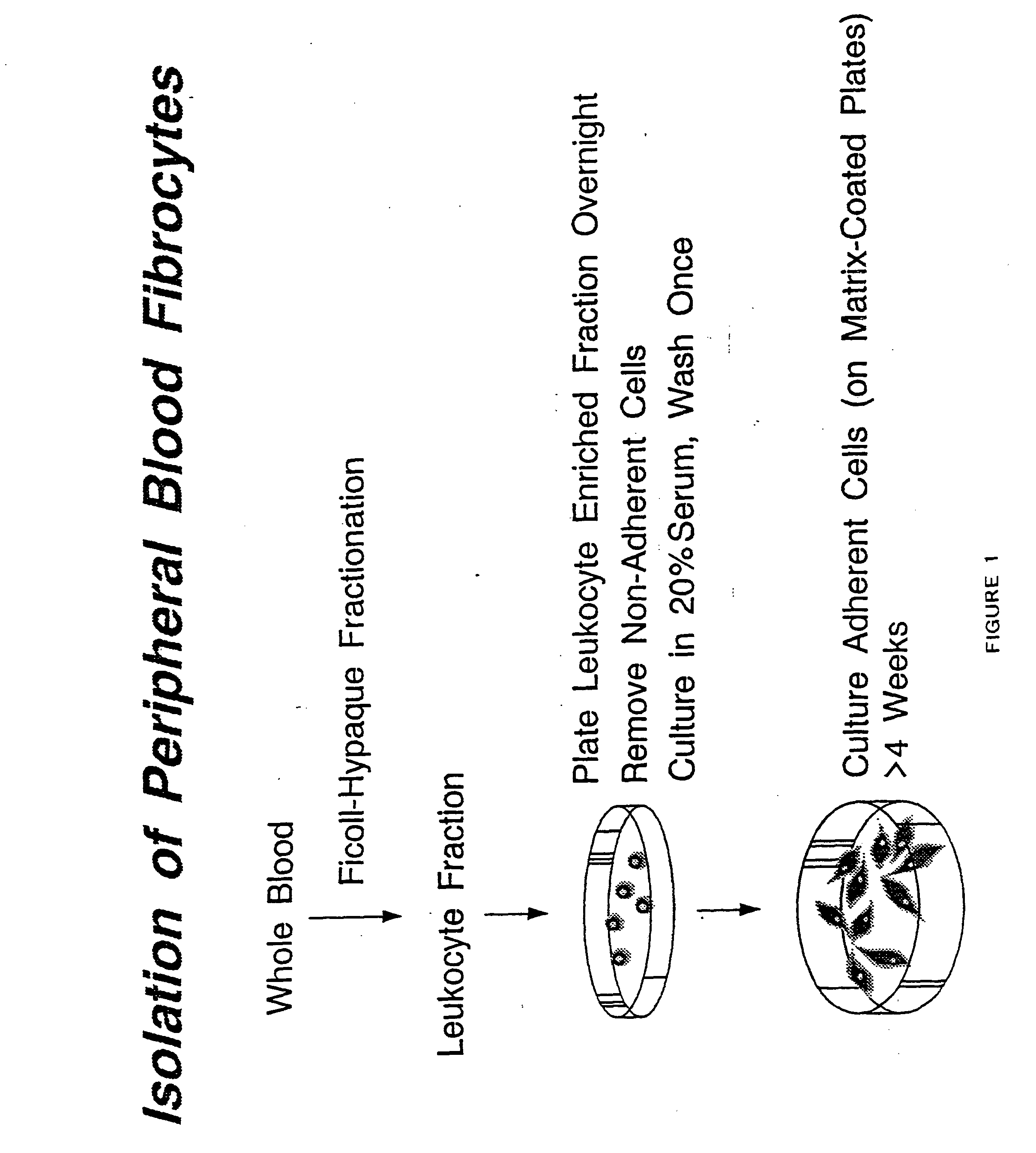
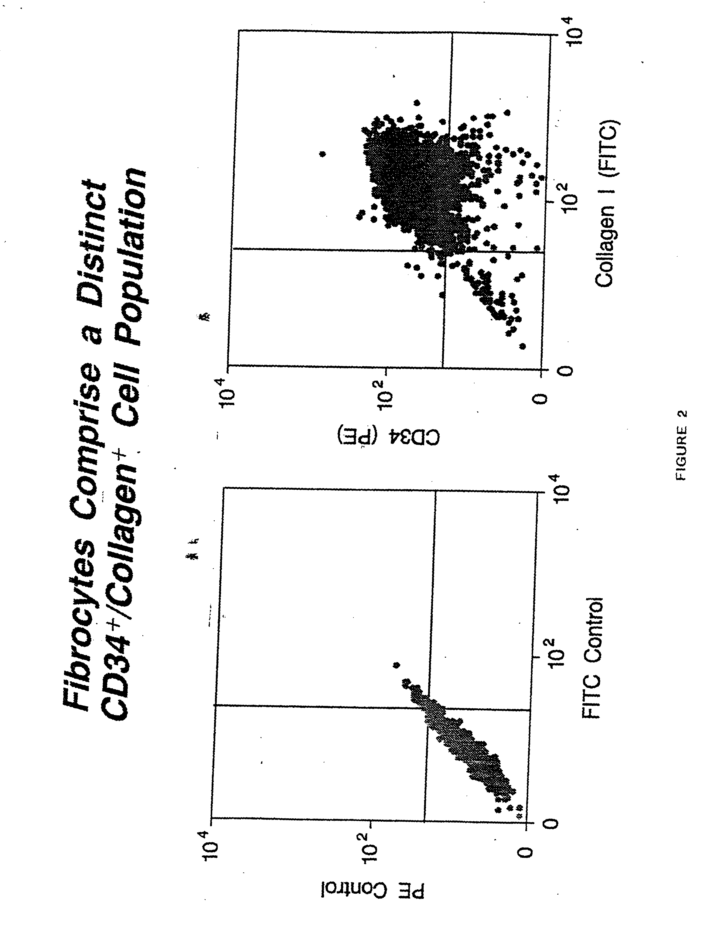
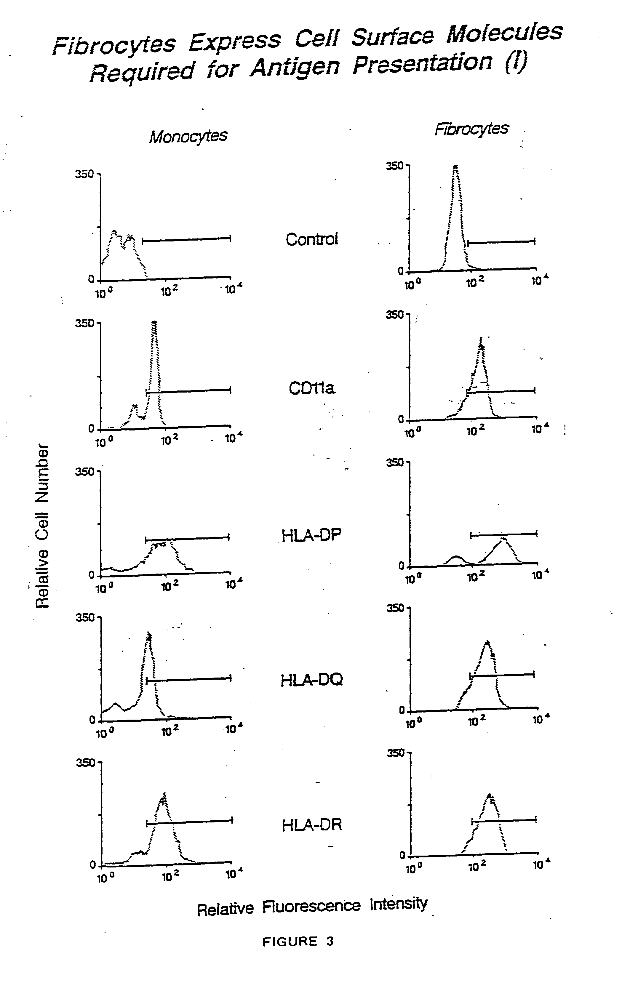
[0051] Use of the fibrocyte for tumor, viral, fungal, bacterial or parasitic immunization has distinct advantages over the use of other APCs (antigen presenting cells). First, unlike dendritic cells, fibrocytes can be conveniently separated from blood and the separation involves straightforward means, without the need for complicated selection, such as a selection based on the CD34 antigen. Second, fibrocytes constitute upwards to 0.3% of the circulating PBL's, a frequency much higher than dendritic cells. Third, fibrocytes are potent APCs, at least equivalent to dendritic cells and superior to monocytes. Fourth, fibrocytes do not require sophisticated and complex cocktails of cytokines and growth factors (such as GM-CSF, IL-3, TNF, IL-1 Flt-3, etc.) for ex vivo expansion, as required for dendritic cells and other APCs. Fifth, fibrocytes (unlike dendritic cells) divide with rapid doubling times ex vivo, which allows for harvesting large numbers of cells useful for immunotherapy and allowing cell banking of fibrocytes for later use.

Problems solved by technology

However, this first signal is insufficient to activate the T-cell, at least as measured by induction of IL-2 synthesis and secretion.

Method used


Image

  • Fibrocyte-based vaccine formulations
  • Fibrocyte-based vaccine formulations
  • Fibrocyte-based vaccine formulations

Examples


example 2

[0056] This example illustrates a comparison of the antigen presenting qualities of fibrocytes to be approximately equivalent to dendritic cells. Various concentrations of mitomycin C-treated fibrocytes were co-cultured with allogeneic T cells for 96 hours and the T cell proliferative activity assessed by the incorporation of [.sup.3H]thymidine into DNA. Fibrocytes were found to induce a significant T cell activation response (5.times.10.sup.3 fibrocytes+2.times.10.sup.5 T cells=70,321.+-.10,111 cpm, versus <300 cpm for T cells or fibrocytes alone, n=3 experiments, P<0.001).

[0057] The fibrocytes were examined for their capacity to present soluble antigen in an autologous, T cell proliferation assay. T cells were purified from the peripheral blood of tetanus toxoid-immunized individuals and stimulated with tetanus toxoid in vitro together with fibrocytes as APCs. To assess the functional capacity of fibrocytes to present antigen, we examined the ability of purified human fibrocytes t...

example 3

[0063] This example illustrates that mouse fibrocytes induced a significant antigen-dependent T cell proliferation response when co-incubated with primed, autologous T cells. T cells were purified from the peripheral blood of BALB / c mice immunized with the HIV proteins p24 and gp120 and stimulated in vitro with p24 or gp120 utilizing fibrocytes as APCs (FIG. 8). BALB / c mice were immunized by i.p. injection of 100 mg of native p24 or gp120 (purified from HIV-1.sub.IIIB infected H9 cells, Advanced Biotechnologies, Columbia, Md.) emulsified at 1 mg / ml with Freund's complete adjuvant. After 14 days, the T cells were purified from spleens by chromatography over a T cell enrichment column (R&D Systems, Minneapolis, Minn.). Mouse fibrocytes were purified from peripheral blood as described above and treated with 25 mg / ml mitomycin C (Sigma) in RPMI medium containing 10% fetal calf serum (RPMI / 10% FCS) for 30 minutes and then washed 5 times with RPMI / 10% FCS. For each assay, the T cells (2.t...

example 4

[0067] This example illustrates antigen-pulsed fibrocytes were not simply transferring antigen to other host APC types. This example provides the results of experiments in which antigen-pulsed fibrocytes from two parent mouse strains were injected into F.sub.1 offspring mice. The T cell reactivity of F.sub.1 offspring was confined predominantly to antigens presented by one of the parental strains, and the priming and re-stimulation APCs must necessarily share the same haplotype (Inaba et al., J. Exp. Med. 172:631-640, 1990; Sprent, J. Exp. Med. 147:1159, 1978; Sprent, J. Exp. Med. 147:1142, 1978). DBA-2 x C3H / HeJ F.sub.1 mice (H-2 .sup.dxk) were injected with pulsed fibrocytes from either parent (DBA-2, H-2.sup.d or C3H / HeJ, H-2.sup.k) and, five days later, the proximal popliteal lymph nodes were isolated and depleted of endogenous class II MHC.sup.+ APCs by immunomagnetic selection. The F.sub.1 APC-depleted lymph node cells were cultured with F.sub.1 (H-2.sup.dxk) or parent strain ...


PUM

PropertyMeasurementUnit
Densityaaaaaaaaaa
Immunogenicityaaaaaaaaaa
Antigenicityaaaaaaaaaa

Abstract

There is disclosed a fibrocyte-based vaccine formulations made from isolated fibrocytes. There is further disclosed a method for establishing an immune response against a specific antigen by administering a fibrocyte-based vaccine formulation, such as one made by pulsing fibrocytes in culture with the antigen peptide or protein, or by fusing tumor cells (whole cells or membrane fragments thereof) with fibrocytes.

Description

[0001] The present invention provides fibrocyte-based vaccine formulations made from isolated fibrocytes. The present invention further provides a method for establishing an immune response against a specific antigen by administering a fibrocyte-based vaccine formulation, such as one made by pulsing fibrocytes in culture with the antigen peptide or protein or transfecting fibrocytes with genes encoding specific antigenic determinants of peptides or proteins, or by fusing tumor cells (whole cells or membrane fragments thereof) with fibrocytes.[0002] The immune response involves a) recognition of a specific antigen by a lymphocyte, b) elaboration of specific cellular and humoral effectors, leading to c) elimination of the antigen by specific effector cells such as T-lymphocytes and antibodies derived from B-cells, which mediate cellular and humoral immune responses, respectively. The cellular immune response begins with the recognition of antigen on the surface of an antigen-presentin...

Claims


Application Information

Patent Timeline
IPC IPC(8): A61K48/00A61K35/12A61K38/00A61K39/00A61P31/00A61P31/04A61P31/12A61P33/00A61P35/00A61P37/04
CPCA61K39/00A61K2039/5158A61K2039/5156A61K2039/515A61P31/00A61P31/04A61P31/12A61P33/00A61P35/00A61P37/04
Inventor RICE, GLENN C.BUCALA, RICHARD J.
Owner RICE GLENN C
Learn More