Methods and compositions for the enhancement of wound healing

a technology of compositions and wound healing, applied in the field of methods and compositions for the enhancement of wound healing, can solve the problems of diabetes often encountering considerable difficulties in healing ulcers and controlling infection, and achieve the effects of stimulating dermal wound healing, reducing the time required for closure, and enhancing the invasion of human neonatal fibroblasts

Inactive Publication Date: 2004-05-20
RGT UNIV OF MICHIGAN
View PDF1 Cites 3 Cited by
  • Summary
  • Abstract
  • Description
  • Claims
  • Application Information

AI Technical Summary

Benefits of technology

0051] About 1.times.10.sup.5 human microvascular cells, neonatal fibroblasts, or undifferentiated human keratinocytes were suspended in medium containing 5% fetal calf serum (FCS), mixed with varying concentrations of the MMP 7 switch peptide, Ac-PRCGVPDVAEY-NH.sub.2 (Gaire et al., (1994) J. Biol. Chem. 269: 2032-2040), function-blocking anti human MMP 1 monoclonal antibody (Birkedal-Hansen et al., (1988) Biochemistry 27; 6751-6758), or total human IgG, and placed on the surfaces of SU-ECM in vitro invasion substrates. SU-ECM invasion substrates are made as described from sea urchin embryos, and contain a blastocoelic cavity enclosed by an intact basement membrane and associated extracellular matrix (Livant et al., (1995) Cancer Research 55, 5085-5093). After 4 hours of incubation (the time necessary to observe maximal invasion percentages), the invasion percentage in each assay was determined by microscopic examination of the SU-ECM invasion substrates and their associated cells at 400-fold magnification. Invasion percentages are the ratios of the numbers of cells located on the internal surfaces of SU-ECM to the total numbers of cells in contact with either the internal or the external surfaces of SU-ECM, and multiplied by 100.
0052] The results are shown in FIGS. 1-6. FIG. 1 shows induction of human microvascular cell invasion by the MMP 7 switch peptide, Ac-PRCGVPDVAEY-NH.sub.2. The peptide used is the MMP 7 switch peptide, Ac-PRCGVPDVAEY-NH.sub.2. The peptide concentrations used are as follows: 2, 6, 20, and 60 ng / ml. FIG. 2 shows that Ac-PRCGVPDVAEY-NH.sub.2 peptide-induced microvascular cell invasion is inhibited by blocking anti MMP 1 COMY4A2 monoclonal antibody, but not by total serum IgG. FIG. 3 shows that invasion of human neonatal fibroblasts is enhanced by the MMP 7 switch peptide, Ac-PRCGVPDVAEY-NH.sub.2. FIG. 4 shows that Ac-PRCGVPDVAEY-NH.sub.2 peptide-induced fibroblast invasion is inhibited by blocking anti MMP 1 COMY4A2 monoclonal antibody, but not by total serum IgG. FIG. 5 shows the induction of human keratinocyte invasion by the MMP 7 switch peptide, Ac-PRCGVPDVAEY-NH.sub.2. The peptide used is the MMP 7 switch peptide, Ac-PRCGVPDVAEY-NH.sub.2. The peptide concentrations used are as follows: 2, 6, 20, and 60 ng / ml. FIG. 6 shows that Ac-PRCGVPDVAEY-NH.sub.2 peptide-induced keratinocyte invasion is inhibited by blocking anti MMP 1 COMY4A2 monoclonal antibody, but not by total serum IgG. In conclusion, the MMP 7 switch peptide induces dose-dependent invasion by human fibroblasts, keratinocytes, and microvascular cells. The inhibition was blocked by a MMP 1-specific monoclonal antibody, but not by serum IgG.
0053] Each treatment group consisted of 10 mice with duplicate, dermal wounds of 12.5 to 16.5 mm.sup.2 made in the shoulder area. Complete methods were as described (Livant et al., (2000) J. Clin. Invest. 105: 1537-1545). Treated animal were treated with 10 .mu.g switch (PRC) sequence from MMP2 (Ac-PRCGNPDVANY-NH.sub.2). Wound areas were measured by integration of digital images.
0054] The results are shown in FIG. 7. One treatment with the PRC peptide just after wounding decreased the time required for closure of all db / db wound by 50%, relative to untreated db / db mice. The closure of PRC-treated wounds appears to be as rapid as that of db / + non-diabetic mice.
0055] This example described methods useful in performing some embodiments of the present invention.
0056] Cell culture and SU-ECM invasion assays. In SU-ECM in vitro invasion substrates, a basement membrane surrounds a blastocoel, in which invading cells localize shortly after suspension and placement on the outer surfaces. Even in the presence of serum, these invasion substrates have been shown to be free of background invasion by normal cells (Livant et al., (1995) Cancer Res. 55: 5085-5093; Livant et al., (2000) J. Clin. Invest. 105: 1537-1545; Livant et al., (2000) Cancer Res. 60: 309-320). These invasion substrates have been used to define the invasion-promoting activity of the PHSRN sequence from the fibronectin cell-binding domain, a previously unanticipated function of this sequence. Ac-PHSRN-NH.sub.2 peptide has been shown to be a potent topical agent for stimulating dermal wound healing in obese diabetic C57BL6Ksdb / db mice (Livant et al., (2000) J. Clin. Invest. 105: 1537-1545). Because they are naturally serum-free, the SU-ECM invasion substrates have been used to define a potent inhibitor of plasma fibronectin-induced invasion by metastatic prostate cancer cells; and this inhibitor has been shown to be an effective and non-toxic anti-tumorigenic and anti-metastatic agent in Copenhagen rats (Livant et al. (2000) Cancer Res. 60: 309-320), as well as in nude mice bearing human prostate cancer tumors (Windsor et al., (1991) Biochem. 30: 641-647; 38).

Problems solved by technology

As a result of impaired wound healing, diabetics often encounter considerable difficulties in healing ulcers and controlling infection, especially in the lower extremities.

Method used


Image

  • Methods and compositions for the enhancement of wound healing
  • Methods and compositions for the enhancement of wound healing
  • Methods and compositions for the enhancement of wound healing

Examples


example 2

Enhancement of Wound Healing in Diabetic Mice

A. Methods

[0053] Each treatment group consisted of 10 mice with duplicate, dermal wounds of 12.5 to 16.5 mm.sup.2 made in the shoulder area. Complete methods were as described (Livant et al., (2000) J. Clin. Invest. 105: 1537-1545). Treated animal were treated with 10 .mu.g switch (PRC) sequence from MMP2 (Ac-PRCGNPDVANY-NH.sub.2). Wound areas were measured by integration of digital images.

B. Results

[0054] The results are shown in FIG. 7. One treatment with the PRC peptide just after wounding decreased the time required for closure of all db / db wound by 50%, relative to untreated db / db mice. The closure of PRC-treated wounds appears to be as rapid as that of db / + non-diabetic mice.

example 3

Methods

[0055] This example described methods useful in performing some embodiments of the present invention.

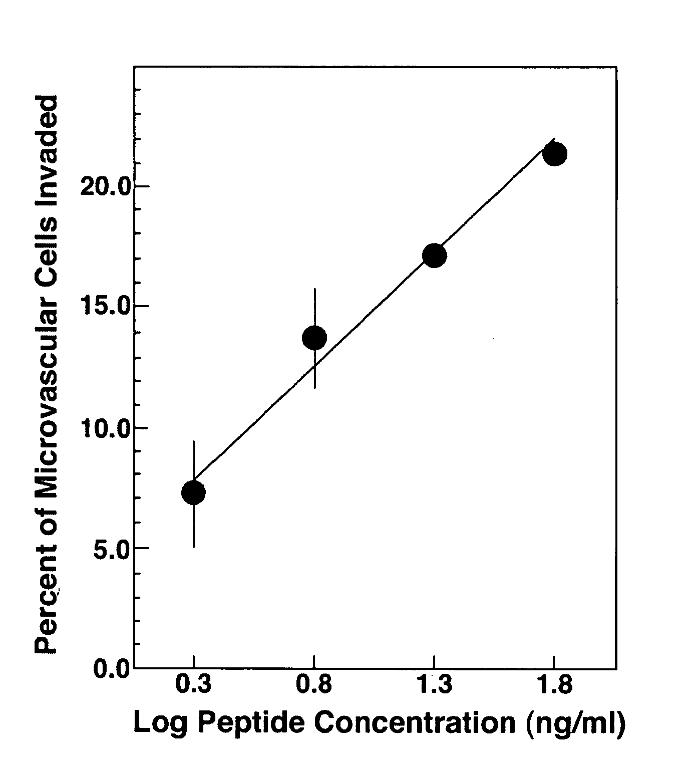
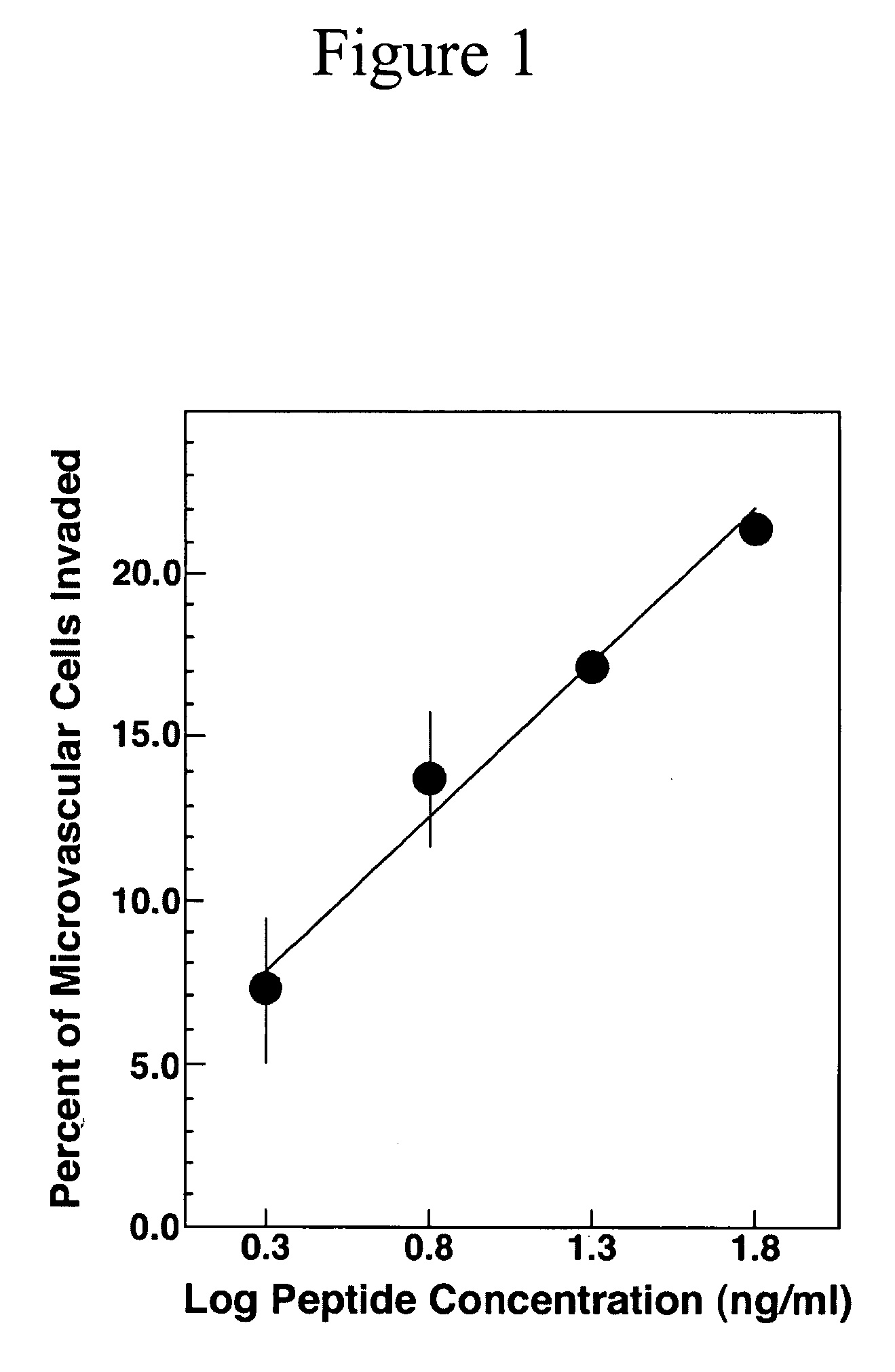
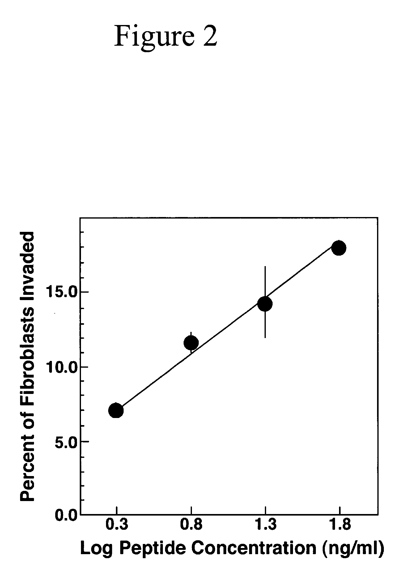
[0056] Cell culture and SU-ECM invasion assays. In SU-ECM in vitro invasion substrates, a basement membrane surrounds a blastocoel, in which invading cells localize shortly after suspension and placement on the outer surfaces. Even in the presence of serum, these invasion substrates have been shown to be free of background invasion by normal cells (Livant et al., (1995) Cancer Res. 55: 5085-5093; Livant et al., (2000) J. Clin. Invest. 105: 1537-1545; Livant et al., (2000) Cancer Res. 60: 309-320). These invasion substrates have been used to define the invasion-promoting activity of the PHSRN sequence from the fibronectin cell-binding domain, a previously unanticipated function of this sequence. Ac-PHSRN-NH.sub.2 peptide has been shown to be a potent topical agent for stimulating dermal wound healing in obese diabetic C57BL6Ksdb / db mice (Livant et al., (2000) J. Clin. Invest. 1...

example 4

Testing the Effects of the MMP 1 Cysteine Switch Peptide on Dermal Wound Healing in Obese Diabetic Mice and in Their Heterozygous, Non-Diabetic Littermates

[0061] Biopsy punches 4 mm in diameter (Henry Schein, Inc., Port Washington N.Y.) are used to wound C57Bl6 KsJ db / db and C57Bl6KsJ db / + mice as previously described (Livant et al., (2000) J. Clin. Invest. 105: 1537-1545). Db / db mice and their non-diabetic, db / + littermates are obtained at 6 to 8 weeks of age from Jackson Laboratories (Bar Harbor Me.).

[0062] Db / db mice are aged at least an additional 2 weeks prior to use in wound healing experiments, to make certain that they display the impaired wound healing phenotype characteristic of this strain. Treatment groups consist of 10 to 20 dermal wounds each, or 5 to 10 db / db or db / + mice with duplicate dermal wounds. Anesthetized mice are wounded once or twice on the upper back by pinching the skin away from the underlying fascia and muscle, and pushing the biopsy punch through the s...


PUM

PropertyMeasurementUnit
concentrationsaaaaaaaaaa

Abstract

The present invention relates to methods and compositions for wound healing, and in particular, methods and compositions to promote and enhance wound healing. In particular, the present invention provides MMP derived peptides for use in enhancing wound healing.

Description

[0001] The present invention claims priority to U.S. Provisional Patent Application Serial No. 60 / 396,366, filed Jul. 17, 2002, the disclosure of which is herein incorporated by reference in its entirety.[0002] The present invention relates to methods and compositions for wound healing, and in particular, methods and compositions to promote and enhance wound healing.[0003] The primary goal in the treatment of wounds is to achieve wound closure. Open cutaneous wounds represent one major category of wounds and include burn wounds, neuropathic ulcers, pressure sores, venous stasis ulcers, and diabetic ulcers. Open cutaneous wounds routinely heal by a process which comprises six major components: i) inflammation, ii) fibroblast proliferation, iii) blood vessel proliferation, iv) connective tissue synthesis v) epithelialization, and vi) wound contraction. Wound healing is impaired when these components, either individually or as a whole, do not function properly. Numerous factors can aff...

Claims


Application Information

Patent Timeline
Patent Type & Authority Applications(United States)
IPC IPC(8): A61K38/48
CPCA61K38/4886
Inventor LIVANT, DONNA L.
Owner RGT UNIV OF MICHIGAN
Why Eureka
Try Eureka