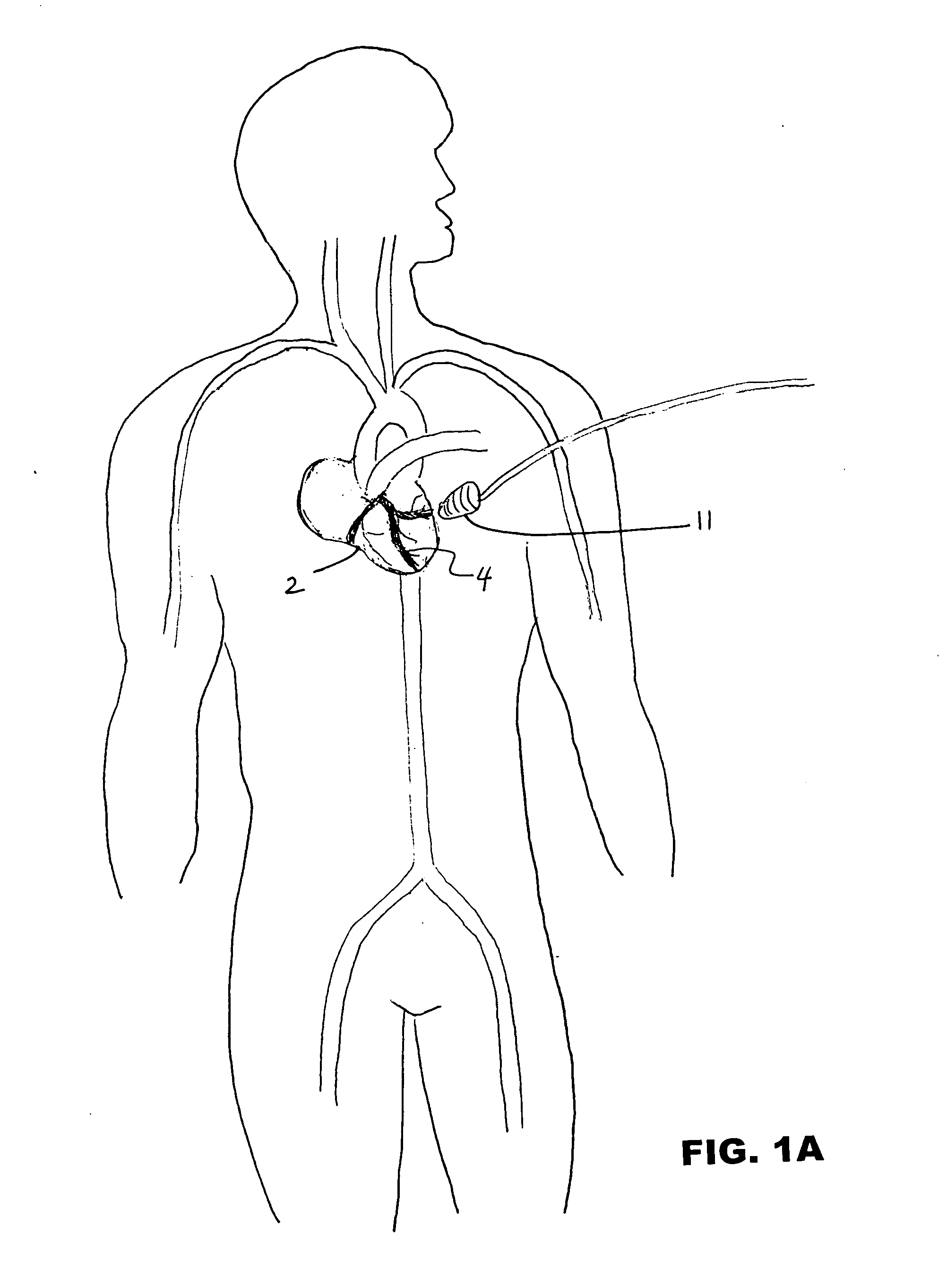
[0006] Microcirculatory damage is a crucial pathogenic mechanism in the induction and propagation of damage in ischemia-reperfusion (I / R)-induced injury associated with decreased perfusion, especially in acute myocardial infarction. The present invention relates to methods and devices for stimulating reperfusion so as to minimize microcirculatory damage after an ischemic event. In a first method, a patient is selected having a tissue with compromised perfusion, such as a myocardial infarction. An ultrasound transducer is applied to a location near the heart, e.g., on the chest above the heart, preferably in the area of the coronary occlusion. It may be desirable to apply a gel to enhance transmission of transcutaneous ultrasound.
[0007] The transducer is activated to initiate exposure of the myocardium and coronary arteries to ultrasound. The exposure to ultrasound causes local vasodilatation of the coronary arteries through shear stress-stimulated production of nitric oxide and / or attenuation of oxygen free radical species thus determining vasodilatation and protecting the endothelial barrier during reperfusion. Ultrasound treatment is likely to be most effective if initiated within 30 minutes of the myocardial infarction. Ultrasound treatment may be accompanied by injecting an anticlotting agent into the patient, the anticlotting agent being any of aspirin, tissue plasminogen activator, and / or streptokinase.
[0008] Exposure to ultrasound will be maintained for a duration of time, usually 0.5 to 10 minutes, more preferably 5 to 10 minutes, in some cases longer than 10 minutes, and in other cases up to 15 minutes or more than 15 minutes. In certain cases it may be desirable to confirm that local vasodilatation has been stimulated. Measuring enhancement of perfusion can confirm that local vasodilatation has been stimulated. Angiography, electrocardiogram, diagnostic ultrasound, and measuring blood levels of creatine kinase can be used to measure enhancement of perfusion in the coronary arteries. Typically, a baseline coronary blood flow is measured before treating the ischemic condition. Then, after ultrasound exposure, coronary blood flow is measured again and compared with the baseline measurement to determine the level of enhancement. In certain cases, ultrasound exposure will be repeated a second time, or more, until normal blood flow to the ischemic tissues is established.
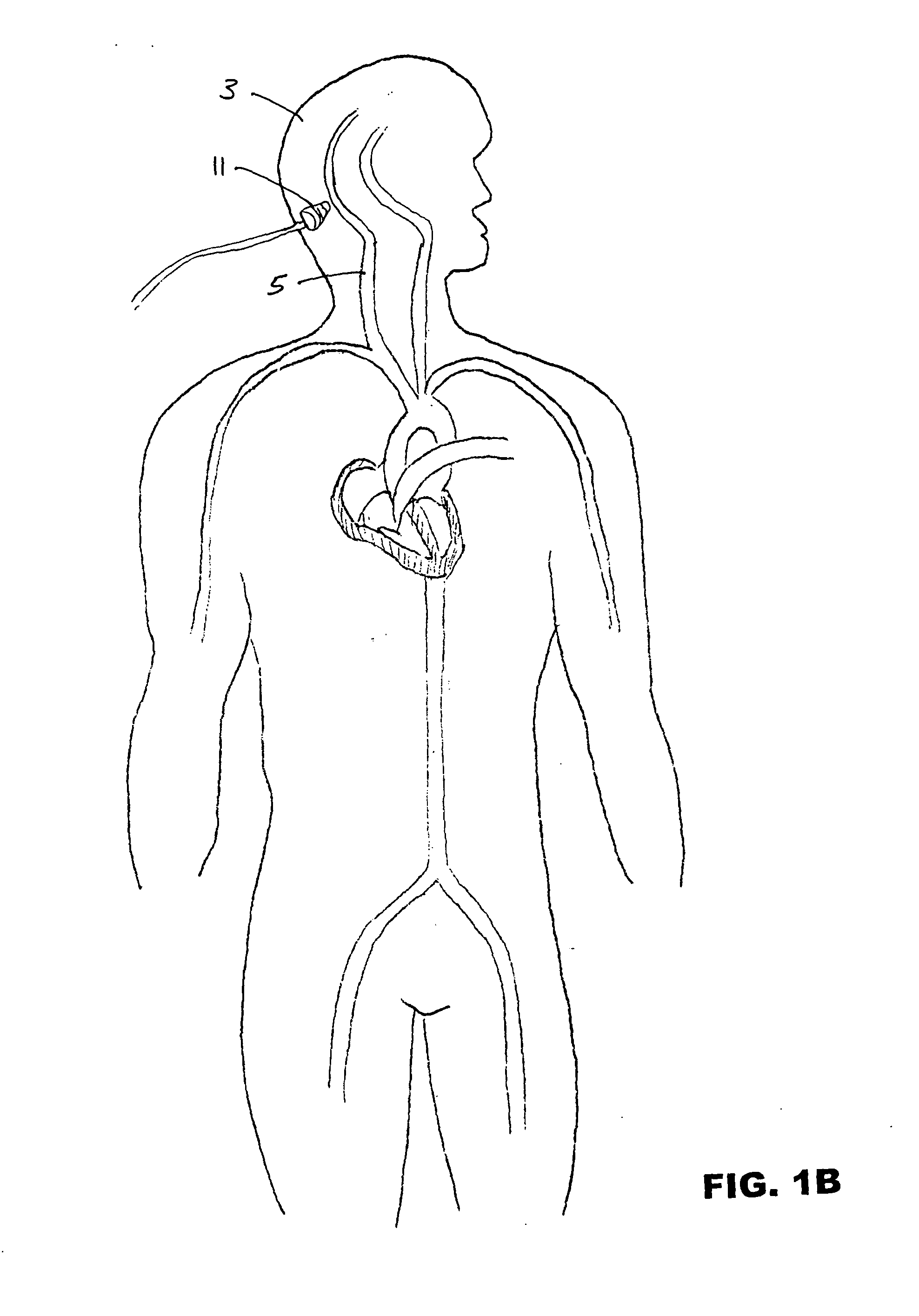
[0009] In another method, a patient is selected having a cerebral tissue with compromised perfusion, e.g., a stroke. An ultrasound transducer is applied to a location near the head and / or neck, preferably in the area of the cerebral vascular occlusion. A gel may be used to enhance transcutaneous ultrasound. The transducer is activated to initiate exposure of the head or neck to ultrasound. Cerebral blood flow is enhanced by dilating one or more of the right brachiocephalic trunk, left common carotid artery, left subclavian artery, right common carotid artery, right subclavian artery, left internal carotid artery, left middle cerebral artery, left anterior cerebral artery, right internal carotid artery, anterior cerebral arteries, anterior communicating artery, right posterior communicating artery, left posterior communicating artery, right posterior cerebral artery, left posterior cerebral artery, left vertebral artery, right vertebral artery, basilar artery, femoral artery, brachial artery, a carotid bulb, and any other arteries of the head and neck that provide cerebral perfusion. In certain methods it is desirable to use transcranial Doppler or carotid Doppler to confirm the enhancement of cerebral blood flow during or after application of ultrasound therapy.
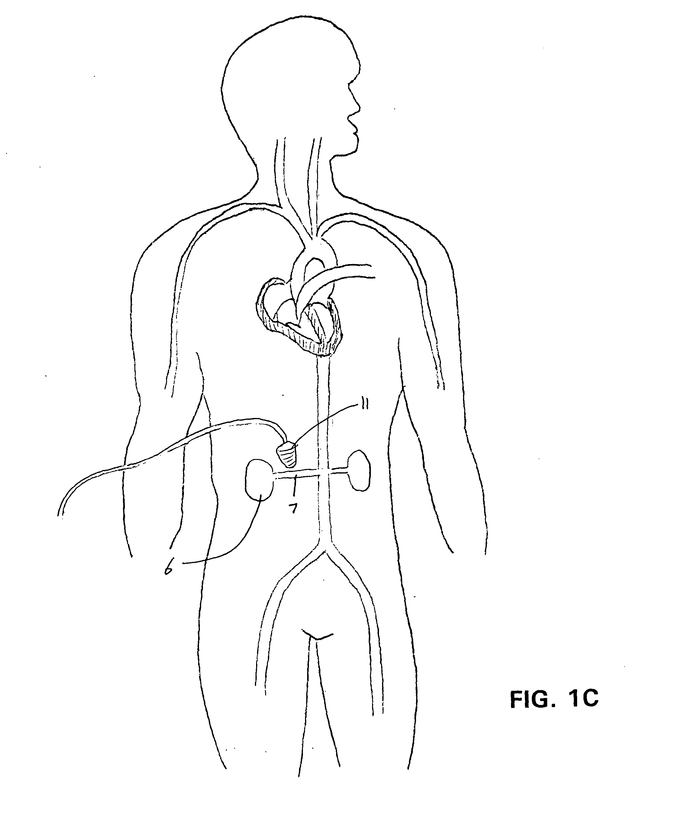
[0010] In another method, a patient is selected having a tissue or organ transplant, e.g., a kidney, liver, heart, or lung transplant, or a tissue or skin graft. An ultrasound transducer is applied to a location near the transplanted tissue. A gel may be used to enhance transcutaneous ultrasound. The transducer is activated to initiate exposure of the transplanted tissue to ultrasound. Local vasodilatation is stimulated to enhance early reperfusion and minimize oxygen free radical injury. In certain methods it is desirable to confirm the enhancement of blood flow during or after application of ultrasound therapy using angiogram or ultrasound with Doppler.

