[0010] The methods and systems of the present invention utilize a 3D
structured light scanner and geometry based
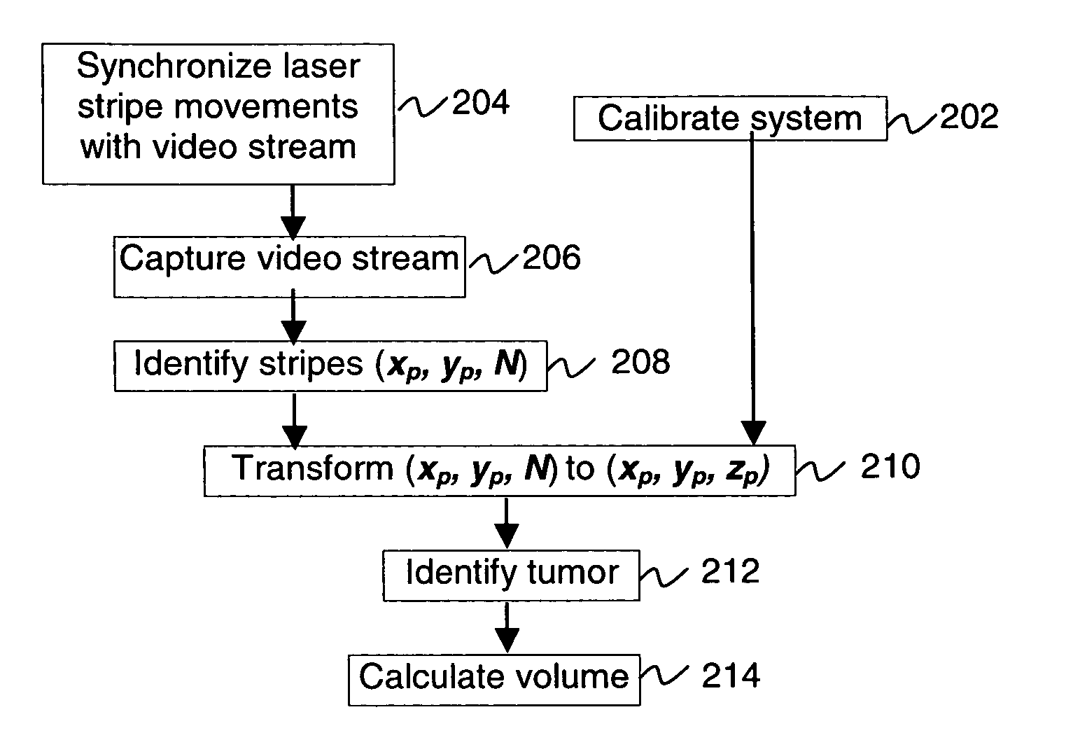
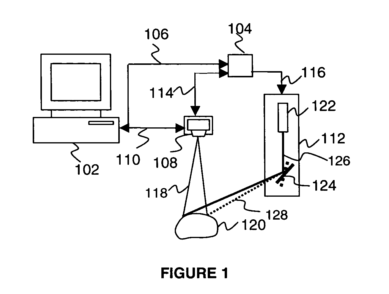
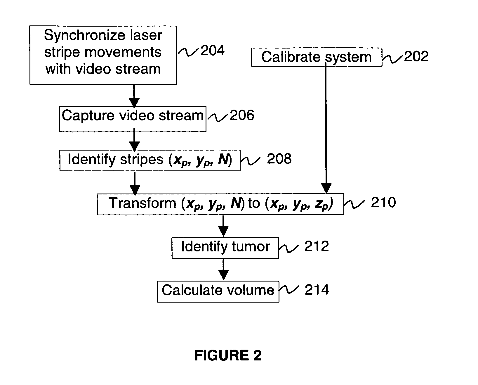
feature recognition logic to obtain shape, size and / or volume of the animal surface features observed in clinical studies. Such features may include tumors, lesions, sores, and wounds and any other suitable feature on the surface of animals, plants and humans, and any other suitable surface. The invention may obtain a number of 2D images of the surface feature, such as a tumor, with patterns of
structured light projected on the feature. The
parallax between a camera and a
light source is used to compute a 3D
point cloud of the feature's surface. The point cloud is a collection of (x, y, z) coordinates of the surface feature points.
Recognition algorithm separates the feature points from the healthy points by minimizing the standard deviation of the point cloud points from the suggested shape. After the separation is achieved, the feature size and volume may be calculated.
[0012] In this way, the invention provides for fast, non-contact, automated measurements of external features such as tumors, lesions, sores, wounds shape, size and volume on the surface of animals, plants and humans.
[0013] The invention also provides time savings, which are extremely important when measuring, for example, tumor progress using
large animal sets. For example, the 3D
scanner of the present invention is many times faster than CAT methods. Compared to caliper method, the 3D
scanner measurements are also faster. In addition, the 3D scanner method may be completely automated (i.e., measurement data may be electronically transferred to a computer from a measuring device, eliminating extra time needed to
record and transfer paper recorded data to computers).
[0014] The invention may further be totally non-invasive (i.e., no part of the device touches the animal). This leads to three important consequences, for example, for making tumor
volume measurements: i) animals are not stressed as much, which less affects the
tumor growth; ii) there is no need to sterilize the device; and iii) tumors are not deformed by the
measuring equipment. Only the 3D scanner light touches the animals. The 3D scanner method does not involve injecting contrast agents or radioactive substances into animals. Since it takes a very short time, around a few seconds, to complete the measurement, the 3D scanner method is less stressful to animals and preferably does not require anesthetizing them.
[0016] Further in accordance with the invention, certain embodiments also include: basing the separation and calculation upon the feature portion of the surface having a predetermined feature shape and the non-feature portion of the surface having predetermined non-feature shape; the: predetermined feature shape and the predetermined non-feature shape being described by a finite set of parameters; performing successive approximations to find the set of parameters; selecting an arbitrary initial choice of the set of parameters for the successive approximations; calculating distances between coordinates in the set and each of the predetermined feature shape and the predetermined non-feature shape; assigning the coordinates in the set to the feature portion of the set when the coordinates are closer to the predetermined feature shape than to the predetermined non-feature shape, and to the non-feature portion of the set when the coordinates are closer to the predetermined non-feature shape than to the predetermined feature shape; finding the set of parameters by minimizing the standard deviation of the feature portion of the set from the predetermined feature shape and by minimizing the standard deviation of the non-feature portion of the set from the predetermined non-feature shape; and calculating the size of the predetermined feature shape based on the set of parameters and estimating the size of the feature portion of the surface to be the size of the predetermined feature shape.


