Incontinence treatment device

a treatment device and incontinence technology, applied in the field of medical electronic devices, can solve the problems of stress incontinence, inability of pelvic muscles to hold back urinary flow from the bladder, discomfort and embarrassment, etc., and achieve the effect of inhibiting involuntary urine flow

Inactive Publication Date: 2005-05-26
ASTORA WOMENS HEALTH
View PDF61 Cites 24 Cited by
  • Summary
  • Abstract
  • Description
  • Claims
  • Application Information

AI Technical Summary

Benefits of technology

[0018] It is a further object of some aspects of the present invention to provide a device and method for enhancing function of muscles, particularly those associated with urine control.
[0080] responsive to the analysis, applying an electrical waveform to the electrode, which stimulates the muscle to contract so as to inhibit involuntary urine flow.

Problems solved by technology

Urinary stress incontinence affects millions of people, causing discomfort and embarrassment, sometimes the point of social isolation.
Stress incontinence results from weakness or inability of pelvic muscles to hold back urinary flow from the bladder when abdominal pressure increases due to everyday events such as coughing, laughing or mild physical exertion.
These products are not sufficiently absorbent to be effective in severe cases.
They are uncomfortable to wear, and cause skin irritation, as well as unpleasant odors.
The disadvantages of surgery are its high cost, need for hospitalization and long recovery period, and high frequency of complications.
The constant concentric pressure that the peripheral cuff exerts on the urethra results in impaired blood supply to tissue in the area, leading to tissue atrophy, urethral erosion and infection.
Furthermore, the constant pressure in the cuff is not always sufficient to overcome transient increases in bladder pressure that may result from straining, coughing, laughing or contraction of the detrusor muscle, for example.
In such cases, urine leakage may result.
This system is complicated, however, and requires manual manipulation of the subdermal pump and cuff control.
Retraining muscles is not possible or fully effective for most patients, however, particularly when there may be neurological damage or other pathologies involved.

Method used


Image

  • Incontinence treatment device
  • Incontinence treatment device
  • Incontinence treatment device

Examples


Embodiment Construction

I. Overview of Preferred Embodiments

[0088] A. General description of stimulator device

[0089] B. Sensing and control functions of the device

[0090] C. Signal processing

[0091] D. Power consumption control

II. Detailed Description of Figures

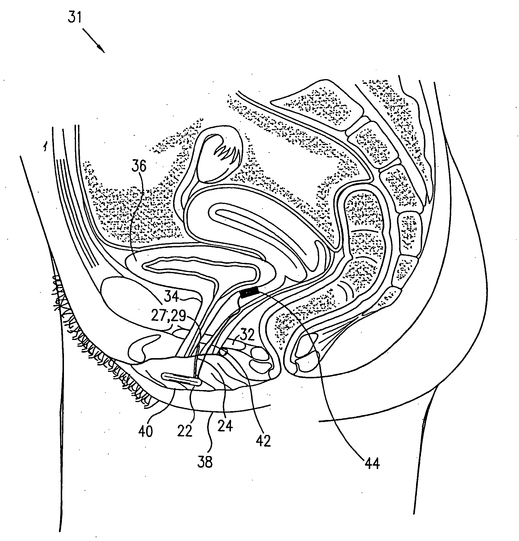
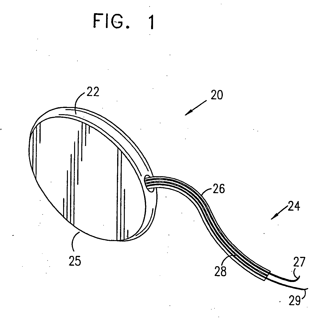
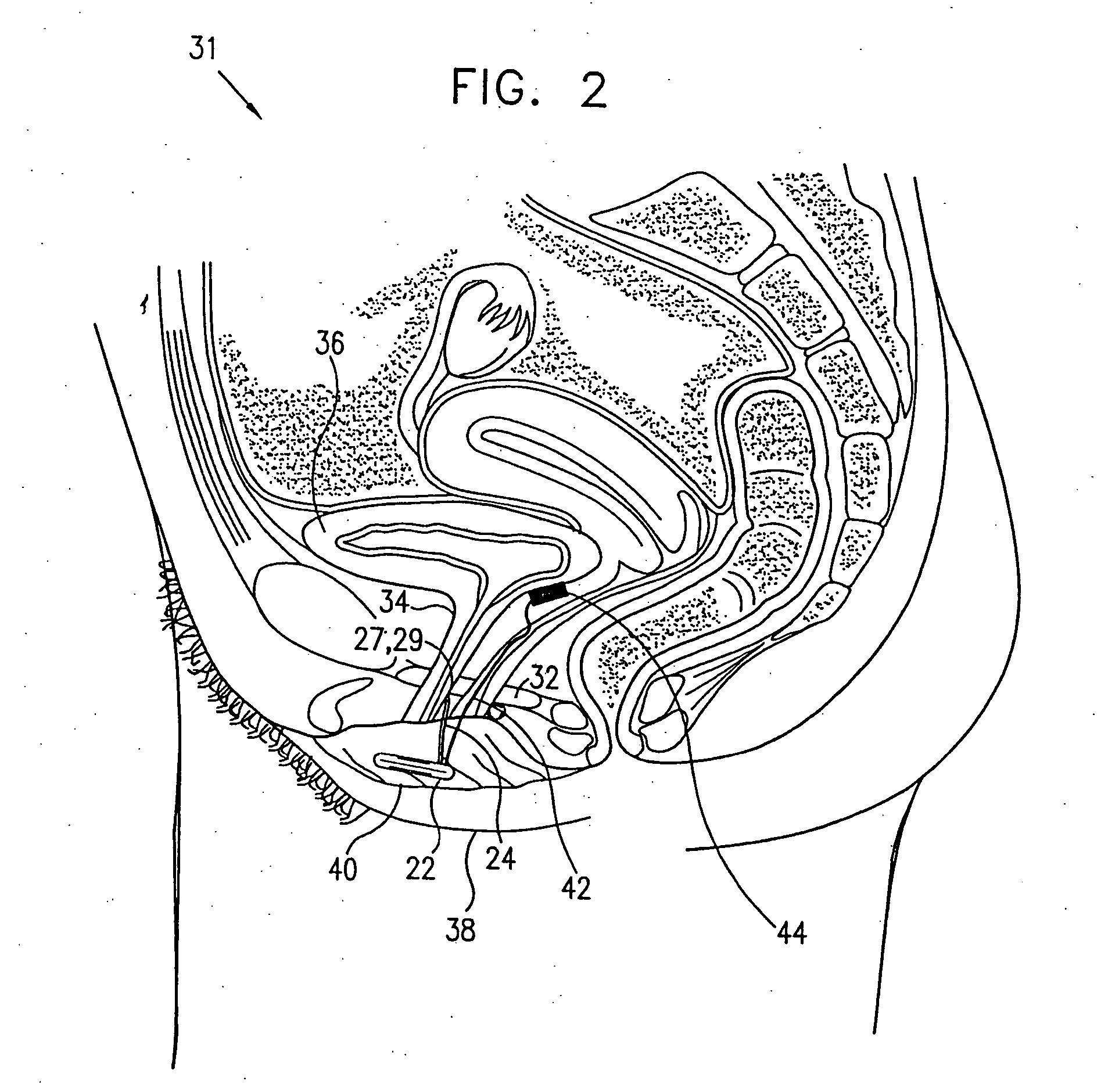
[0092] A. External elements of a stimulator device

[0093] B. Anatomical and surgical considerations

[0094] C. Signal processing [0095] (i) hardware and algorithms [0096] (ii) simulation of a typical EMG [0097] (iii) experimentally measured EMG signals: distinguishing incontinence from voluntary voiding

[0098] D. Muscle stimulation

[0099] E. Provision of power to the control unit

[0100] F. External communication with the control unit

[0101] G. Utilization of other sensors

[0102] H. Reduction of power consumption

I. Overview of Preferred Embodiments

[0103] A. General Description of Stimulator Device

[0104] Various aspects of the present invention are described in this section (I) and in greater detail in the following section (II). As described w...


PUM


Abstract

A device and method for treatment of urinary stress incontinence. At least one electrode is implanted in a pelvic muscle of a patient. A control unit receives signals indicative of abdominal stress in the patient and responsive thereto applies an electrical waveform to the electrode which stimulates the muscle to contract, so as to inhibit involuntary urine flow through the patient's urethra due to the stress.

Description

CROSS-REFERENCE TO RELATED APPLICATIONS [0001] This application is a continuation-in-part of U.S. patent application Ser. No. 09 / 167,244, which is incorporated herein by reference.FIELD OF THE INVENTION [0002] The present invention relates generally to medical electronic devices, and specifically to implantable electrical muscle stimulators. BACKGROUND OF THE INVENTION [0003] Urinary stress incontinence affects millions of people, causing discomfort and embarrassment, sometimes the point of social isolation. Stress incontinence results from weakness or inability of pelvic muscles to hold back urinary flow from the bladder when abdominal pressure increases due to everyday events such as coughing, laughing or mild physical exertion. Muscles involved in controlling the urinary flow include primarily the urethral sphincter and the levator ani, with the cooperation of fibromuscular extensions along the urethra and other muscles in the general region of the pelvic diaphragm. In the United...

Claims


Application Information

Patent Timeline
Patent Type & Authority Applications(United States)
IPC IPC(8): A61FA61F2/00A61F2/02A61N1/18A61N1/36
CPCA61B5/04882A61B2018/00523A61N1/378A61F2250/0001A61N1/36007A61F2/0036A61B5/391
Inventor GROSSCOHEN, EHUDNISSENKORN, ISRAELLIFSCHITZ, DAVID
Owner ASTORA WOMENS HEALTH
Why Eureka
Try Eureka