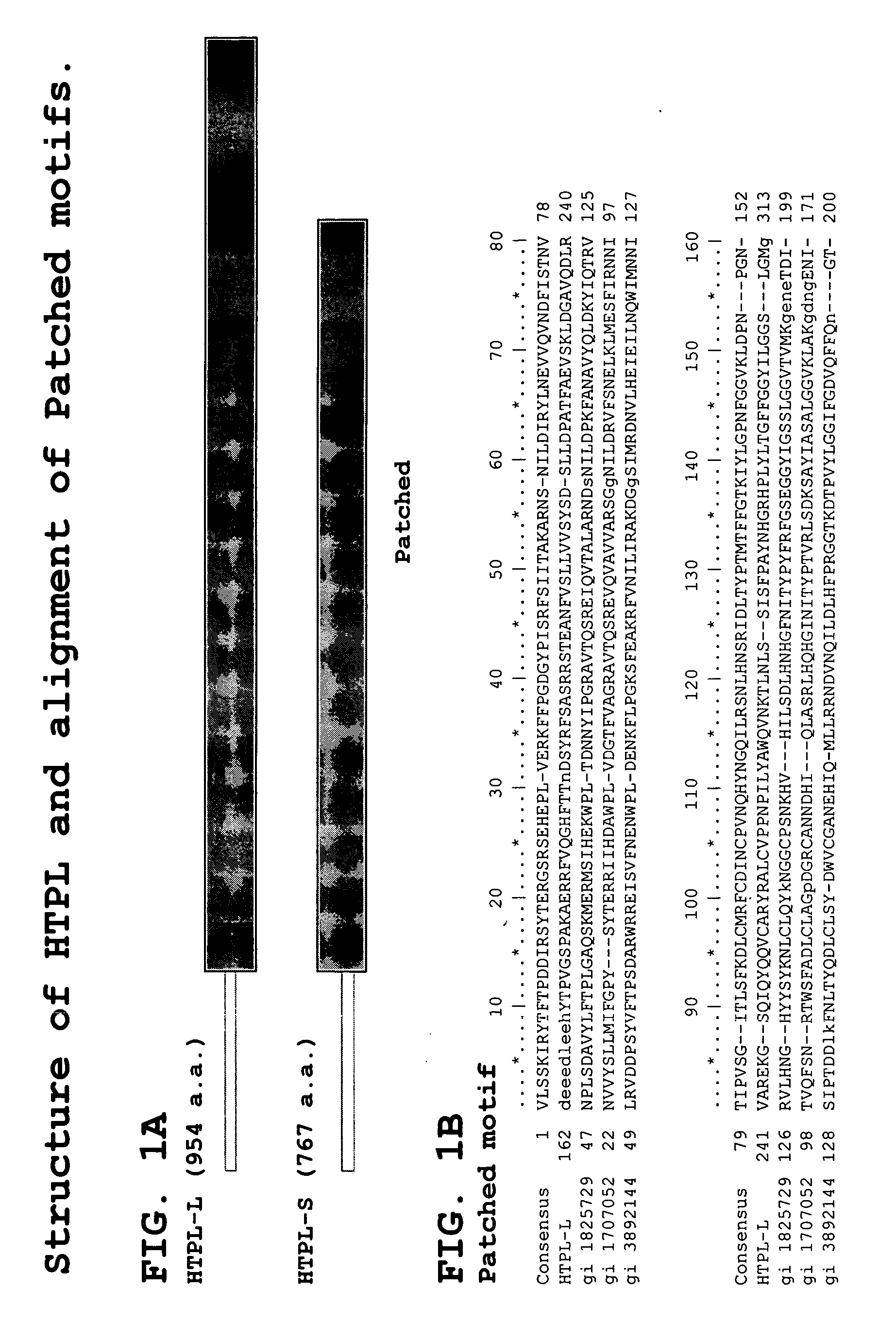
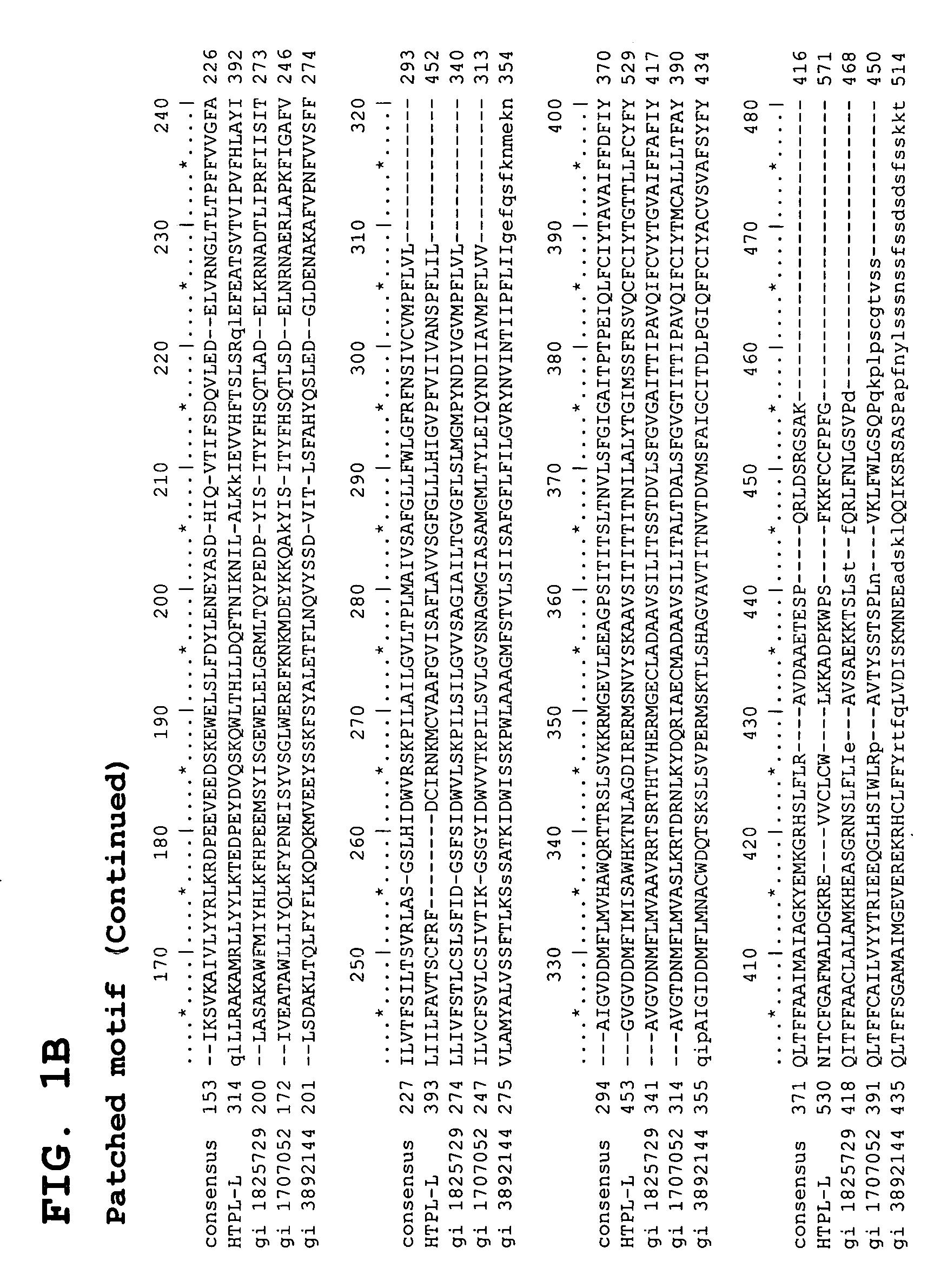
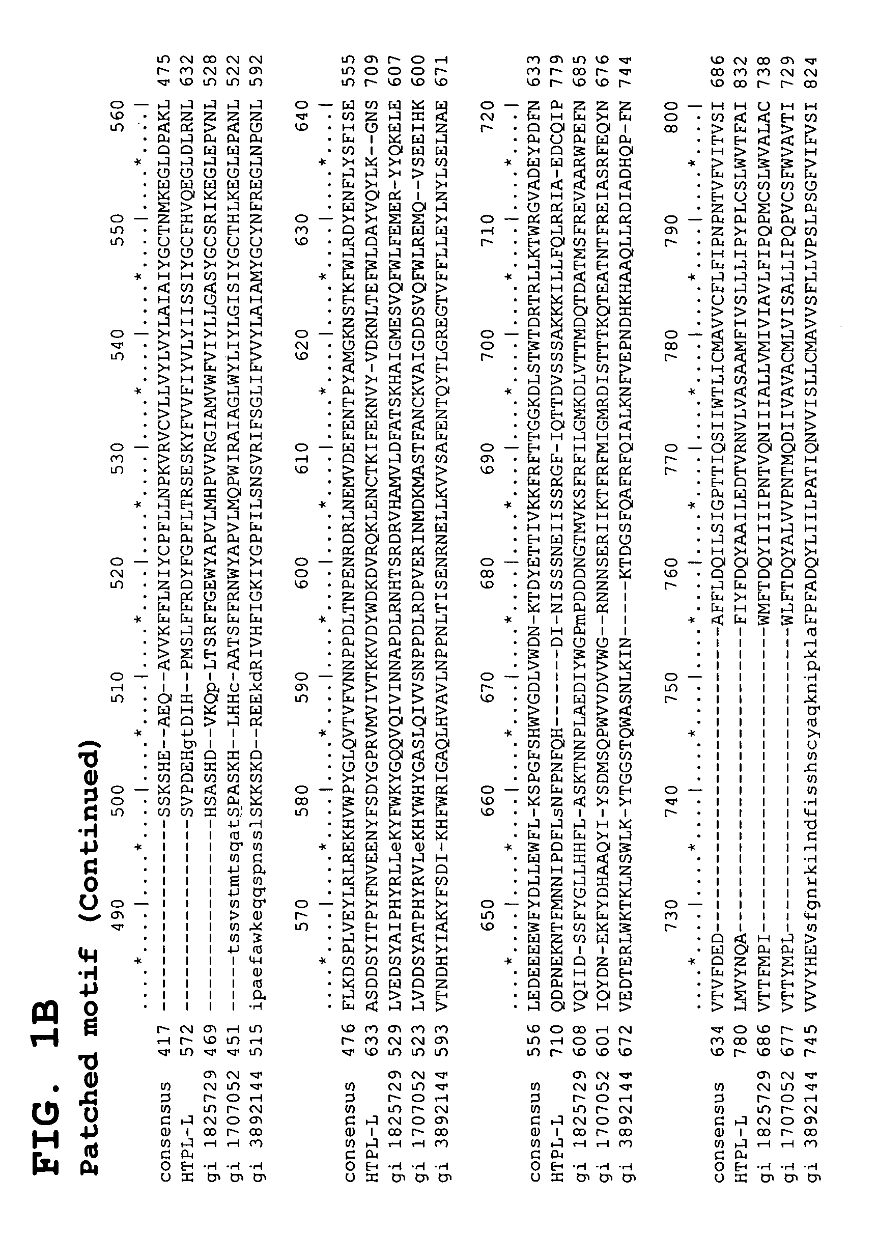
Human testis expressed patched like protein
Inactive Publication Date: 2005-06-16
ZHANG JIAN
1 Cites 0 Cited by
AI-Extracted Technical Summary
Problems solved by technology
For example, mutations in SSD of SCAP result in the disruption of cholesterol homeostasis.
Although Patched-2 has yet to be demonstrated ...
Method used
[0073] As a consequence, any nucleic acid sequence presented herein may contain errors introduced by erroneous incorporation of nucleotides during polymerization, by erroneous base calling by the automated sequencer (although such sequencing errors have been minimized for the nucleic acids directly determined herein, unless otherwise indicated, by the sequencing of each of the complementary strands of a duplex DNA), or by similar errors accessioned into the public database. Such errors can readily be identified and corrected by resequencing of the genomic locus using standard techniques.
[0115] The uncharged nature of the PNA backbone provides PNA/DNA and PNA/RNA duplexes with a higher thermal stability than is found in DNA/DNA and DNA/RNA duplexes, resulting from the lack of charge repulsion between the PNA and DNA or RNA strand. In general, the Tm of a PNA/DNA or PNA/RNA duplex is 1° C. higher per base pair than the Tm of the corresponding DNA/DNA or DNA/RNA duplex (in 100 mM NaCl).
[0137] The nucleic acids of the present invention can be attached covalently to a surface of the support substrate or applied to a derivatized surface in a chaotropic agent that facilitates denaturation and adherence by presumed noncovalent interactions, or some combination thereof.
[0164] Isothermal amplification approaches, such as rolling circle amplification, are also now well-described. See, e.g., Schweitzer et al., Curr. Opin. Biotechnol. 12(1):21-7 (2001); U.S. Pat. Nos. 6,235,502, 6,221,603, 6,210,884, 6,183,960, 5,854,033, 5,714,320, 5,648,245, and international patent publications WO 97/19193 and WO 00/15779, the disclosures of which are incorporated herein by reference in their entireties. Rolling circle amplification can be combined with other techniques to facilitate SNP detection. See, e.g., Lizardi et al., Nature Genet. 19(3):225-32 (1998).
[0252] As yet another example, insect cells are often chosen for high efficiency protein expression. Where the host cells are from Spodoptera frugiperda—e.g., Sf9 and Sf21 cell lines, and expresSF™ cells (Protein Sciences Corp., Meriden, Conn., USA)—the vector replicative strategy is typically based upon the baculovirus life cycle. Typically, baculovirus transfer vectors are used to replace the wild-type AcMNPV polyhedrin gene with a heterologous gene of interest. Sequences that flank the polyhedrin gene in the wild-type genome are positioned 5′ and 3′ of the expression cassette on the transfer vectors. Following cotransfection with AcMNPV DNA, a homologous recombination event occurs between these sequences resulting in a recombinant virus carrying the gene of interest and the polyhedrin or p10 promoter. Selection can be based upon visual screening for lacZ fusion activity.
[0266] As another example, vectors for expressing proteins of the present invention in mammalian cells will include a promoter active in mammalian cells. Such promoters are often drawn from mammalian viruses—such as the enhancer-promoter sequences from the immediate early gene of the human cytomegalovirus (CMV), the enhancer-promoter sequences from the Rous sarcoma virus long terminal repeat (RSV LTR), and the enhancer-promoter from SV40. Often, expression is enhanced by incorporation of polyadenylation sites, such as the late SV40 polyadenylation site and the polyadenylation signal and transcription termination sequences from the bovine growth hormone (BGH) gene, and ribosome binding sites. Furthermore, vectors can include introns, such as intron II of rabbit β-globin gene and the SV40 splice elements.
[0269] As another example of inducible elements, hormone response elements, such as the glucocorticoid response element (GRE) and the estrogen response element (ERE), can confer hormone inducibility where vectors are used for expression in cells having the respective hormone receptors. To reduce background levels of expression, elements responsive to ecdysone, an insect hormone, can be used instead, with coexpression of the ecdysone receptor.
[0270] Expression vectors can be designed to fuse the expressed polypeptide to small protein tags that facilitate purification and/or visualization.
[0280] Alternatively, the GFP-like chromophore can be selected from GFP-like chromophores modified from those found in nature. Typically, such modifications are made to improve recombinant production in heterologous expression systems (with or without change in protein sequence), to alter the excitation and/or emission spectra of the native protein, to facilitate purification, to facilitate or as a consequence of cloning, or are a fortuitous consequence of research investigation.
[0283] Toward the other end of the emission spectrum, EBFP (“enhanced blue fluorescent protein”) and BFP2 contain four amino acid substitutions that shift the emission from green to blue, enhance the brightness of fluorescence and improve solubility of the protein, Heim et al., Curr. Biol. 6:178-182 (1996); Cormack et al., Gene 173:33-38 (1996). EBFP is optimized for expression in mammalian cells whereas BFP2, which retains the original jellyfish codons, can be expressed in bacteria; as is further discussed below, the host cell of production does not affect the utility of the resulting fusion protein. The GFP-like chromophores from EBFP and BFP2 can usefully be included in the fusion proteins of the present invention, and vectors containing these blue-shifted variants are available from Clontech Labs (Palo Alto, Calif., USA).
[0286] Fusions to the IgG Fc region increase serum half life of protein pharmaceutical products through interaction with the FcRn receptor (also denominated the FcRp receptor and the Brambell receptor, FcRb), further described in international patent application nos. WO 97/43316, WO 97/34631, WO 96/32478, WO 96/18412.
[0288] Stable expression is readily achieve...
Benefits of technology
[0008] The present invention solves these and other needs in the art by providing isolated nucleic acids that encode human testis expres...
Abstract
The invention provides isolated nucleic acids that encode HTPL, including two isoforms, and fragments thereof, vectors for propagating and expressing HTPL nucleic acids, host cells comprising the nucleic acids and vectors of the present invention, proteins, protein fragments, and protein fusions of the novel HTPL isoforms, and antibodies thereto. The invention further provides transgenic cells and non-human organisms comprising human HTPL nucleic acids, and transgenic cells and non-human organisms with targeted disruption of the endogenous orthologue of the human HTPL gene. The invention further provides pharmaceutical formulations of the nucleic acids, proteins, and antibodies of the present invention, and diagnostic, investigational, and therapeutic methods based on the HTPL nucleic acids, proteins, and antibodies of the present invention.
Application Domain
Peptide/protein ingredientsAntibody mimetics/scaffolds +13
Technology Topic
AntibodyProtein Fragment +7
Image
Examples
- Experimental program(8)
Example
EXAMPLE 1
Identification and Characterization of cDNAs Encoding HTPL Proteins
[0511] Predicating our gene discovery efforts on use of genome-derived single exon probes and hybridization to genome-derived single exon microarrays—an approach that we have previously demonstrated will readily identify novel genes that have proven refractory to mRNA-based identification efforts—we identified an exon in raw human genomic sequence that is particularly expressed in human adrenal, adult and fetal liver, bone marrow, brain, kidney, lung, placenta and prostate.
[0512] Briefly, bioinformatic algorithms were applied to human genomic sequence data to identify putative exons. Each of the predicted exons was amplified from genomic DNA, typically centering the putative coding sequence within a larger amplicon that included flanking noncoding sequence. These genome-derived single exon probes were arrayed on a support and expression of the bioinformatically predicted exons assessed through a series of simultaneous two-color hybridizations to the genome-derived single exon microarrays.
[0513] The approach and procedures are further described in detail in Penn et al., “Mining the Human Genome using Microarrays of Open Reading Frames,”Nature Genetics 26:315-318 (2000); commonly owned and copending U.S. patent application Ser. No. 09/864,761, filed May 23, 2001, Ser. No. 09/774,203, filed Jan. 29, 2001, and Ser. No. 09/632,366, filed Aug. 3, 2000, the disclosures of which are incorporated herein by reference in their entireties.
[0514] Using a graphical display particularly designed to facilitate computerized query of the resulting exon-specific expression data, as further described in commonly owned and copending U.S. patent application Ser. No. 09/864,761, filed May 23, 2001, Ser. No. 09/774,203, filed Jan. 29, 2001 and 09/632,366, filed Aug. 3, 2000, the disclosures of which are incorporated herein by reference in their entireties, one exon was identified that is expressed in all the human tissues tested; subsequent analysis revealed that the exon represent a gene.
[0515] Table 1 summarizes the microarray expression data obtained using genome-derived single exon probe corresponding to exon four. The probe was completely sequenced on both strands prior to its use on a genome-derived single exon microarray; sequencing confirmed the exact chemical structure of the probe. An added benefit of sequencing is that it placed us in possession of a set of single base-incremented fragments of the sequenced nucleic acid, starting from the sequencing primer's 3′ OH. (Since the single exon probe was first obtained by PCR amplification from genomic DNA, we were of course additionally in possession of an even larger set of single base incremented fragments of the single exon probe, each fragment corresponding to an extension product from one of the two amplification primers.).
[0516] Signals and expression ratios are normalized values measured and calculated as further described in commonly owned and copending U.S. patent application Ser. No. 09/864,761, filed May 23, 2001, Ser. No. 09/774,203, filed Jan. 29, 2001, Ser. No. 09/632,366, filed Aug. 3, 2000, and U.S. provisional patent application No. 60/207,456, filed May 26, 2000, the disclosures of which are incorporated herein by reference in their entireties. TABLE 1 Expression Analysis Genome-Derived Single Exon Microarray Amplicon 86654, Exon 4 (TISSUE) Signal Ratio Adrenal 1.02 1.06 adult liver 0.95 1.00 bone marrow 1.63 1.01 Brain 1.20 1.27 fetal liver 1.22 −1.22 Kidney 0.90 1.07 Lung 0.96 −1.11 Placenta 1.02 −1.05 Prostate 0.85 1.18
[0517] As shown in Table 1, low level expression of exon four was seen in adrenal, adult and fetal liver, bone marrow, brain, kidney, lung, placenta and prostate. Low level expression of HTPL in these tissues was further confirmed by RT-PCR analysis (see below).
[0518] Rapid Amplification of cDNA ends (RACE, Clontech Laboratories, Palo Alto, Calif.) and direct RT-PCR were used to obtain the coding region of the human cDNA sequence. The final pair of primers used to amplify the 3.3 kb transcript were: 62NF3 (5′-CAGGAAACCGTCTGGTGGGATCTC-3′; SEQ ID NO: 4801) and 62487Rend (5′-CTGAGACGGAGTCTCATTCTTGTCACC-3′; SEQ ID NO: 4802).
[0519] Human testis Marathon ready cDNA (Clontech Laboratories) was used to clone the entire coding region. The PCR parameters were as follows: 94° C. 1 min; (94° C. 10 seconds; 64° C. 30 seconds; 72° C. 3 min.) for 35 cycles. The PCR reaction composition was as follows: 5 ul of cDNA; 5 ul of 10× amplification buffer; 1 ul dNTP (10 M); 3 ul of primer pairs; 1 ul of Taq polymerase Mix and 35 ul double distilled water.
[0520] The PCR product was cloned into a pGEM-Teasy vector and inserts of multiple clones were sequenced on both strands using a MegaBACE™ automatic sequencer (Amersham Biosciences, Sunnyvale, Calif.). Single base pair sequence changes were identified between two groups of clones, and each group was recognized as one isoform. The presence of two isoforms was confirmed by the presence of two genomic clones (see below), in the public database, with the same single base pair sequence changes between them. For reasons described below, we named the two isoforms HTPL-L and HTPL-S. Sequencing both strands provided us with the exact chemical structure of the cDNA, which are shown in FIG. 3 and FIG. 4 and further presented in the SEQUENCE LISTING as SEQ ID NOs: 1 and 4, and placed us in actual physical possession of the entire set of single-base incremented fragments of the sequenced clones, starting at the 5′ and 3′ termini.
[0521] As shown in FIG. 3, the HTPL-L cDNA spans 3296 nucleotides and contains an open reading frame from nucleotide 78 through and including nt 2942 (inclusive of termination codon), predicting a protein of 954 amino acids with a (posttranslationally unmodified) molecular weight of 107.6 kD. The clone appears full length, with the reading frame opening starting with a methionine and terminating with a stop codon.
[0522] As shown in FIG. 4, the HTPL-S cDNA span 3298 nucleotides and contains an open reading frame from nucleotide 78 through and including nt 2381 (inclusive of termination codon), predicting a protein of 767 amino acids with a (posttranslationally unmodified) molecular weight of 86.9 kD. The clone appears full length, with the reading frame opening starting with a methionine and terminating with a stop codon.
[0523] BLAST query of genomic sequence identified one BAC, spanning 16.7 kb, which constitutes the minimum set of clones encompassing the cDNA sequences. Based upon the known origin of the BAC (GenBank accession number AC005875.2), the HTPL gene can be mapped to human chromosome 10p12.1.
[0524] Comparison of the cDNA and genomic sequences identified four exons for HTPL. Exon organization for HTPL is listed in Table 2. TABLE 2 HTPL Exon Structure Exon no. cDNA range genomic range BAC accession 1 1-1166 100221-99056 AC005875.2 2 1167-1288 97823-97702 3 1289-1434 89251-89106 4 1435-3296 85101-83240 (HTPL-L) 1435-3298 85101-83238 (HTPL-S)
[0525]FIG. 2 schematizes the exon organization of the HTPL gene.
[0526] At the top is shown the bacterial artificial chromosome (BAC), with GenBank accession number (AC005875.2), that span the HTPL locus. The genome-derived single-exon probe first used to demonstrate expression from this locus is shown below the BAC and labeled “500”. The 500 bp probe includes sequence drawn solely from exon four.
[0527] As shown in FIG. 2, two HTPL isoforms have been identified. Both isoforms contain four exons, with a few single base pair differences between them (FIG. 3 and FIG. 4). Indeed, BAC AC005875.2 contains the same exonic sequence as HTPL-L, while BAC AL355493.1 contains the same exonic sequence as HTPL-S. The longer isoform, HTPL-L, encodes a protein of 954 amino acids that has a predicted molecular weight, prior to any post-translational modification, of 107.6 kD. One of the single base pair changes in the shorter form (HTPL-S) introduces a premature stop codon at position 2379 of the HTPL-S cDNA. HTPL-S, therefore, encodes a shortened protein of 767 amino acids that has a predicted molecular weight, prior to any post-translational modification, of 86.9 kD. Both cDNA clones appear full length, with the open reading frame starting with a methionine and terminating with a stop codon.
[0528] As further discussed in the examples herein, expression of HTPL was assessed using hybridization to genome-derived single exon microarrays. Microarray analysis of the fourth exon showed low level expression in all tissues tested, including adrenal, adult liver, bone marrow, brain, fetal liver, kidney, lung, placenta and prostate. This was confirmed by RT-PCR. RT-PCR also detected strong expression in testis and weak expression in colon and skeletal muscle (see Example 3).
[0529] The sequence of the HTPL cDNA was used as a BLAST query into the GenBank nr and dbEst databases. The nr database includes all non-redundant GenBank coding sequence translations, sequences derived from the 3-dimensional structures in the Brookhaven Protein Data Bank (PDB), sequences from SwissProt, sequences from the protein information resource (PIR), and sequences from protein research foundation (PRF). The dbEst (database of expressed sequence tags) includes ESTs, short, single pass read cDNA (mRNA) sequences, and cDNA sequences from differential display experiments and RACE experiments.
[0530] BLAST search identified a single human EST (AW665031.1), a single mouse EST (AV280614.1), and a single pig EST (AW436721.1) as having sequence closely related to HTPL.
[0531] Globally, the human HTPL proteins resemble a putative mouse transcript (GenBank accession: BAB29848, the HTPL-L protein with 66% amino acid identity and 78% amino acid similarity over the entire open reading frame).
[0532] Motif searches using Pfam (http://pfam.wustl.edu), SMART (http://smart.embl-heidelberg.de), and PROSITE pattern and profile databases (http://www.expasy.ch/prosite), identified several known domains shared with Patched, including the Patched domain and the Sterol-sensing domain.
[0533]FIG. 1 shows the domain structure of HTPL-L and HTPL-S as well as the alignment of the Patched domain of HTPL-L with that of other protein.
[0534] As schematized in FIG. 1, HTPL shares an overall structural organization with the Patched protein. The shared structural features strongly imply that HTPL plays a role similar to that of Patched, in male germ cell development, and is a potential tumor suppressor.
[0535] Like Patched, HTPL-L contains a Patched domain (http://pfam.wustl.edu/hmmsearch.shtml), a Sterol-sensing domain (SSD, http://motif.genome.ad.jp/) and twelve transmembrane domains (http://smart.embl-heidelberg.de/smart/show_motifs.pl). The Patched domain in HTPL-L covers amino acid sequences 166-952 of HTPL-L. The SSD domain in HTPL-L covers amino acid sequences 383-540 of HTPL-L. The presence of these domains in HTPL-L suggest that HTPL-L, like Patched and other Patched domain containing proteins, is involved in the Hedgehog signaling pathway (see background section). Because of the premature stop of protein translation, HTPL-S contains a partial Patched domain, a complete Sterol-sensing motif and seven transmembrane domains. The presence of these domains in HTPL-S suggests that HTPL-S is also involved in the Hedgehog signaling pathway.
[0536] Other signatures of the newly isolated HTPL proteins were identified by searching the PROSITE database (http://www.expasy.ch/tools/scnpsitl.html), and the list below is for both HTPL-L and HTPL-S unless specified otherwise. These include seven N-glycosylation sites (192-195, 275-278, 279-282, 530-533, 678-681, 692-695 and 737-740), one cAMP- and cGMP-dependent protein kinase phosphorylation site (201-204), seven protein kinase C phosphorylation sites (194-196, 200-202, 508-510, 561-563, 662-664, 746-748, and 759-761; plus one for HTPL-L at 800-802), twelve Casein kinase II phosphorylation sites (19-22, 36-39, 62-65, 79-82, 190-193, 215-218, 219-222, 225-228, 230-233, 572-575, 597-600, and 740-743), two tyrosine kinase phosphorylation site (329-335, and 681-688; plus one for HTPL-L at 887-893), four N-myristoylation sites (307-312, 418-4223, 504-509, and 535-540; plus one for HTPL-L at 935-940), and a single amidation site at 541-544.
[0537] Possession of the genomic sequence permitted search for promoter and other control sequences for the HTPL gene. A putative transcriptional control region, inclusive of promoter and downstream elements, was defined as 1 kb around the transcription start site, itself defined as the first nucleotide of the HTPL cDNA clone. The region, drawn from sequence of BAC AC005875.2, has the sequence given in SEQ ID NO: 23, which lists 1000 nucleotides before the transcription start site.
[0538] Transcription factor binding sites were identified using a web based program (http://motif.genome.ad.jp/), including binding sites for homeo domain factor Nkx-2.5/Csx (625-631 bp), for USF (891-898 bp) and for CdxA (399-405 and 612-618 bp, with numbering according to SEQ ID NO: 23), amongst others.
[0539] We have thus identified a newly described human gene, HTPL, which shares certain protein domains and an overall structural organization with Patched. The shared structural features strongly imply that the HTPL protein plays a role similar to Patched, in the hedgehog signaling pathway, functioning as a potential tumor suppressor and in germ cell development. This makes the HTPL proteins and nucleic acids clinically useful diagnostic markers and potential therapeutic agents for male infertility and cancer.
Example
EXAMPLE 2
Preparation and Labeling of Useful Fragments of HTPL
[0540] Useful fragments of HTPL are produced by PCR, using standard techniques, or solid phase chemical synthesis using an automated nucleic acid synthesizer. Each fragment is sequenced, confirming the exact chemical structure thereof.
[0541] The exact chemical structure of preferred fragments is provided in the attached SEQUENCE LISTING, the disclosure of which is incorporated herein by reference in its entirety. The following summary identifies the fragments whose structures are more fully described in the SEQUENCE LISTING: [0542] SEQ ID NO: 1 (nt, full length HTPL-L cDNA) [0543] SEQ ID NO: 2 (nt, cDNA ORF of HTPL-L) [0544] SEQ ID NO: 3 (aa, full length HTPL-L protein) [0545] SEQ ID NO: 4 (nt, full length HTPL-S cDNA) [0546] SEQ ID NO: 5 (nt, cDNA ORF of HTPL-S) [0547] SEQ ID NO: 6 (aa, full length HTPL-S protein) [0548] SEQ ID NO: 7 (nt, (nt 1-2021) portion of HTPL-L) [0549] SEQ ID NO: 8 (nt; 5′ UT portion of SEQ ID NO: 7) [0550] SEQ ID NO: 9 (nt, coding region of SEQ ID NO: 7) [0551] SEQ ID NO: 10 (aa, (aa 1-648) CDS entirely within SEQ ID NO: 9) [0552] SEQ ID NO: 11 (nt, (nt 2637-3041) portion of HTPL-L) [0553] SEQ ID NO: 12 (nt, coding region of SEQ ID NO: 11) [0554] SEQ ID NO: 13 (nt, 3′ UT portion of SEQ ID NO: 11) [0555] SEQ ID NO: 14 (aa, (aa 854-954) CDS entirely within SEQ ID NO: 12) [0556] SEQ ID NO: 15-18 (nt, exons 1-4 of HTPL-L) [0557] SEQ ID NO: 19-22 (nt, 500 bp genomic amplicons centered about exons 1-4 of HTPL-L) [0558] SEQ ID NO: 23 (nt, 1000 bp putative promoter of HTPL) [0559] SEQ ID NOs: 24-2028 (nt, 17-mers scanning SEQ ID NO: 7) [0560] SEQ ID NOs: 2029-4025 (nt, 25-mers scanning SEQ ID NO: 7) [0561] SEQ ID NOs: 4026-4414 (nt, 17-mers scanning SEQ ID NO: 11) [0562] SEQ ID NOs: 4415-4795 (nt, 25-mers scanning SEQ ID NO: 11) [0563] SEQ ID NO: 4796 (nt, (nt 1-2021) portion of HTPL-S) [0564] SEQ ID NO: 4797 (nt, 5′ UT portion of SEQ ID NO: 4796) [0565] SEQ ID NO: 4798 (nt, coding region of SEQ ID NO: 4796) [0566] SEQ ID NO: 4799 (aa, (aa 1-648) CDS entirely within SEQ ID NO: 4798) [0567] SEQ ID NO: 4800 (nt, (nt 2637-3041) portion of HTPL-S) [0568] SEQ ID NO: 4801 (nt, primer 62NF3 for cloning of HTPL) [0569] SEQ ID NO: 4802 (nt, primer 62487Rend for cloning of HTPL) [0570] SEQ ID NO: 4803 (nt, primer 62487Pu for RT-PCR analysis of HTPL) [0571] SEQ ID NO: 4804 (nt, primer 62487Pd for RT-PCR analysis of HTPL)
[0572] Upon confirmation of the exact structure, each of the above-described nucleic acids of confirmed structure is recognized to be immediately useful as a HTPL-specific probe.
[0573] For use as labeled nucleic acid probes, the above-described HTPL nucleic acids are separately labeled by random priming. As is well known in the art of molecular biology, random priming places the investigator in possession of a near-complete set of labeled fragments of the template of varying length and varying starting nucleotide.
[0574] The labeled probes are used to identify the HTPL gene on a Southern blot, and are used to measure expression of HTPL mRNA on a northern blot and by RT-PCR, using standard techniques.
Example
EXAMPLE 3
Expression Analysis of HTPL by RT-PCR
[0575] The Advantage 2 PCR amplification kit and PCR cDNA of different human tissues were obtained from Clontech Laboratories Inc. (Palo Alto, Calif.). The PCR parameters were set-up as follows, 94° C. 15 seconds; 59° C. 30 seconds; 72° C. 40 seconds for 35 cycles. The PCR composition is as follows: 0.5 ul of cDNA; 2.5 ul of 10× amplification buffer; 0.5 ul dNTP (10 M); 1 ul of primer pairs (10 M each; SEQ ID NO: 4803 and SEQ ID NO: 4804); 0.5 ul of Advantage polymerase Mix in 25 ul reaction mixture. The amplified DNA products were resolved in 1.2% agarose gel in TAE buffer. The gel was scanned using Typhoon™ Imaging System (Amersham Biosciences). HTPL is strongly expressed in testis, weakly expressed in skeletal muscle, bone marrow, lung, liver, kidney, colon and placenta, while hardly expressed in brain, heart and uterus (FIG. 5).
PUM
Property | Measurement | Unit |
Fraction | 0.95 | fraction |
Fraction | 0.65 | fraction |
Fraction | 0.99 | fraction |
Description & Claims & Application Information
We can also present the details of the Description, Claims and Application information to help users get a comprehensive understanding of the technical details of the patent, such as background art, summary of invention, brief description of drawings, description of embodiments, and other original content. On the other hand, users can also determine the specific scope of protection of the technology through the list of claims; as well as understand the changes in the life cycle of the technology with the presentation of the patent timeline. Login to view more.
Similar technology patents
Programmable microcontroller architecture (mixed analog/digital)
InactiveUS7221187B1excellent interfaceneed be solve
Owner:MONTEREY RES LLC
Classification and recommendation of technical efficacy words
- need be solve
Programmable microcontroller architecture (mixed analog/digital)
InactiveUS7221187B1excellent interfaceneed be solve
Owner:MONTEREY RES LLC