Method and apparatus for phase-sensitive magnetic resonance imaging

a phase-sensitive magnetic resonance and imaging technology, applied in the field of medical imaging, can solve the problems of not always realizing the potential for increasing tissue contrast by ir imaging, reducing or even reversed the contrast in an ir image, and lacking the true reliable and automated phase correction method, etc., to achieve the effect of maintaining consistency and maintaining polarity consistency

Active Publication Date: 2005-07-28
BOARD OF RGT THE UNIV OF TEXAS SYST
View PDF12 Cites 52 Cited by
  • Summary
  • Abstract
  • Description
  • Claims
  • Application Information

AI Technical Summary

Benefits of technology

[0020] The method may also provide steps for maintaining polarity consistency of the phase-corrected inversion recover image or for maintaining consistency for water and fat identification for two-point Dixon chemical shift imaging. In one embodiment, the method provides steps for calculating a coil-correlation index for polarity consistency among different coils. In other embodiments, the method provides steps for calculating a ...

Problems solved by technology

The underlying reason is that some unwanted error phase almost always accompanies the desired phase.
Although many methods have been developed to remove the error phase, a truly reliable and automated phase correction method is still lacking.
The potential for increased tissue contrast by IR imaging, however, is not always realized because conventional MR image reconstruction preserves only the magnitude of the MR signals and may actually lead to reduced or even reversed contrast in an IR image.
The main challenge in PSIR image reconstruction is a phase-correction process to separate the intrinsic signal phase in the complex image from other phase errors, which are common in an MR image.
However, these approaches reduce data acquisition efficiency.
Further, spatial misregistration between the actual and calibration scans due to patient motion can also be problematic.
As such, pixels with large phase variation, such as in regions of low signal-to-noise ratio (SNR) or along tissue boundaries may corrupt the phase correction process.
The actual threshold value, however, can be critical.
If the value selected is too small, phase correction cannot reach beyond the regions defined by the threshold value and may thus be terminated prematurely.
Alternatively, if the value selected is too large, errors in phase...

Method used


Image

  • Method and apparatus for phase-sensitive magnetic resonance imaging
  • Method and apparatus for phase-sensitive magnetic resonance imaging
  • Method and apparatus for phase-sensitive magnetic resonance imaging

Examples


example 1

Reconstructing Images Using a 2-point Dixon Technique

[0085] As shown in FIGS. 6A-6D and FIGS. 7A-7B, the resulting images are from the reconstruction and phase correcting methods. The images are from abdominal examinations using 1.5 Tesla MRI scanners and a four-channel torso phased-array receiver coil. In one embodiment, the scanning protocol may include: a sequence repetition time of 150 ms, an echo time (TE) of 2.2 ms for opposed-phase images, an TE of 4.7 ms for in-phase images, a receiver bandwidth of ±62.5 KHz, an acquisition matrix of 256×192 pixels, a field of view of 36×cm, a slice thickness of 5-7 mm, and a gap of 0-1 mm. The protocol may generate approximately 20 slices collected within 20s. Thus, the entire abdomen may be scanned in one or two breath holds.

[0086] In one embodiment of the invention, the reconstruction of the water-only and fat-only images may be performed offline using an IBM ThinkPad PC with an Intel processor (e.g., x86 family operating at approximat...

example 2

Phase Sensitive Inversion Recovery Imaging of Multi-slice and Multi-coil Data

[0093] As noted above, phase-sensitive inversion recover (PSIR) image reconstruction scheme uses both the magnitude and the phase information of the signals for phase correction. The phase correction component may be extended and optimized from a region growing scheme for Dixon water and fat chemical shift imaging. The methods provided by example require neither selection of an empirical phase threshold nor explicit use of a special phase filter. Further, the global polarity of images from different receiver coils and from different slices may be made consistent by exploiting the inter-coil and inter-slice correlation intrinsic to the original complex images.

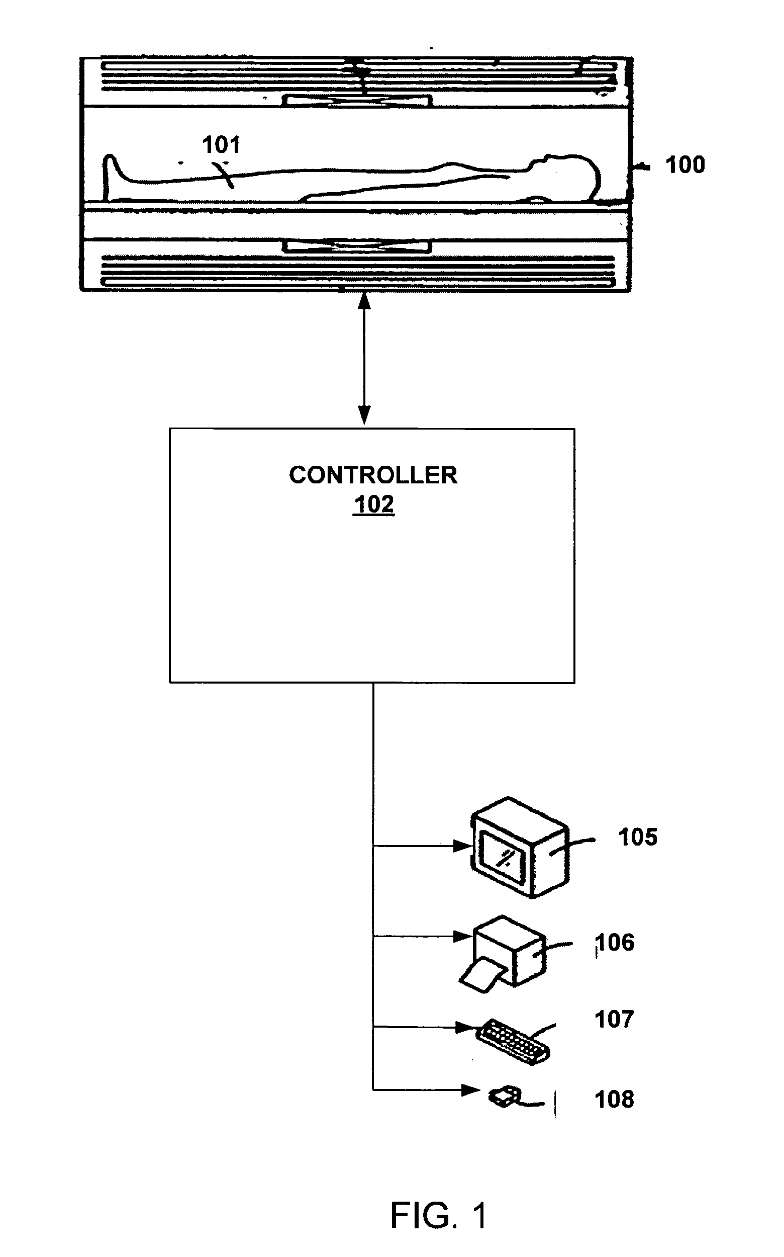
[0094] Referring to FIG. 8, a system 800 including MR scanner 801 such as the General Electric Signa 3.0 Tesla whole-body MR scanner (GE Healthcare, Waukesha, Wis.) using an IR fast spin-echo pulse sequence and head coil 802 such as an 8-channel phas...

example 3

Improving Signal-to-Noise in Multi-slice and Multi-coil Data

[0099] To increase the signal-to-noise ratio (SNR) and / or increase imaging speed, data acquisition in MR imaging is increasingly performed using multiple receiver coils. For image reconstruction methods, such as phase sensitive inversion recovery (PSIR), the images from the multiple receiver coils contain both positive and negative values after a successful phase correction. In order to produce a final image, the images from the different receiver coils must be combined.

[0100] According to an embodiment of the present invention, the polarity of the images from one slice of all the different receiver coils is checked for consistency. Referring to FIGS. 12A-12H, the images are from one particular slice but from 8 different receiver coils. These images are a result of a phase correction, but only FIGS. 12B, 12E, and 12H have the correct polarities. As such, the polarities of the images are altered for consistency. The sum o...


PUM


Abstract

Systems and methods are described for phase-sensitive magnetic resonance imaging using an efficient and robust phase correction algorithm. A method includes obtaining a plurality of MRI data signals, such as two-point Dixon data, one-point Dixon data, or inversion recovery prepared data. The method further includes implementing a phase-correction algorithm, that may use phase gradients between the neighboring pixels of an image. At each step of the region growing, the method uses both the amplitude and phase of pixels surrounding a seed pixel to determine the correct orientation of the signal for the seed pixel. The method also includes using correlative information between images from different coils to ensure coil-to-coil consistency, and using correlative information between two neighboring slices to ensure slice-to-slice consistency. A system includes a MRI scanner to obtain the data signals, a controller to reconstruct the images from the data signals and implement the phase-correction algorithm, and an output device to display phase sensitive MR images such as fat-only images and/or water-only images.

Description

[0001] This patent application claims priority to, and incorporates by reference in its entirety, U.S. Provisional Patent Application Ser. No. 60 / 525,143 filed on Nov.26, 2003.BACKGROUND OF THE INVENTION [0002] 1. Field of the Invention [0003] The invention relates generally to the field of medical imaging. More particularly, the invention relates to phase sensitive magnetic resonance imaging using an efficient and robust image processing algorithm. [0004] 2. Discussion of the Related Art [0005] Magnetic resonance imaging (MRI) has proven useful in the diagnosis of many diseases such as hepatic steatosis, cancer, multiple sclerosis, sports related injury, and bone marrow disorders. MRI provides unique imaging capabilities which are not attainable in any other imaging method. For example, MRI can provide detailed images of soft tissues, abnormal tissues such as tumors, and other structures which cannot be readily imaged using techniques like X-rays. Further, MRI operates without expo...

Claims


Application Information

Patent Timeline
IPC IPC(8): A61B5/05G06T
CPCG01R33/4828G01R33/5659G01R33/56563G01R33/5602
Inventor MA, JINGFEI
Owner BOARD OF RGT THE UNIV OF TEXAS SYST
Why Eureka
Try Eureka