[0009] The method described here is a laboratory safe method for prion isolation using
guanidine thiocyanate lyses of the sample followed by
phenol /
chloroform purification of the proteins. Following
precipitation, the proteins are dissolved in SDS and submitted to
sodium dodecyl suphate
polyacrylamide gel electrophoresis (SDS PAGE). The gel is transferred to Immuno-
blot membrane and Western blotted using
monoclonal antibody against recombinant prion protein (we have found MAB 3F4 to work with human CJD cases but other monoclonals have worked with animal TSE cases suggesting that the nature of the
antibody is not a
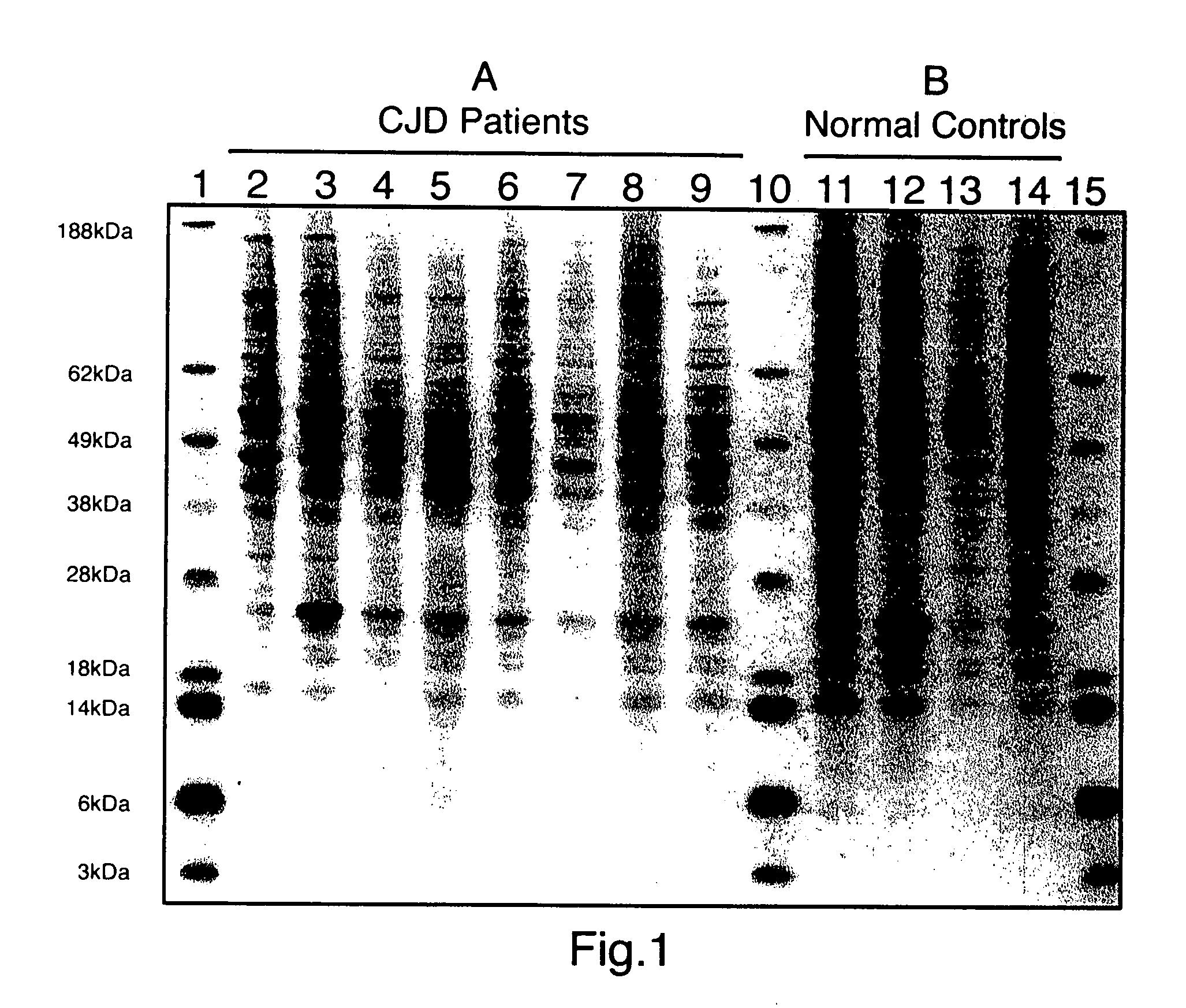
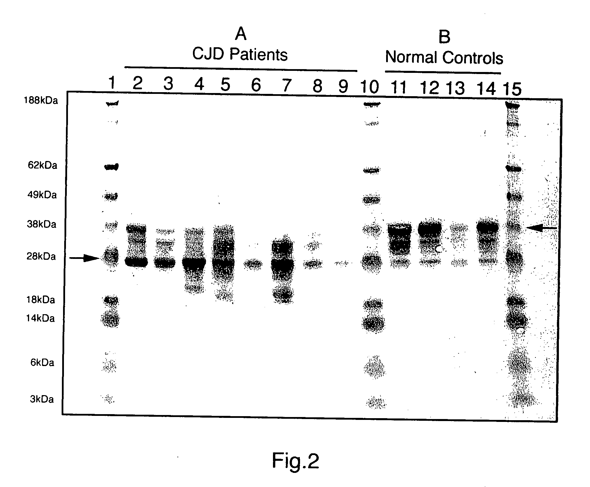
limiting factor). Using this method, the typical normal prion immunoblot pattern was seen in all normal brains examined (15 / 15), whereas all CJD brains (18 / 18) showed a clearly distinct immunoblot pattern. Representative samples of CJD-infected and control samples are shown in FIG. 1. CJD diseased brains could be easily differentiated from normals by this method. We found that normal sheep brains (5) could be easily differentiated from scrapie infected sheep brains (5) using this method with
monoclonal antibody specific for
sheep prion (FIG. 5). This method is safe because the guanidine salts effectively kill the
infectivity of the TSE samples, and, thus, this method can be used in both diagnostic and research laboratories. Since guanidine salts stabilize biomolecules for days to weeks, TSE samples can be procured in the field and added to the guanidine lyses buffer well before the
assay is done making it applicable to any situation where there is problem in transfer. Diagnosis is available within 4-5 hours after the samples are submitted to the laboratory. The method also provides the prion isoforms in the same preparatory way allowing direct comparison research.
[0010] The advantages of this method over current methodology are: 1) This method for detection of PrPres is simple, is not
time consuming or
technician labor intensive, and does not require special equipment; 2) This method is not based upon the use of proteinase-K to detect the prion isoforms and thus is not prone to the danger of false positives due to the eccentricities involved in using proteinase-K
digestion (Buschmann et al., 1998). 3)
Guanidine is efficient in killing the
infectivity of the sample so that the methodology can be carried out in a
routine laboratory situation without danger or concern to the technicians (Manuelidis, 1997). This procedure should alleviate the current fear by many diagnostic laboratories in their reluctance to
handle processing of CJD cases; 4) procurement of tissues is always optimum since the sample is immediately dissolved in the guanidine lyses buffer and can be stored at
room temperature, with the ability to obtain both
nucleic acid and proteins several days later. We propose a kit for obtaining the samples at nominal cost, which would contain 20 vials each containing 800 microliter of
guanidine thiocyanate lyses solution, 20 disposable scalpels and
forceps for obtaining the sample, and instructions for transport; 5) This method negates the need for formalin fixation and thus samples can be optimally examined by Western
blot rather than by error prone
immunohistochemistry; 6) This modification should remove the occurrence of inconclusive cases, especially since we have found 100% correlation of
positive test with CJD diagnosis and Western
blot can be used in preference to error prone
immunohistochemistry; 7) this method allows the ability to investigate normal or abnormal prion isoforms by direct comparison since proteinase-K is not used in the procedure and this method results in
immunoprecipitation of both prion isoforms. The demonstration of both prion isoforms in the same preparation lends itself to
automation wherein ratios of prion isoforms can be determined. In summary, a new method for prion extraction has been developed which is simple and can be applied to large numbers of samples. The method is safe in that guanidine salts effectively destroy infectivity so that this method can be adapted to most research laboratories.

