
c-Met kinase binding proteins

a technology of kinase and protein, applied in the field of c-met kinase binding proteins, can solve the problems of reducing the tumorigenic potential of human tumour cells, misexpression often correlates with poor prognosis, and downregulation of met or hgf expression in human tumour cells

Inactive Publication Date: 2006-01-12
AMGEN MOUNTAIN VIEW
View PDF2 Cites 345 Cited by
  • Summary
  • Abstract
  • Description
  • Claims
  • Application Information

AI Technical Summary

Benefits of technology

[0169] A significant advantage of the present invention is that known ligands, or unknown ligands can be used to select the monomer domains and / or multimers. No prior information regarding ligand structure is required to isolate the monomer domains of interest or the multimers of interest. The monomer domains and / or multimers identified can have biological activity, which is meant to include at least specific binding affinity for a selected or desired ligand, and, in some instances, will further include the ability to block the binding of other compounds, to stimulate or inhibit metabolic pathways, to act as a signal or messenger, to stimulate or inhibit cellular activity, and the like. Monomer domains can be generated to function as ligands for receptors where the natural ligand for the receptor has not yet been identified (orphan receptors). These orphan ligands can be created to either block or activate the receptor top which they bind.
[0170] A single ligand can be used, or optionally a variety of ligands can be used to select the monomer domains and / or multimers. A monomer domain of the present invention can bind a single ligand or a variety of ligands. A multimer of the present invention can have multiple discrete binding sites for a single ligand, or optionally, can have multiple binding sites for a variety of ligands.
[0171] In some embodiments, the multimer comprises monomer domains with specificities for different proteins. The different proteins can be related or unrelated. Examples of related proteins including members of a protein family or different serotypes of a virus. Alternatively, the monomer domains of a multimer can target different molecules in a physiological pathway (e.g., different blood coagulation proteins). In yet other embodiments, monomer domains bind to proteins in unrelated pathways (e.g., two domains bind to blood factors, two other domains bind to inflammation-related proteins and a fifth binds to serum albumin). In another embodiment, a multimer is comprised of monomer domains that bind to different pathogens or contaminants of interest. Such multimers are useful as a single detection agent capable of detecting for the possibility of any of a number of pathogens or contaminants.
[0172] In some embodiments, the multimers of the invention bind to the same or other multimers to form aggregates. Aggregation can be mediated, for example, by the presence of hydrophobic domains on two monomer domains, resulting in the formation of non-covalent interactions between two monomer domains. Alternatively, aggregation may be facilitated by one or more monomer domains in a multimer having binding specificity for a monomer domain in another multimer. Aggregates can also form due to the presence of affinity peptides on the monomer domains or multimers. Aggregates can contain more target molecule binding domains than a single multimer.
[0173] Multimers with affinity for both a cell surface target and a second target may provide for increased avidity effects. In some cases, membrane fluidity can be more flexible than protein linkers in optimizing (by self-assembly) the spacing and valency of the interactions. In some cases, multimers will bind to two different targets, each on a different cell or one on a cell and another on a molecule with multiple binding sites. See. e.g., FIGS. 16 and 17.
[0174] In some embodiments, the monomers or multimers of the present invention are linked to another polypeptide to form a fusion protein. Any polypeptide in the art may be used as a fusion partner, though it can be useful if the fusion partner forms multimers. For example, monomers or multimers of the invention may, for example, be fused to the following locations or combinations of locations of an antibody:

Problems solved by technology

First, mouse and human cell lines that ectopically overexpress HGF and / or Met become tumorigenic and metastatic in athymic nude mice.
Secondly, downregulation of Met or HGF expression in human tumour cells decreases their tumorigenic potential.
Third, a large number of studies show that HGF and / or Met are frequently expressed in carcinomas, in other types of human solid tumours and in their metastases, and that HGF and / or Met over- or misexpression often correlates with poor prognosis.
Fourth, unequivocal evidence that implicates Met in human cancer is provided by the activating mutations that have been discovered in both sporadic and inherited forms of human renal papillary carcinomas.

Method used


Image

  • c-Met kinase binding proteins
  • c-Met kinase binding proteins
  • c-Met kinase binding proteins

Examples


example 1

[0339] This example describes selection of monomer domains and the creation of multimers.

[0340] Starting materials for identifying monomer domains and creating multimers from the selected monomer domains and procedures can be derived from any of a variety of human and / or non-human sequences. For example, to produce a selected monomer domain with specific binding for a desired ligand or mixture of ligands, one or more monomer domain gene(s) are selected from a family of monomer domains that bind to a certain ligand. The nucleic acid sequences encoding the one or more monomer domain gene can be obtained by PCR amplification of genomic DNA or cDNA, or optionally, can be produced synthetically using overlapping oligonucleotides.

[0341] Most commonly, these sequences are then cloned into a cell surface display format (i.e., bacterial, yeast, or mammalian (COS) cell surface display; phage display) for expression and screening. The recombinant sequences are transfected (transduced or tran...

example 2

[0345] This example describes the selection of monomer domains that are capable of binding to Human Serum Albumin (HSA).

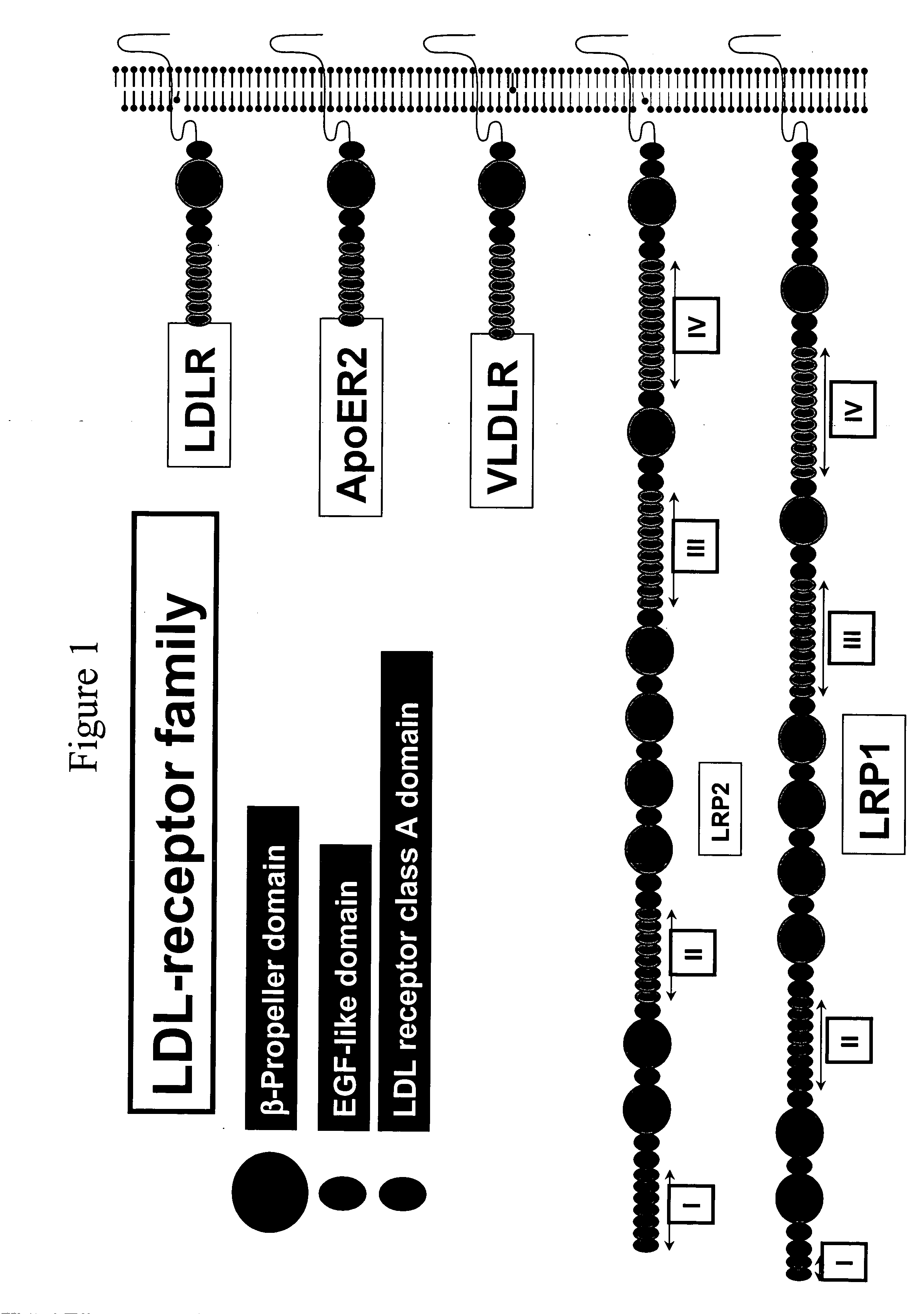
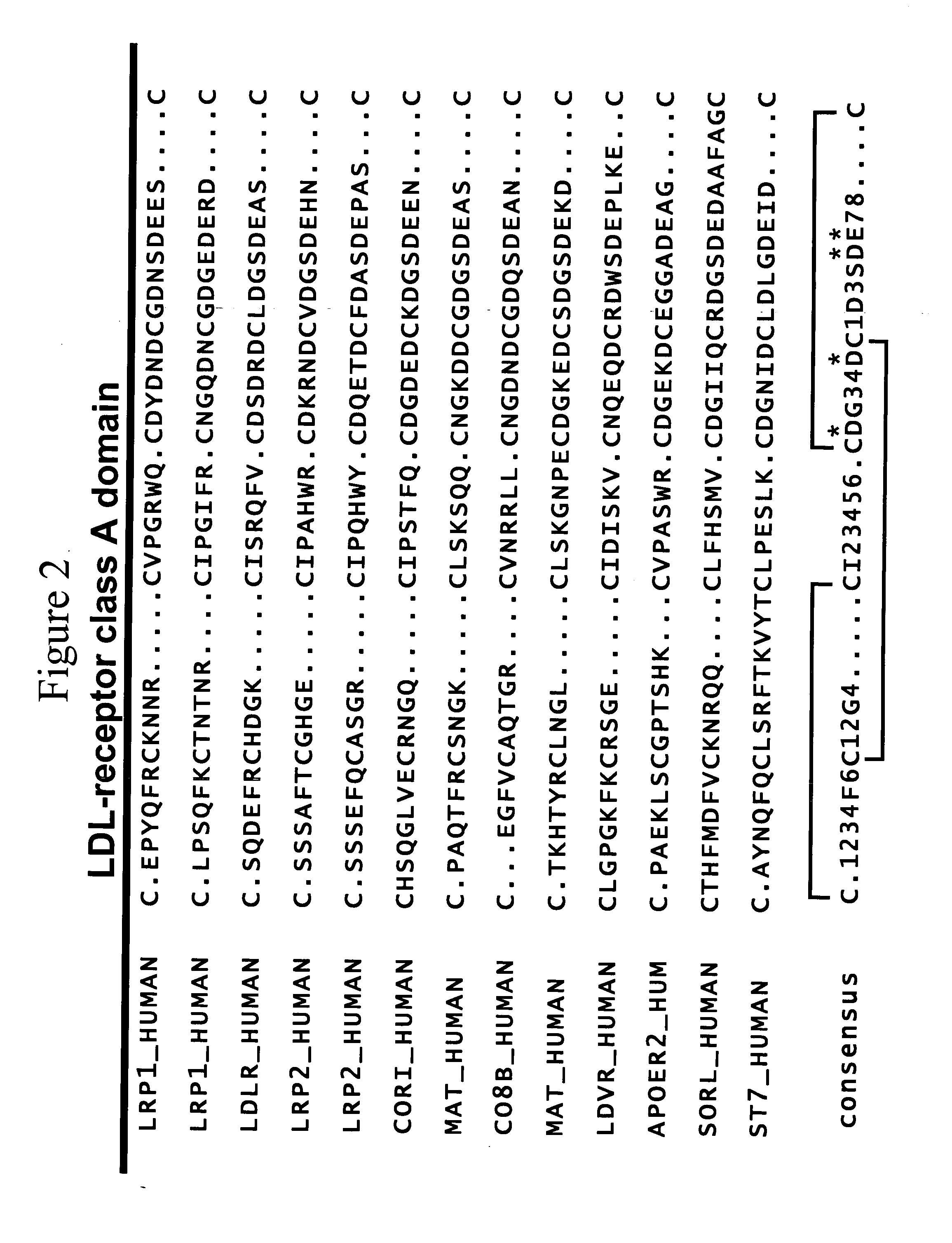
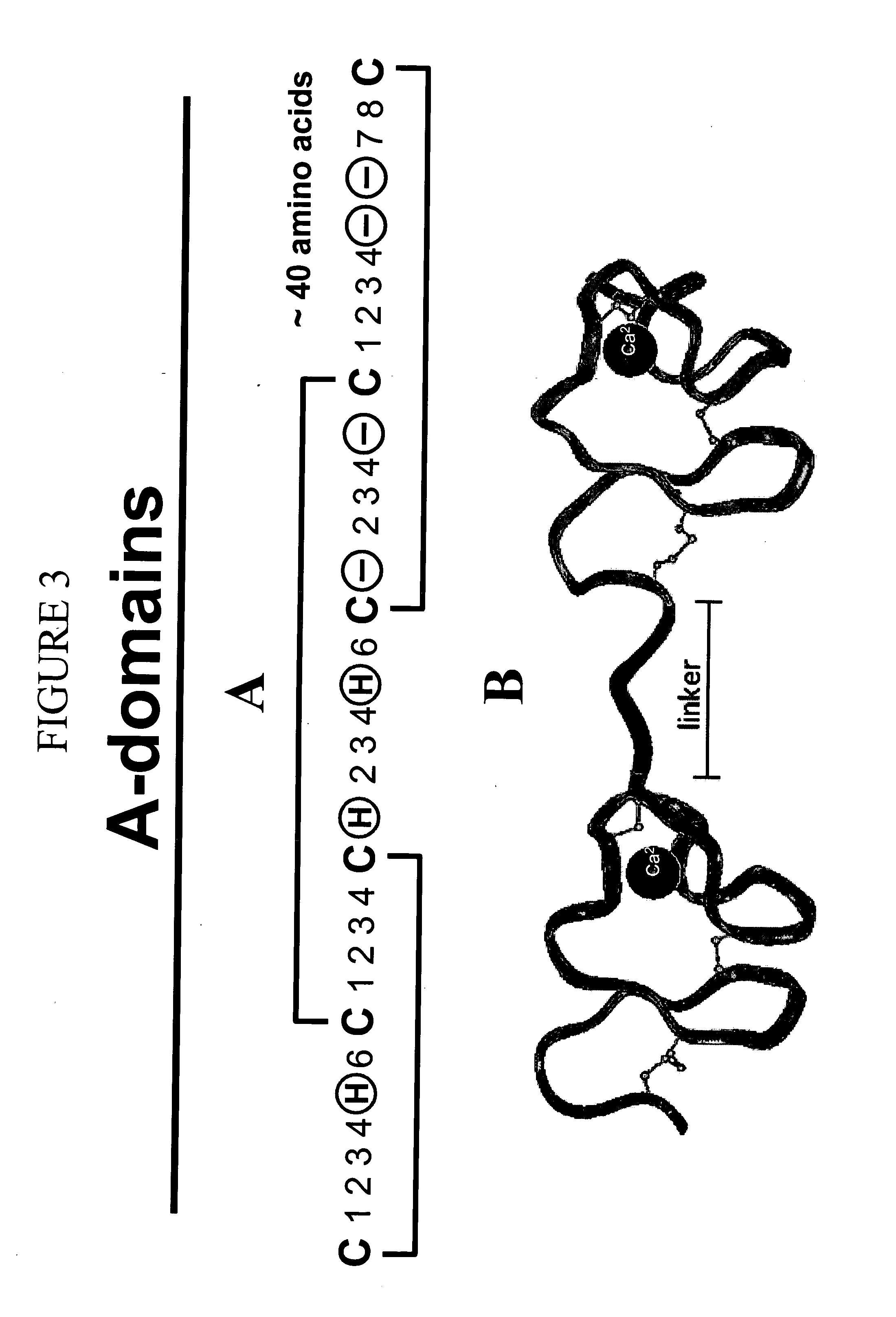
[0346] For the production of phages, E. coli DH10B cells (Invitrogen) were transformed with phage vectors encoding a library of LDL receptor class A-domain variants as a fusions to the pIII phage protein. To transform these cells, the electroporation system MicroPulser (Bio-Rad) was used together with cuvettes provided by the same manufacturer. The DNA solution was mixed with 100 μl of the cell suspension, incubated on ice and transferred into the cuvette (electrode gap 1 mm). After pulsing, 2 ml of SOC medium (2% w / v tryptone, 0.5% w / v yeast extract, 10 mM NaCl, 10 mM MgSO4, 10 mM MgCl2) were added and the transformation mixture was incubated at 37 C for 1 h. Multiple transformations were combined and diluted in 500 ml 2xYT medium containing 201 g / m tetracycline and 2 mM CaCl2. With 10 electroporations using a total of 10 μg ligated DNA 1.2×10⁸ independent clones...

example 3

[0351] This example describes the determination of biological activity of monomer domains that are capable of binding to HSA.

[0352] In order to show the ability of an HSA binding domain to extend the serum half life of a protein in vivo, the following experimental setup was performed. A multimeric A-domain, consisting of an A-domain which was evolved for binding HSA (see Example 2) and a streptavidin binding A-domain was compared to the streptavidin binding A-domain itself. The proteins were injected into mice, which were either loaded or not loaded (as control) with human serum albumin (HSA). Serum levels of a-domain proteins were monitored.

[0353] Therefore, an A-domain, which was evolved for binding HSA (see Example 1) was fused on the genetic level with a streptavidin binding A-domain multimer using standard molecular biology methods (see Maniatis et al.). The resulting genetic construct, coding for an A-domain multimer as well as a hexahistidine tag and a HA tag, were used to ...


PUM

PropertyMeasurementUnit
molecular weightaaaaaaaaaa

Abstract

Polypeptides comprising monomer domains that bind to c-Met, or portions thereof, are provided.

Description

BACKGROUND OF THE INVENTION [0001] Hepatocyte Growth Factor / Scatter Factor (HGF / SF) is a mesenchyme-derived pleiotropic factor, which regulates cell growth, cell motility, and morphogenesis of various types of cells and mediates epithelial-mesenchymal interactions responsible for morphogenic tissue interactions during embryonic development and organogenesis. Although HGF was originally identified as a potent mitogen for hepatocytes, it has also been identified as an angiogenic growth factor. [0002] Met was first identified in the 1980s as an oncogene and is the receptor for HGF. The proto-oncogene c-Met, was found to encode a receptor tyrosine kinase. In response to HGF treatment a range of activities are observed: phosphorylation of receptor, docking of signaling intermediates Gab-1 / Grb2, culminating in activation of kinases such as P13K, ERK1 and 2, and AKT. These activities aid in cell growth, survival, migration, and neovascularisation. [0003] Inappropriate expression or signali...

Claims


Application Information

Patent Timeline
Patent Type & Authority Applications(United States)
IPC IPC(8): G01N33/53A61K38/17C07K14/47
CPCA61K38/00C07K14/47C07K14/705G01N2500/00G01N33/574G01N2333/4753G01N2333/82G01N33/566A61P35/00A61P35/02C07K7/08G01N33/543
Inventor STEMMER, WILLEM P.C.PERLROTH, D. VICTORSATYAL, SANJEEV
Owner AMGEN MOUNTAIN VIEW
Learn More