Contrast-enhanced ocular imaging

a contrast-enhanced, ocular imaging technology, applied in the field of medical devices and methods for ocular imaging, can solve the problems of blindness if untreated, patients may suffer substantial, irreversible vision loss, and associated with significant side effects

Inactive Publication Date: 2006-02-16
GLAUKOS CORP
View PDF1 Cites 212 Cited by
  • Summary
  • Abstract
  • Description
  • Claims
  • Application Information

AI Technical Summary

Benefits of technology

[0027] Some embodiments relate to a method for treating glaucoma of an eye comprising steps of providing a trabecular stent, wherein the stent comprises a first terminal and a second terminal having a lumen connecting the first and second terminals. Contrast agent may be infused retrogradely through an episcleral vein into an existing aqueous outflow pathway of the eye. The pathway may then be imaged using a contrast agent-enhanced imaging apparatus, and the stent may be injected ab externally toward the imaged pathway and advanced stent through the trabecular meshwork. The second terminal of the stent preferably is sized and shaped to be received within a portion of the existing aqueous outflow pathway, and the...

Problems solved by technology

Glaucoma causes pathological changes in the optic nerve, visible on the optic disk, and it causes corresponding visual field loss, resulting in blindness if untreated.
Patients may suffer substantial, irreversible vision loss prior to diagnosis and treatment.
However, there are secondary open-angle glaucomas which may include edema or swelling of the trabecular spaces (e.g., from corticosteroid use), abnormal pigment dispersion, or diseases such as hyperthyroidism that produce vascular congestion.
However, these drug therapies for glaucoma are sometimes associated with significant side effects, such as headache, blurred vision, allergic reactions, death from cardiopulmonary complications, and potential interactions with other drugs.
However, long-term review of surgical results showed only limited success in adults.
In retrospect, these procedures probably failed due to cellular repair and fibrosis mechanisms and a process of “filling in.” Filling in is a detrimental effect of collapsing and closing in of the created opening in the trabecular mesh...

Method used


Image

  • Contrast-enhanced ocular imaging
  • Contrast-enhanced ocular imaging
  • Contrast-enhanced ocular imaging

Examples


Embodiment Construction

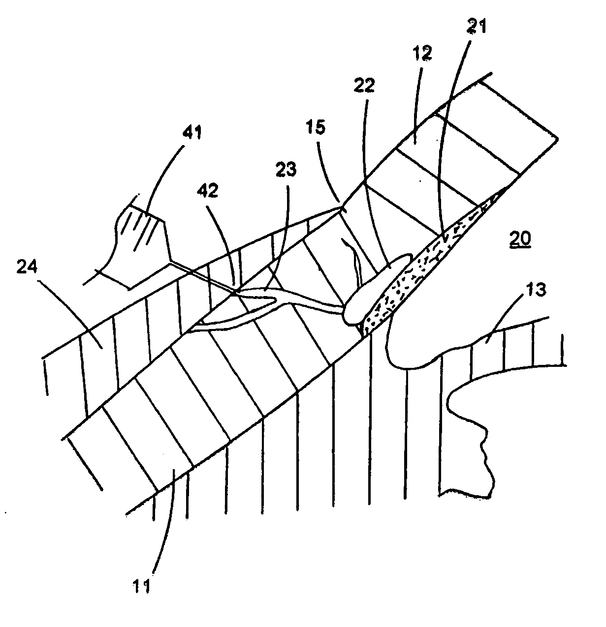
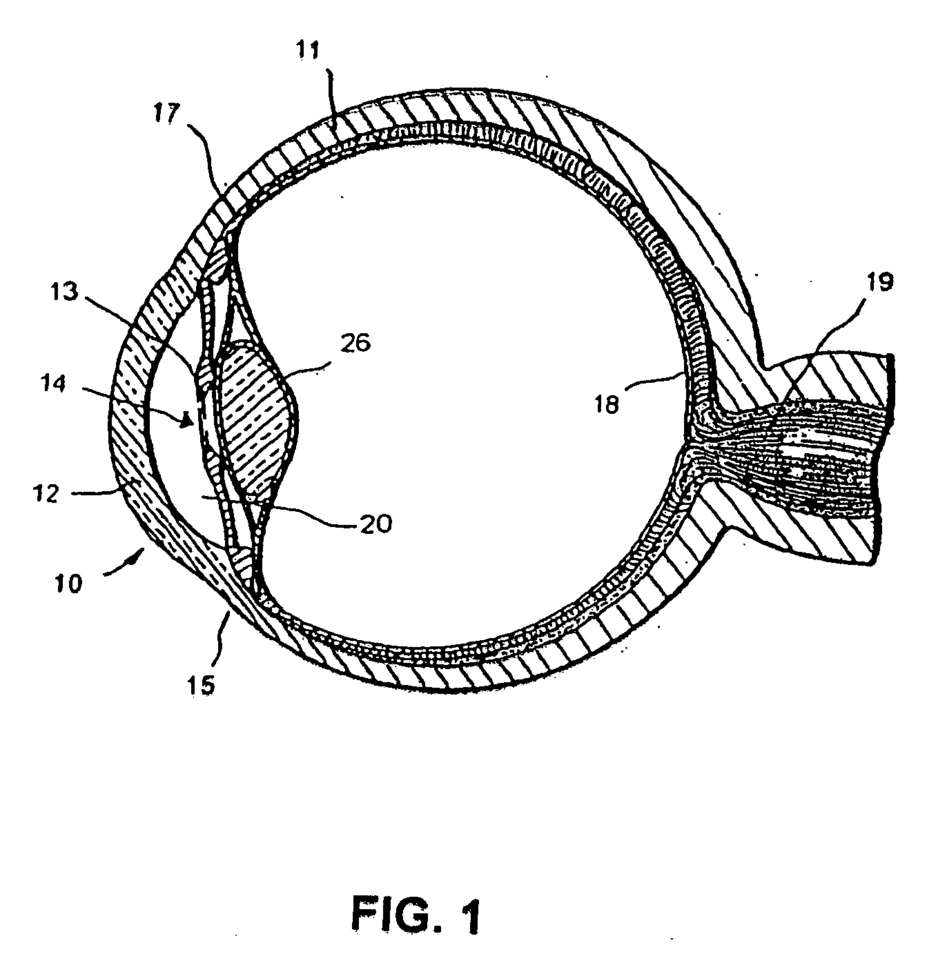
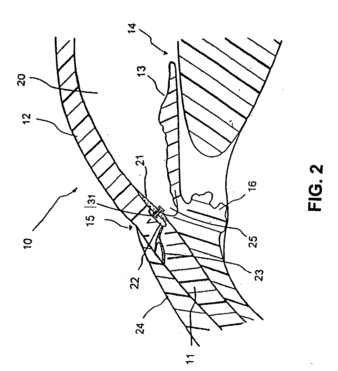
[0034] The preferred embodiments of the invention described herein relate particularly to surgical implantation of a trabecular stent for reduction of intraocular pressure ab externally with assistance of enhanced magnetic resonance imaging techniques. While the description sets forth various embodiment specific details, it will be appreciated that the description is illustrative only and should not be construed in any way as limiting the invention. Furthermore, various applications of the invention, and modifications thereto, which may occur to those who are skilled in the art, are also encompassed by the general concepts described herein.

[0035] The trabecular meshwork and juxtacanilicular tissue together provide the majority of resistance to the outflow of aqueous and, as such, are logical targets for the treatment of glaucoma. Various embodiments of glaucoma devices and methods are disclosed herein for treating glaucoma by an ab externo procedure, with respect to trabecular mesh...


PUM


Abstract

The invention relates generally to medical devices and methods for ocular imaging and, more particularly, to devices and methods for increasing contrast in an eye in which an imaging contrast agent is introduced into an aqueous humor outflow channel. For example, in one embodiment, the outflow channel may be Schlemm's Canal, or in another embodiment, the outflow channel may be an episcleral vein. Also disclosed are methods for implanting a trabecular stent via an ab extemo procedure with assistance of enhanced magnetic resonance imaging to restore a part or all of the normal physiological function of directing aqueous outflow for maintaining a normal intraocular pressure in an eye.

Description

BACKGROUND OF THE INVENTION [0001] 1. Field of the Invention [0002] The invention relates generally to medical devices and methods for ocular imaging and, more particularly, to devices and methods for increasing imaging contrast in an eye. [0003] 2. Description of the Related Art [0004] The human eye is a specialized sensory organ capable of light reception and able to receive visual images. The trabecular meshwork serves as a drainage channel and is located in the anterior chamber angle formed between the iris and the cornea. The trabecular meshwork maintains a balanced pressure in the anterior chamber of the eye by draining aqueous humor from the anterior chamber. [0005] About two percent of the United States population suffers from glaucoma. Glaucoma is a group of eye diseases encompassing a broad spectrum of clinical presentations, etiologies, and treatment modalities. Glaucoma causes pathological changes in the optic nerve, visible on the optic disk, and it causes corresponding...

Claims


Application Information

Patent Timeline
IPC IPC(8): A61F9/007
CPCA61F9/00781A61F9/0017
Inventor TU, HOSHENG
Owner GLAUKOS CORP
Why Eureka
Try Eureka