Isolation of endothelial progenitor cell subsets and methods for their use

Inactive Publication Date: 2006-02-16
MEDTRONIC INC
View PDF16 Cites 13 Cited by
  • Summary
  • Abstract
  • Description
  • Claims
  • Application Information

AI Technical Summary

Problems solved by technology

The shortcomings of this approach are technical limitations and include restrictions in determining the i

Method used


Image

  • Isolation of endothelial progenitor cell subsets and methods for their use
  • Isolation of endothelial progenitor cell subsets and methods for their use
  • Isolation of endothelial progenitor cell subsets and methods for their use

Examples


example 1

Identification of Seven CPC Subsets by Four-Parameter Flow Cytometry

[0066] The phenotypic heterogeneity of endothelial progenitor cells (EPC) based on patterns of combined expression of three circulating progenitor cells (CPC) markers, CD34, CD133 and KDR was analyzed using four-parameter (three-color) flow cytometric analysis.

[0067] Mononuclear cells were isolated from heparinized blood by lymphoprep density gradient centrifugation (Nycomed, Oslo, Norway). Because the number of circulating progenitor cells (PC), irrespective of phenotype, is low, lineage-negative (Lin−) cells, i.e. uncommitted, potential PCs, were enriched from total peripheral blood mononuclear cells (MNC) by high-speed flow cytometry sorting, whereas Lin+ cells were discarded (FIG. 6A). Total MNC were stained with a cocktail of phycoerythrin (PE)-labeled monoclonal antibodies (moAbs) against CD3 (T cells), CD14 (monocytes), CD19 (B cells), CD16 / 56 (NK cells) and CD31 (mature endothelial cells) (all from IQ Corp...

example 2

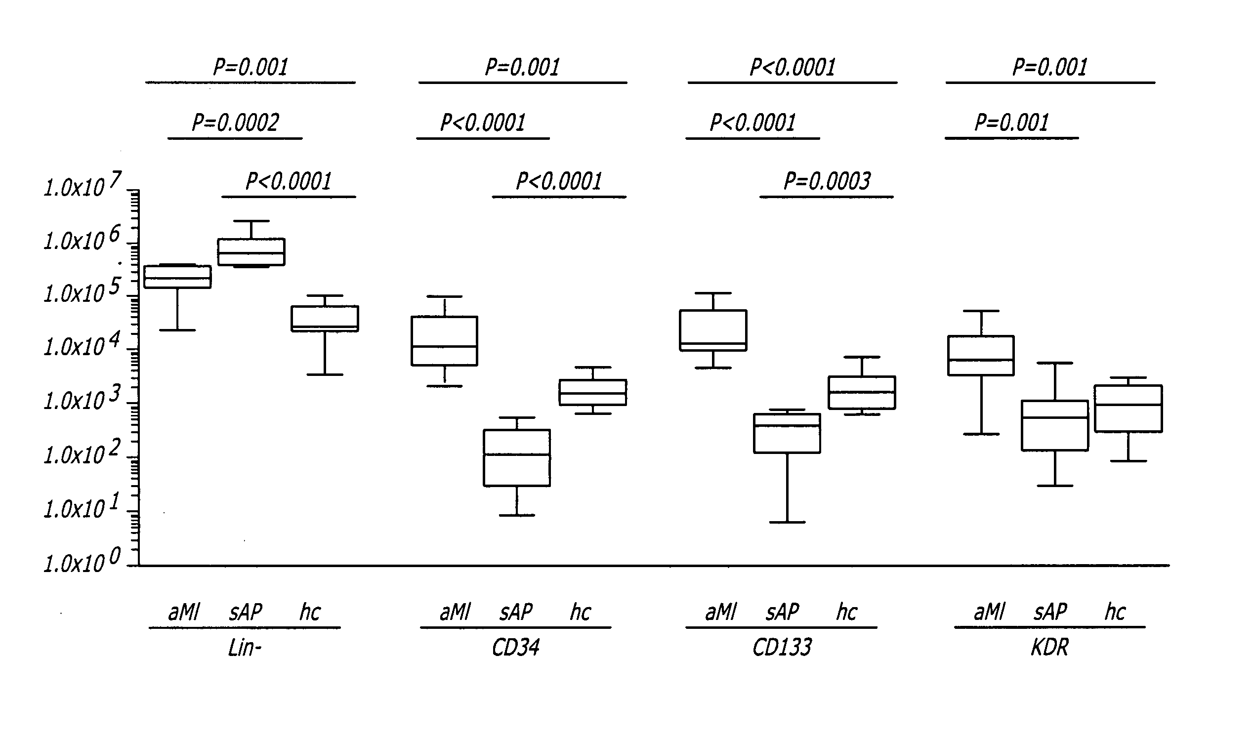
CPC Numbers in aMI Patients and Healthy Controls

[0073] Ten aMI patients and nine healthy control volunteers were compared with regard to EPC numbers to determine whether the number of cells in the seven CPC subsets correlated with the event of aMI. A possible correlation between CPC numbers and aMI is reflected in the number of Lin− cells (within which CPC were detected). Numbers of Lin− cells were compared between the two subject groups. In aMI patients the number of Lin− cells averaged 2.6×10⁵ cells / mL blood (range 0.2-4.7×10⁵ cells / mL blood), which was significantly higher (P=0.001) than in healthy controls (mean 0.5×10⁵ cells / mL blood, range 0.04-1.4×10⁵ cells / mL blood) (FIG. 7), equivalent with a 5.2-fold higher number of Lin− cells in aMI patients as compared to controls.

[0074] The numbers of CPC expressing CD34, CD133 or KDR were compared in aMI patients and healthy controls. CPC numbers in all three subsets were significantly higher in aMI patients than in healthy controls...

example 3

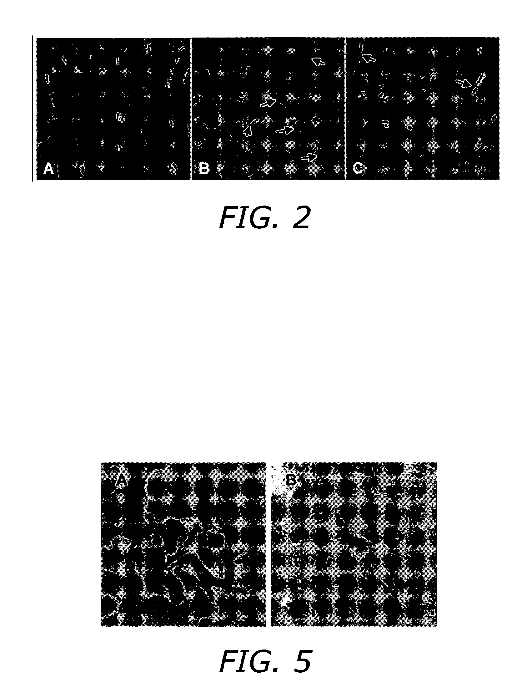
Blood Vessel-Forming Activity of CPC Subsets

[0078] This experiment investigated the behavior of CPC subsets in vivo, primarily with respect to differentiation into mature endothelial cells (EC). CPC expressing either CD34, CD133 or KDR were sorted in 200 μL Matrigel® and supplemented with 10 ng basic fibroblast growth factor (b-FGF, Chemicon, Temecula, Calif.) and 12 U heparin (Leo Pharma, Ballerup, Denmark) at 5,000 to 15,000 cells per implant and implanted subcutaneously in nude mice. Bare Matrigel® contains the b-FGF and heparin supplement. After 14 days, the Matrigel® pellets were explanted, partly snap-frozen in liquid nitrogen for immunohistochemistry, or fixed in 2% paraformaldehyde in 0.1 M sodium phosphate buffer, dehydrated and embedded in resin (Technovit 8100, Heraeus Kulzer, Wehrheim, Germany). For overall morphologic evaluation, 2 μm sections of resin-embedded Matrigel® pellets were stained with toluidin blue. Morphologic analysis of the implants showed that by 14 day...


PUM


Abstract

A method is provided for the isolation of endothelial progenitor cells from a source of progenitor cells by isolating a population of lineage-negative cells and further isolating CD34+ cells from the lineage-negative population by fluorescence-activated cell sorting. Isolated populations of endothelial progenitor cells and therapeutic compositions containing CD34+ cells for the induction of blood vessels, induction of angiogenic responses in surrounding blood vessels and the chemotaxis of inflammatory cells are also provided.

Description

RELATED APPLICATIONS [0001] The application claims priority under 35 U.S.C. §119(e) to U.S. Provisional Patent Application 60 / 601,188 filed Aug. 13, 2004.FIELD OF THE INVENTION [0002] The present invention relates generally to methods of isolating endothelial progenitor cells for the treatment of cardiovascular disease. BACKGROUND OF THE INVENTION [0003] The development of new blood vessels in response to tissue ischemia constitutes a natural host reaction intended to maintain tissue perfusion required for physiologic organ function. This natural angiogenesis is impaired in advanced age, diabetes and hypercholesterolemia. In each of these conditions, there is a reduction in endogenous expression of vascular endothelial growth factor (VEGF) and exogenous VEGF administration leads to enhanced neovascularization. [0004] Ischemic tissue injury triggers a series of events, including mobilization and recruitment of circulating progenitor cells (CPC) to the injury site. In models of post-i...

Claims


Application Information

Patent Timeline
IPC IPC(8): G01N33/567C12N5/08A61K35/12C12N5/074
CPCA61K2035/124C12N5/0692C12N2501/115G01N33/566C12N2533/90G01N33/5064G01N33/5073C12N2501/91A61P43/00A61P9/10
Inventor POPA, ELIANE R.HARMSEN, MARTIN C.VAN DER STRATE, BARRY W.A.VAN LUYN, MARJA J.A.HENDRIKS, MARC
Owner MEDTRONIC INC
Why Eureka
Try Eureka