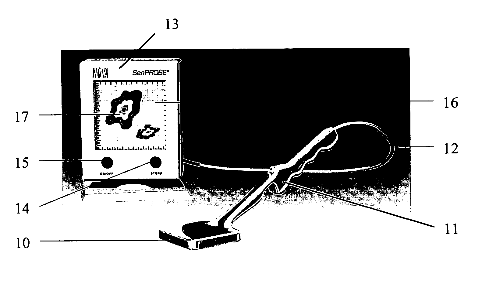
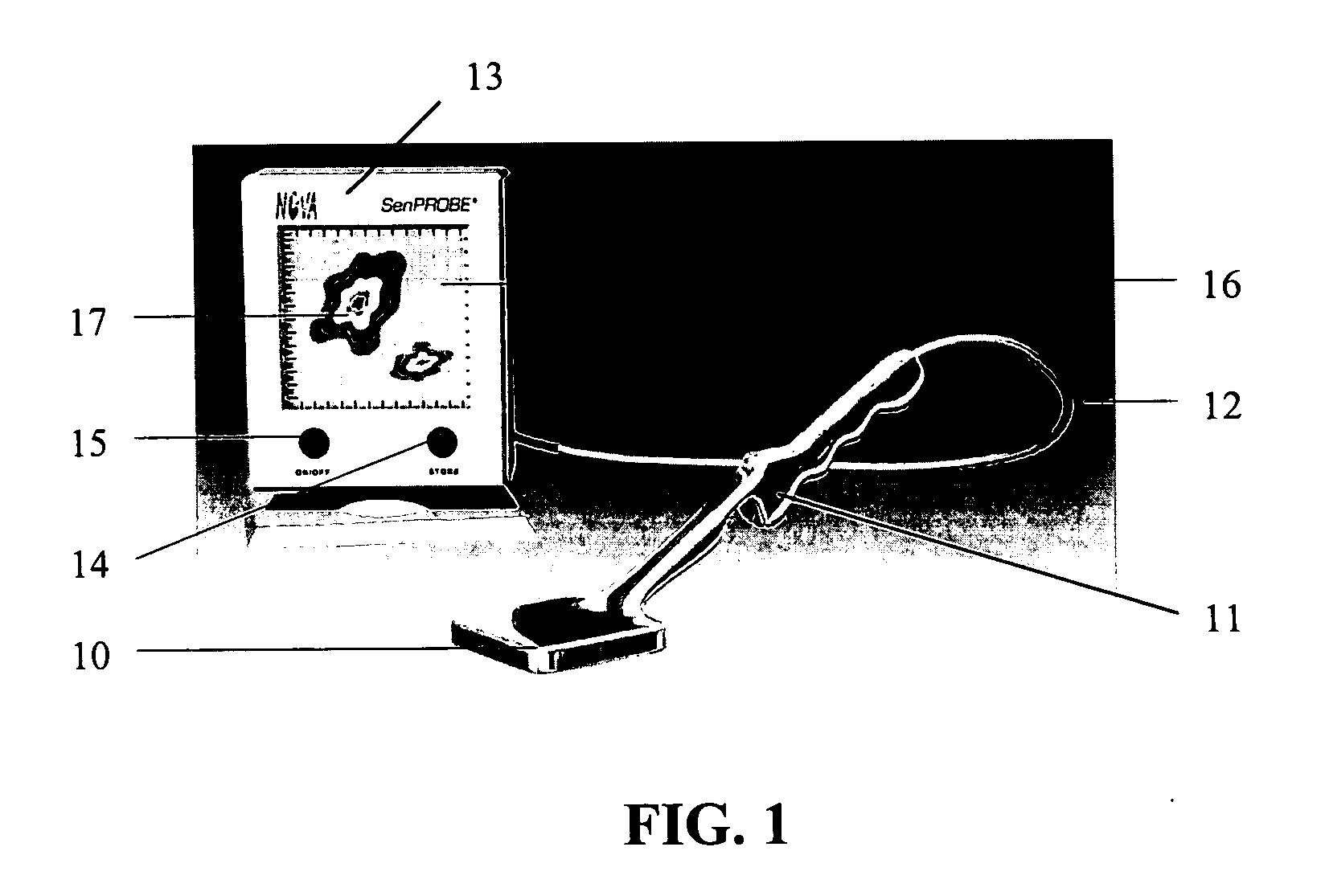
Imaging probe

a portable, imaging probe technology, applied in the field of imaging probes, can solve the problems of limited use, inaccurate location of sentinel nodes, and inability to provide accurate information on the extent of tumors, and achieve the effects of high sensitivity, high sensitivity, and high sensitivity of the reported system

Active Publication Date: 2006-02-16
NOVA R&D
View PDF3 Cites 37 Cited by
  • Summary
  • Abstract
  • Description
  • Claims
  • Application Information

AI Technical Summary

Benefits of technology

[0010] A small, compact, portable solid state imaging probe with a built in high sensitivity tiny gamma camera as shown in FIG. 1 is discussed here as a probe to locate sentinel lymph nodes. It can also be used as a high sensitivity tool for scintimammography. The high sensitivity of the reported system is due to the very short distance to the source as the probe will be used making direct contact with the tissue, even inside a surgical cut; high Z solid state CdZnTe detector material with high quantum efficiency; and high energy resolution, about 5% to 10%, to discriminate against scattered photons and other background.
[0011]FIG. 1 is showing a drawing of the SenPROBE with one Image / Reset button and the separate LCD monitor with On / Off and Store buttons displaying two active sentinel lymph node sites. FIG. 2. is a drawing of the SenPROBE showing the internal components; honeycomb collimator (at the bottom) which is removable and interchangeable for higher sensitivity or higher spatial resolution. On top of the collimator there are the CdZnTe pixel detectors mounted on a circuit board. On the other side of the circuit board the new front-end chips will be mounted directly on the circuit board without bulky packaging to achieve the small thickness required. The data acquisition and display electronics will be housed in the color LCD monitor. The collimator is shown here as integrated into the probe. However, in practice the collimator will be easily exchanged or removed in the operating room. This will allow trade off between sensitivity and spatial resolution.
[0016] 4. An integrated circuit is developed specifically for this applications. The noise is expected to be lower and energy resolution higher. The new chip will enable compact and portable design of the imaging probe.
[0026] The detector has a handle for holding the medical imaging system. The medical imaging system is also made compact and portable.

Problems solved by technology

However, they are limited in use as they do not provide an image, just a crude count rate from a 1 cm2 area detector.
Therefore, locating the sentinel node is not very accurate and it does not provide accurate information on the extent of the tumor.
The large size of the gamma camera makes it difficult to position optimally relative to the breast.
Not usable during surgery due to the large size, low sensitivity and low spatial resolution.

Method used


Image

  • Imaging probe
  • Imaging probe
  • Imaging probe

Examples


Embodiment Construction

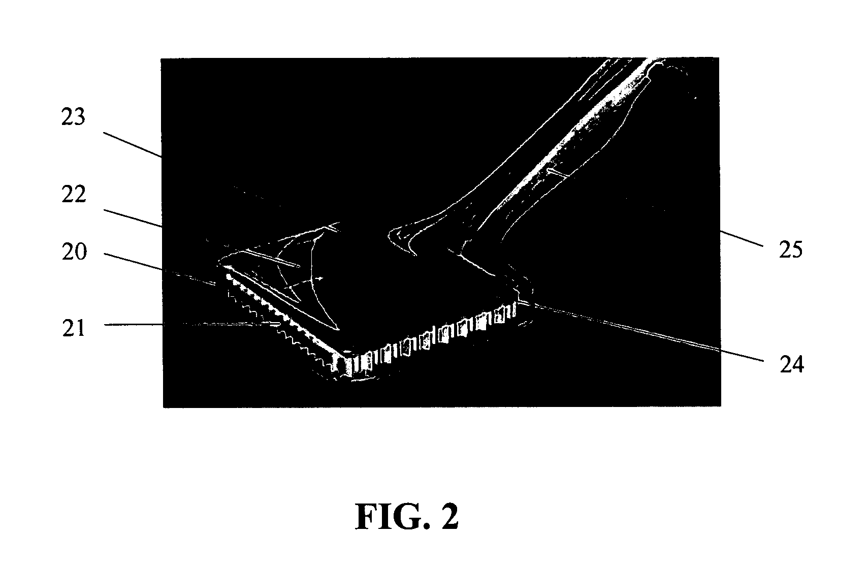
[0035] For this application, we plan to use detectors 24, 30, 82 with a thickness of 3 to 5 mm, which is well suited for photons from 99mTc, the radionuclide most commonly used in radiopharmaceuticals. The pixel sizes will be selected from 1 to 3 mm. One side of these detectors have two-dimensional array of pixels (electrodes) normally as anodes and the other side is a single plane electrode, normally used as cathode. Another embodiment would be to make the pixels as cathodes and the backside electrode to function as anode. A bias voltage is applied between the anode and cathode where the electrons generated by an x-ray or a gamma ray are collected at the anode(s). In the main embodiment the two dimensional pixelated side faces the printed circuit board.

[0036] The energy resolution of our current CdZnTe pixel detectors (FIG. 3) 30 with 4×8 pixels and 3 x 3 mm pixel pitch, read out by the RENA chip, has been measured using 57Co, 139Ce, and 241 Am sources. Sample energy spectra are s...


PUM


Abstract

The design of a compact, handheld, solid-state and high-sensitivity imaging probe and a micro imager system is reported. These instruments can be used as a dedicated tool for detecting and locating sentinel lymph nodes and also for detecting and imaging radioactive material. The reported device will use solid state pixel detectors and custom low-noise frontend / readout integrated circuits. The detector will be designed to have excellent image quality and high spatial resolution. The imaging probes have two different embodiments, which are comprised of a pixelated detector array and a highly integrated readout system, which uses a custom multi-channel mixed signal integrated circuit. The instrument usually includes a collimator in front of the detector array so that the incident photons can be imaged. The data is transferred to an intelligent display system. A hyperspectral image can also be produced and displayed. These devices are designed to be portable for easy use.

Description

CROSS REFERENCE TO PROVISIONAL AND PARENT PATENT APPLICATION [0001] This application claims the benefit of the filing date of U.S. Provisional Patent Application No. 60 / 330,597 filed Oct. 25, 2001 and the parent U.S. patent application Ser. No. 10 / 279,003 filed Oct. 24, 2002, now U.S. Pat. No. 6,940,070, the disclosure of which are incorporated herein by reference.FIELD OF INVENTION [0002] The focus of this work is to develop an enhanced portable imaging probe for detecting and locating sentinel lymph nodes during breast cancer surgery. It may also be used for scintimammography: diagnosis and accurate location of breast cancer tumors and their spread to surrounding tissue, especially axillary lymph nodes. It is expected to improve and expedite the sentinel node detection and locating, and enhance breast and other cancer surgery. [0003] The instruments described can also be used for many different applications. In medical imaging, for example, they can be used for many types of x-ray...

Claims


Application Information

Patent Timeline
Patent Type & Authority Applications(United States)
IPC IPC(8): A61B5/05A61B6/00G01T1/161G01T7/00
CPCA61B6/4258A61B6/508G01T7/00G01T1/161
Inventor TUMER, TUMAY O.
Owner NOVA R&D
Why Eureka
Try Eureka