Retinal cell grafts and instrument for implanting

a technology of retinal cells and instruments, applied in the field of retinal cell grafts and instruments for implanting, can solve the problems of destroying the cellular polarity and native organization of the donor retinal pigment epithelium, unable to achieve effective reconstruction methods, and unable to achieve the effect of regrowth of photoreceptor axons

Inactive Publication Date: 2006-02-23
HUGHES STEPHEN E
View PDF42 Cites 10 Cited by
  • Summary
  • Abstract
  • Description
  • Claims
  • Application Information

AI Technical Summary

Benefits of technology

[0012] Among the objects of the present invention, therefore, may be noted the provision of a method for preparation of a graft for use in the reconstruction of a dystrophic retina; the provision of such a method which conserves relatively large expanses of the tissue harvested from a donor eye; the provision of such a method in which the polarity and organization of the cells at the time of harvest are maintained in the graft; the provision of a graft for use in the reconstruction of a dystrophic retina; the provision of such a graft which facilitates regrowth of photoreceptor axons by maintaining the polar organization of the photoreceptor and the close proximity of their postsynaptic targets with the adjacent outer plexiform layer upon transplantation; the provision of a surgical tool for use in the transplantation method which allows appropriate retinotopic positioning and which protects photoreceptors or other grafted tissue from damage prior to and as the surgical device is positioned in the eye; and the provision of a method for transplantation of grafts to the subretinal area of an eye.
[0018] The present invention is further directed to an instrument for the implantation of an intact planar cellular structure between the retina and supporting tissues in an eye. The instrument comprises an elongate supporting platform for holding the planar cellular structure. The platform has a distal end for insertion into an eye, and a proximal end. The distal edge of the platform is convexly curved for facilitating the insertion of the platform into the eye and the advancement of the platform between the retina and the supporting tissues. The instrument has a side rail on each side of the platform for retaining the planar cellular structure on the platform, the distal ends of the side rails being rounded, and the distal portions of the side rails tapering toward the distal end of the platform to facilitate the insertion of the instrument between the retina and the supporting tissue.
[0020] The present invention is further directed to a kit for transplantation of a graft to the subretinal area of a host eye. The kit contains a graft comprising a population of cells selected from retinal cells, epithelial tissue and choroidal tissue, the population of cells being maintained in the same organization and cellular polarity as is present in normal tissue of that type. The kit additionally contains a surgical instrument comprising an elongate tube, having a flat, wide cross-section, with a top, a bottom for supporting the planar cellular structure, and opposing sides. The tube has a beveled distal edge facilitating the insertion of the tube into the eye and the advancement of the tube between the retina and the supporting tissues. The surgical instrument also comprises plunger means for ejecting a planar cellular structure from the distal end of the tube.

Problems solved by technology

In an effort to recover what was previously thought to be an irreparably injured retina, researchers have suggested various forms of grafts and transplantation techniques, none of which constitute an effective manner for reconstructing a dystrophic retina.
However, this method destroys the cellular polarity and native organization of the donor retinal pigment epithelium which is desirable for transplants.
While del Cerro reports that the intraocular transplantation of retinal strips can survive, he notes that the procedure has some definite limitations.
For instance, his techniques do not allow for the replacement of just the missing cells (e.g. photoreceptors) but always include a mixture of retinal cells.
Thus, with such a transplant appropriate reconstruction of the dystrophic retina that lacks a specific population of cells (e.g., photoreceptors) is not possible.
Furthermore, no means of isolating and purifying any given population of retinal cells (e.g. photoreceptors) from other retinal cells was demonstrated.
This conclusion is based on the well known optical characteristics of photoreceptors (outer segments act as light guides) and clinical evidence showing that folds or similar, even minor disruptions in the retinal geometry can severely degrade visual acuity.

Method used


Image

  • Retinal cell grafts and instrument for implanting
  • Retinal cell grafts and instrument for implanting
  • Retinal cell grafts and instrument for implanting

Examples


example 1

Experimental Animals

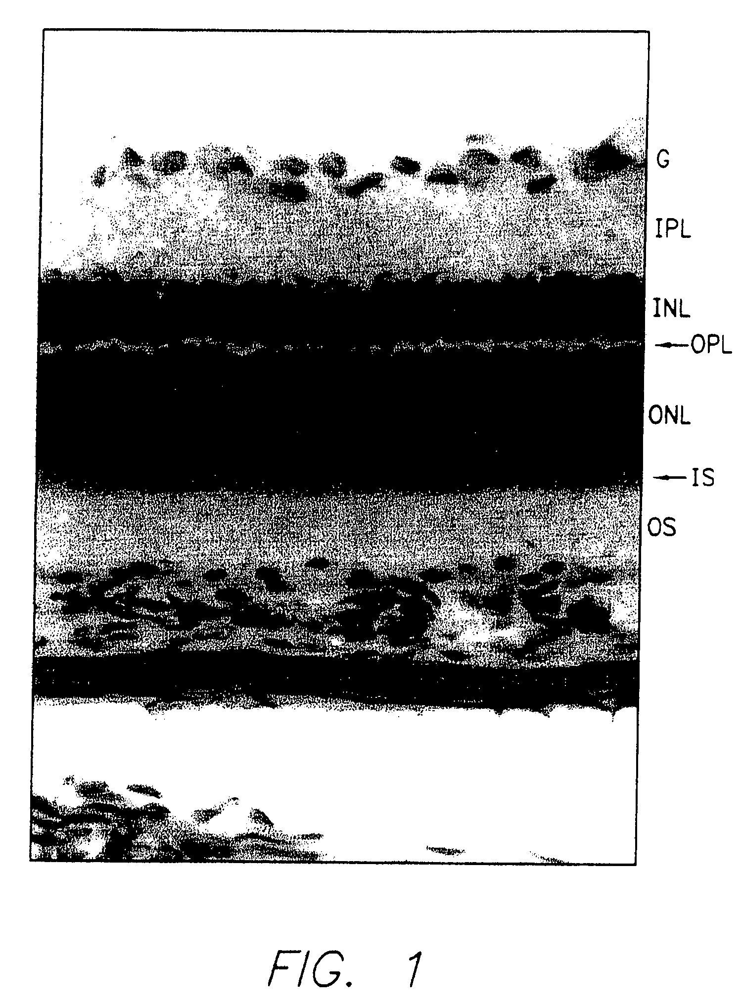
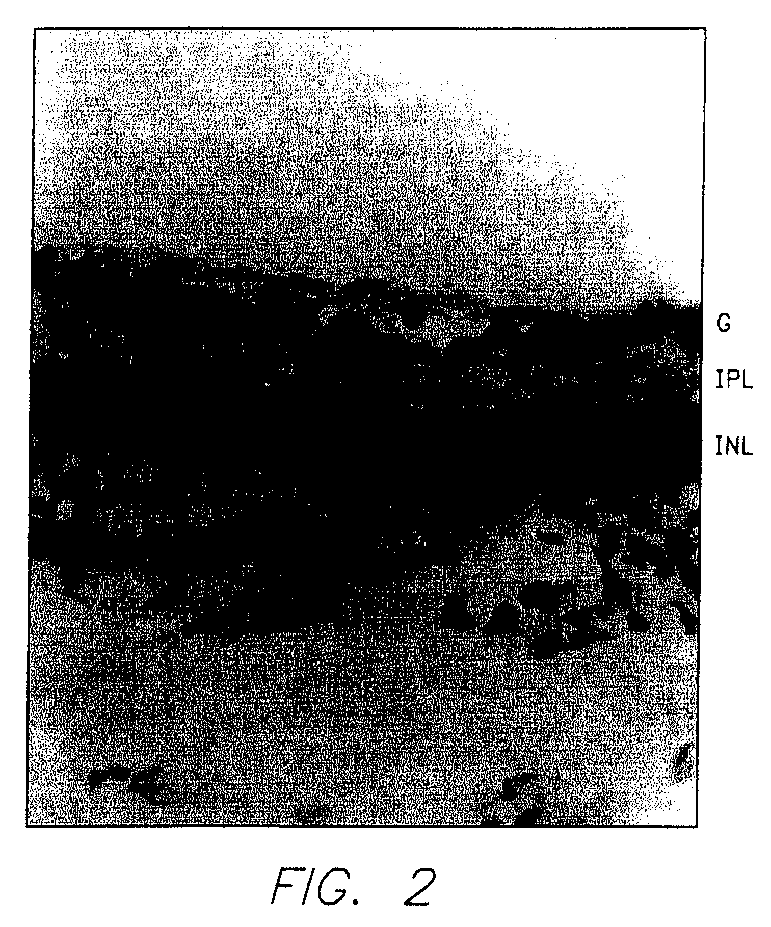
[0092] Adult albino rats (Sprague-Dawley) were exposed to constant illumination averaging 1900 lux for 2 to 4 weeks as described in O'Steen, Exp. Neurol. 27:194 (1970).

[0093] As shown in FIG. 1, this exposure destroys most photoreceptors, eliminating cells of the outer nuclear layer but leaving the remaining neural retina intact. Photoreceptors for transplantation were taken from 8-day-old normal rats of the same strain that had been maintained under colony room illumination (10-20 lux) on a 12 hr / 12 hr light / dark cycle. Experimental animals were anesthesized with ketamine and sodium pentobarbital. A preoperative dose of dexamethasone (10 mg / kg IP) was also administered.

Photoreceptor Preparation

[0094] The retina from the anesthetized 8-day-old rat was removed, flattened with radial cuts and placed with the receptor side down on a gelatin slab secured to the vibratome chuck. Molten gelatin (4-5% solution) was deposited adjacent the retina at the retina / gelat...

example 2

[0109] The procedures of Example 1 were repeated except as noted. Substituted for the Sprague-Dawley rats were the rd mouse and the RCS rat which are afflicted with inherited retinal degeneration. In the rd mouse it is thought that the deficit resides in the photoreceptor whereas in the RCS it is thought that the deficit resides in the pigment epithelium. In these animals, almost all photoreceptors are eliminated while the remaining retina is preserved; either the rd mouse or the RCS rat were blinded by constant illumination as set forth in Example 1. The rd mouse and the RCS respectively received transplants of immature (7-8 day old mouse or rat) and mature rat photoreceptors.

rd Mouse

[0110] The transplantation technique was adapted to the smaller size of the mouse eye. This modification allowed sheets of intact outernuclear layer to be transplanted to the subretinal space of the mouse eye. Neonatal (8 days old) photoreceptors were transplanted from rd control mice to the subreti...

example 3

[0115] The procedures of Example 1 were repeated except as noted.

[0116] Donor photoreceptors were originally harvested at the earliest ontogenetic time in which the photoreceptors could be isolated from other portions of the retina (7-8 days old) since it is generally believed that more embryonic and undifferentiated neural tissue survives transplantation far better than more mature and differentiated tissue. To determine the effect of developmental age on photoreceptor survival and ability to integrate with the host retina, photoreceptors were subsequently transplanted from 8, 9, 12, 15 and 30 day old rats into light damaged adults. These show progressive development and maturation of the photoreceptors including mature outer segments (at 15 and 30 days).

[0117] Using the same criteria as in Example 1, it was found that for all ages tested the transplants survived for as long as examined (2 months) and integrated with the host retina. FIG. 26, panel A, is a photograph of a transpl...


PUM

PropertyMeasurementUnit
lengthaaaaaaaaaa
surface areaaaaaaaaaaa

Abstract

Surgical instruments, surgical techniques, retinal cell grafts, retinal cell and tissue isolation techniques, and a method for transplanting retinal cells, including photoreceptors, and / or retinal pigment epithelium, with the cells in the isolated tissue having their normal cell to cell configuration are disclosed.

Description

PRIORITY INFORMATION [0001] This application is a divisional of Ser. No. 10 / 323,958, filed Dec. 17, 2002, which is a divisional of Ser. No. 09 / 411,122 filed Oct. 4, 1999, now U.S. Pat. No. 6,579,256, which is a divisional of Ser. No. 08 / 322,735, filed Oct. 13, 1994, now U.S. Pat. No. 5,962,027, which is a continuation of Ser. No. 07 / 566,996 filed Aug. 13, 1990, abandoned, which is a continuation in part of Ser. No. 07 / 394,377, filed Aug. 14, 1989, abandoned, all of which are incorporated by reference as if reproduced in full below.BACKGROUND OF THE INVENTION [0002] The present invention relates in general to surgical instruments, surgical techniques, and cell and tissue isolation techniques. More particularly, the present invention is directed to a surgical tool for transplanting retinal cells, epithelium and choroidea within their normal planar configuration, a graft for transplantation to the subretinal region of the eye, a method for preparing such grafts for transplantation, and...

Claims


Application Information

Patent Timeline
Patent Type & Authority Applications(United States)
IPC IPC(8): A61K35/44A61M31/00A61B17/00A61B19/00A61F2/14A61F9/007A61L27/38C12N5/08
CPCA61B19/00A61M2025/0042A61F2/14A61F9/0017A61F9/007A61K35/44A61L27/3604A61L27/3641A61L27/3683A61L27/3804A61L27/3813A61L27/383A61L27/3839A61L27/3891A61L2430/16A61B2017/00969A61B90/00
Inventor HUGHES, STEPHEN E.
Owner HUGHES STEPHEN E
Why Eureka
Try Eureka