
Management of ophthalmologic disorders, including macular degeneration

a technology for ophthalmologic disorders and macular degeneration, applied in the direction of sense disorders, organic chemistry methods, drug compositions, etc., can solve the problems of affecting the phagocytosis of outer segments, age-related diseases of vision, and vision loss and blindness in ageing populations, so as to prevent or reduce accumulation of a2e in rod outer-segment discs, prevent binding, and reduce the effect of a2e production in discs

Inactive Publication Date: 2006-03-30
PRESIDENT & FELLOWS OF HARVARD COLLEGE
View PDF0 Cites 97 Cited by
  • Summary
  • Abstract
  • Description
  • Claims
  • Application Information

AI Technical Summary

Benefits of technology

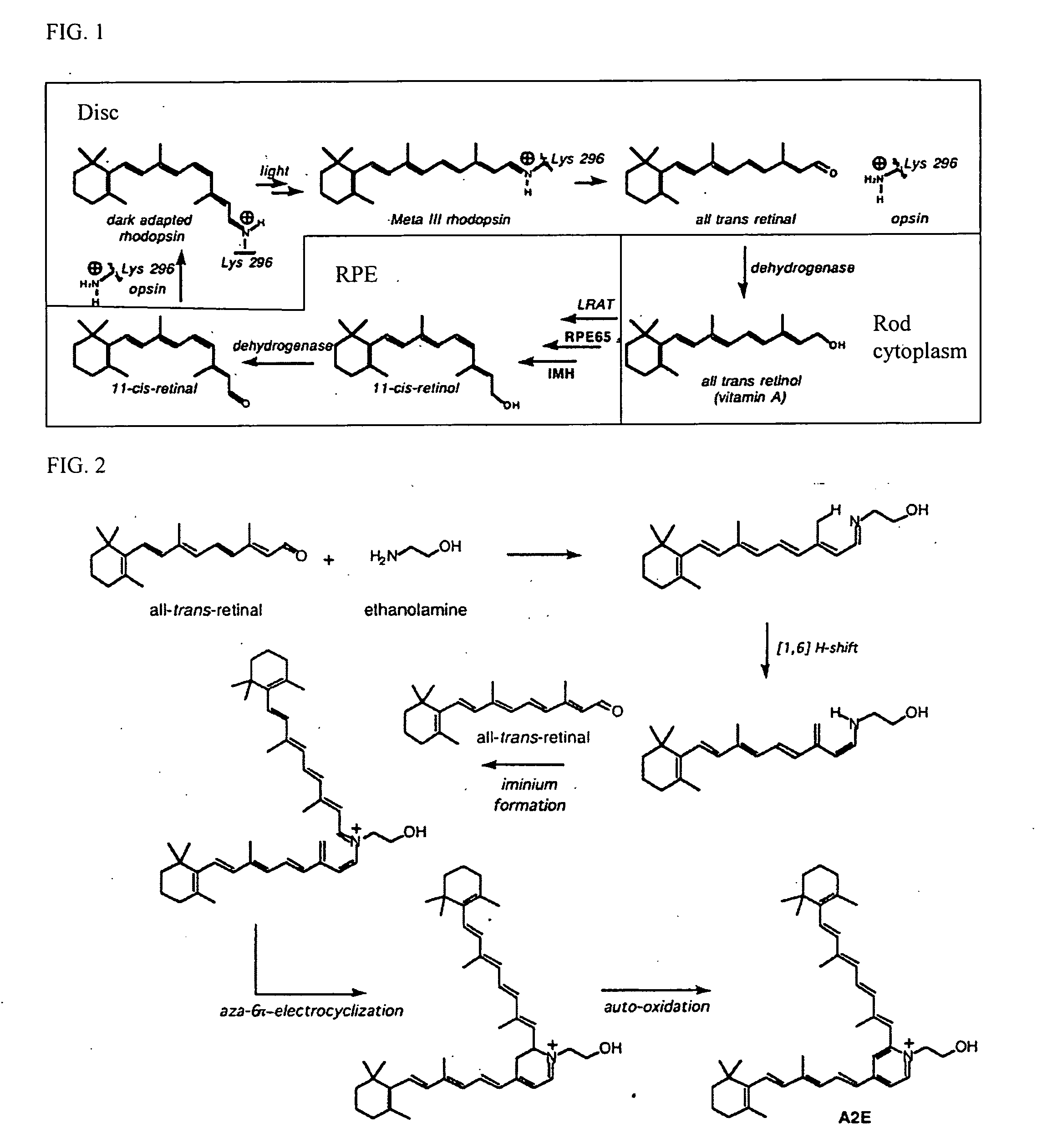
[0011] In one embodiment, the accumulation of A2E in rod outer-segment discs is prevented or reduced. It has been found that A2E production in discs can be reduced by administering a drug that limits the visual cycle. The limitation can be achieved in a number of ways. In one approach, a drug can effectively short-circuit the portion of the visual cycle that generates the A2E precursor, all-trans-retinal. In another approach, a drug can inhibit particular steps in the visual cycle necessary for synthesizing all-trans-retinal. In yet another approach, a drug can prevent binding of intermediate products (retinyl esters) to certain chaperone proteins in the retinal pigment epithelium.

Problems solved by technology

Age related diseases of vision are an ever-increasing health problem in industrial societies.
Age related macular degeneration (AMD) affects millions of persons worldwide and is a leading cause of vision loss and blindness in ageing populations.
Consequently, the retinotoxic compounds in the disc are brought into the RPE, where they impair further phagocytosis of outer segments and cause apoptosis of the RPE.
However, in certain pathological conditions, so much A2E can accumulate in the disc that the RPE is “poisoned” when the outer segment is phagocytosed.
However, defects in RmP can derange this process by impeding removal of all-trans-retinal from the disc.
As noted above, some A2E is formed even under normal conditions; however, its production is greatly increased when its precursors accumulate inside the discs due to the defective transporter, and can thereby cause macular degeneration.
Other forms of macular degeneration may also result from pathologies that result in lipofuscin accumulation.

Method used


Image

  • Management of ophthalmologic disorders, including macular degeneration
  • Management of ophthalmologic disorders, including macular degeneration
  • Management of ophthalmologic disorders, including macular degeneration

Examples


example 1

In Vitro

[0929] Materials: Frozen bovine eyecups devoid of retinas were purchased from W. L. Lawson Co., Lincoln, Nebr. Ammonium bicarbonate, BSA, ethylenediaminetetraacetic acid (EDTA), guanidine HCl, imidazole, DEAE-Sepharose, phenyl-Sepharose CL-4B, all-trans-retinol, all-trans-retinyl palmitate, α-Cyano-4-hydroxycinnamic acid and Trizma® base were from Sigma-Aldrich. Dithiothreitol was from ICN Biomedicals Inc. 11-cis-Retinol and 11-cis-retinyl palmitate were synthesized by following the procedure described elsewhere (Shi et al., 1993). Anagrade™ CHAPS and dodecyl maltoside were from Anatrace. HPLC grade solvents were from Sigma-Aldrich Chemicals. Anti RPE65 (NFITKVNPETLETIK) antibody was obtained from Genmed Inc and anti-LRAT antibody was provided by Prof. Dean Bok (University of California at Los Angeles). rHRPE65 baculovirus was provided by Prof. Jian-Xin Ma (University of South Carolina). Hank's TNM-FH Insect medium was obtained from JRH Biosciences. sf21 cells were laborato...

example 2

Effect on Visual Cycle of Short-Circuiting Drugs in vivo

[1021] Mice were injected intraperitoneally (i.p.) with 50 mg / kg of the compounds listed prepared in 25 microliters DMSO. Positive control mice were injected with 13-cis-retinoic acid (ACCUTANE®) 50 mg / kg in 25 microliters DMSO. Negative control mice were injected with 25 microliters DMSO.

[1022] At predetermined times after administration, mice were exposed to sufficient light to cause complete bleaching of the visual cycle. Electroretinograms (ERG) were then performed in bright light or dim light, and the b-wave amplitude measured. The b-wave amplitude is assumed to be proportional to rhodopsin regeneration and thereby correlate with the functioning of the visual cycle (i.e., the higher the b-wave amplitude, the greater the functioning of the visual cycle).

A. 4-butyl-aniline and ethyl 3-aminobenzoate

[1023] 4-butyl-aniline and ethyl 3-aminobenzoate, were prepared as solutions in DMSO. 7 months old wild type (wt; C57BL / 6J×12...

example 3

Effect on Visual Cycle of Enzyme Inhibitors and RPE65 Antagonists in vivo

[1029] The experiments described in Example 2 were repeated additional compounds.

A. Retinyl palmityl ketone and retinyl decyl ketone

[1030]FIG. 19 shows an experiment with these compounds.

B. All-trans-retinyl palmityl ketone, all-trans-retinyl palmityl ether

[1031] FIGS. 20A-B and 21A-B show, respectively, two experiments with these compounds in dim (A) and bright (B) light.

C. Octyl farnesimide, palmityl farnestimide

[1032]FIG. 22A shows the results of an experiment in dim light using these compounds shortly after administration. FIG. 22B shows the results one week after administration.

D. Farnsyl octyl ketone, farnesyl decyl ketone

[1033] FIGS. 23A-C show experiments performed in dim light using these compounds. The data shown in FIG. 23B was obtained 3 days after administration. The data in FIG. 23C was obtained 8 days after administration.

E. Farnesyl palmityl ketone, farnesyl decyl ketone

[1034]FIG. 24 ...


PUM

PropertyMeasurementUnit
Fractionaaaaaaaaaa
Fractionaaaaaaaaaa
Fractionaaaaaaaaaa

Abstract

A drug may be used in the preparation of a medicament for the treatment or prevention of an ophthalmologic disorder, wherein the drug inihibits, antagonizes, or short-circuits the visual cycle at a step of the visual cycle that occurs outside a disc of a rod photoreceptor cell.

Description

CROSS-REFERENCE TO RELATED APPLICATION [0001] This application is a continuation-in-part of International Application No. PCT / US2005 / 004990, filed Feb. 17, 2005, which claims the benefit of U.S. Provisional patent application Ser. No. 60 / 545,456, filed Feb. 17, 2004; U.S. Provisional patent application Ser. No. 60 / 567,604, filed May 3, 2004; and U.S. Provisional patent application Ser. No. 60 / 578,324, filed Jun. 9, 2004. All aforementioned applications are hereby incorporated herein by reference in their entirety.STATEMENT REGARDING FEDERALLY SPONSORED RESEARCH OR DEVELOPMENT [0002] Support for research leading to subject matter disclosed in this application was provided in part by the National Institutes of Health Grant Nos. R01-EY-04096 and / or R01-EY-015425. Accordingly, the United States Government has certain rights with respect to subject matter of this application.INTRODUCTION [0003] Age related diseases of vision are an ever-increasing health problem in industrial societies. ...

Claims


Application Information

Patent Timeline
IPC IPC(8): A61K31/33A61K31/336A61K31/275C07D203/04C07D303/08
CPCC07B2200/09C07C43/15C07D303/08C07C43/162C07C45/68C07C49/203C07C57/26C07C69/24C07C211/45C07C217/86C07C229/56C07C229/60C07C233/05C07C233/09C07C233/25C07C243/38C07C251/40C07C403/08C07C403/10C07C403/12C07C403/16C07C403/18C07C403/20C07C2101/14C07C2101/16C07D203/08C07C2601/14C07C2601/16A61P27/02
Inventor RANDO, ROBERT R.
Owner PRESIDENT & FELLOWS OF HARVARD COLLEGE
Learn More