Minimally invasive glaucoma surgical instrument and method

a glaucoma and minimally invasive technology, applied in the field of new glaucoma surgical instruments and methods, can solve the problems of disturbing the dynamics of aqueous humor, particularly vulnerable nerves to pressure, and obstruction of trabecular meshwork, and achieve the effect of simple glaucoma treatmen

Inactive Publication Date: 2006-05-18
RGT UNIV OF CALIFORNIA
View PDF12 Cites 221 Cited by
  • Summary
  • Abstract
  • Description
  • Claims
  • Application Information

AI Technical Summary

Benefits of technology

[0023] A preferred embodiment of the present invention involves inserting a surgical instrument through a small corneal incision transcamerally under direct visualization to ablate the trabecular meshwork. The instrument may include a foot plate, such that the instrument can penetrate the trabecular meshwork into Schlemm's canal. The footplate may also act as a protective device for the endothelial cells and collector channels lining the scleral wall of Schlemm's canal. The instrument may also comprise an infusion system and aspiration system. Infusion maintains and deepens the anterior chamber so that easy access of the angle of the eye is obtained to the trabecular meshwork and Schlemm's canal. Infusion also allows fluid to flow out to the collector channels whilst the surgery is being performed, thus keeping the surgical site blood free. Aspiration is designed to remove ablated tissue, gas and bubble formation, and all intraocular debris generated. The aspiration may be directly linked to either a cutting mechanism, such as a guillotine cutting machine, laser probe, a piezo-electric crystal producing sonic or ultrasonic energy, or cautery element. These modalities are capable of substantially complete tissue removal by mechanical means, cautery, vaporization, or other tissue destruction techniques.
[0026] This instrument is advantageous because it combines existing procedures with new technology, providing a simple solution for glaucoma treatment.

Problems solved by technology

Glaucoma is generally associated with elevated intraocular pressure, which is an important risk factor for visual field loss because it causes further damage to optic nerve fibers.
Other causes of glaucoma may be that the nerve is particularly vulnerable to the pressure due to poor local circulation, tissue weakness or abnormality of structure.
The most prevalent type is primary open angle glaucoma in which the aqueous humor has free access to the irridocorneal angle, but aqueous humor drainage is impaired through obstruction of the trabecular meshwork.
Any change in 1), 2), or 3) will disturb aqueous humor dynamics and likely alter intraocular pressure.
This leads to an increase in intraocular pressure.
Acute forms of glaucoma may require peripheral iridectomy surgery to relieve pressure where drug therapy is ineffective and the patient's vision is at immediate risk.
This technique has sometimes resulted in early hypotony on account of a difficulty in controlling the sclerostomy size.
In addition, early and late iris prolapse into the sclerostomy has resulted in abrupt closure of the fistula and eventual surgical failure.
Further, despite its relative simplicity, the disadvantage of this procedure, as well as other types of glaucoma filtration surgery, is the propensity of the fistula to be sealed by scarring.
For example, the implants may utilize a valve mechanism for regulating the flow of aqueous humor from the eyeball; defects in and / or failure of such valve mechanisms could lead to excessive loss of aqueous humor from the eyeball and possible hypotony.
The implants also tend to clog over time, either from the inside by tissue, such as the iris, being sucked into the inlet, or from the outside by the proliferation of cells, for example by scarring.
Additionally, the typical implant insertion operation is complicated, costly and takes a long time and is reserved for complicated glaucoma problems.
There are many problems, however, in effectively treating glaucoma with long term medicinal or surgical therapies.
One problem is the difficulty in devising means to generate pharmacologically effective intraocular concentrations and to prevent extraocular side effects elicited by a systemic administration.
Pharmacological treatment is prohibitively expensive to a large majority of glaucoma patients.
In addition, many people afflicted with the disease live in remote or undeveloped areas where the drugs are not readily accessible.
The drugs used in the treatment often have undesirable side effects and many of the long-term effects resulting from prolonged use are not yet known.
These drainage or filtering procedures, however, often fail by virtue of closure of the passageway resulting from the healing of the very wound created for gaining access to the surgical site.
Failures most frequently result from scarring at the site of the incisions in the conjunctiva and the Tenon's capsule.
These drugs, however, are highly toxic and have undesirable side effects, including scleral melting, hypotony, leaks, and late infections.

Method used


Image

  • Minimally invasive glaucoma surgical instrument and method
  • Minimally invasive glaucoma surgical instrument and method
  • Minimally invasive glaucoma surgical instrument and method

Examples


Embodiment Construction

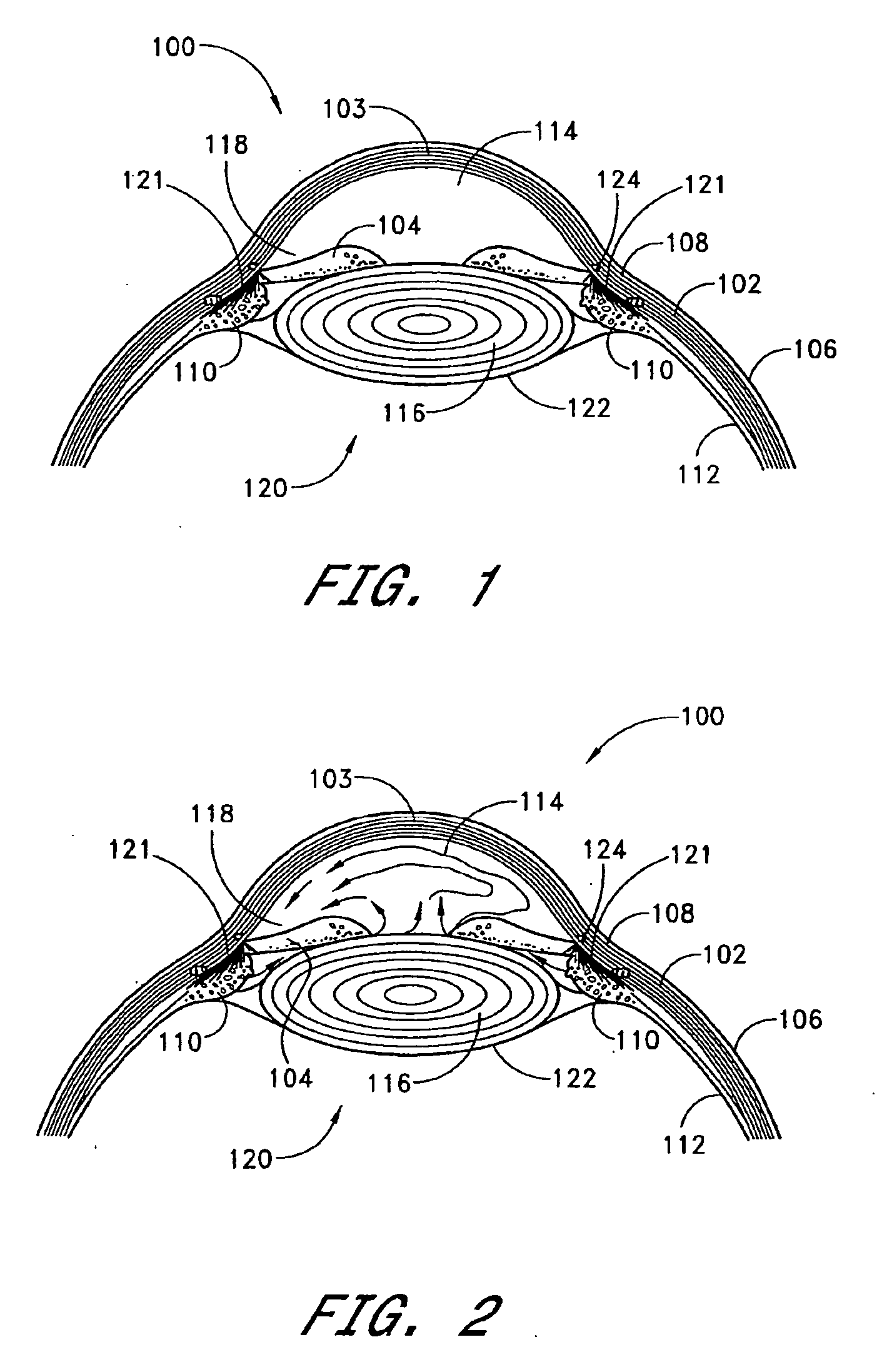
[0071] Referring to FIG. 1, relevant structures of the eye will be briefly described, so as to provide background for the anatomical terms used herein. Certain anatomical details, well known to those skilled in the art, have been omitted for clarity and convenience.

[0072] As shown in FIG. 1, the cornea 103 is a thin, transparent membrane which is part of the outer eye and lies in front of the iris 104. The cornea 103 merges into the sclera 102 at a juncture referred to as the limbus 108. A layer of tissue called bulbar conjunctiva 106 covers the exterior of the sclera 102. The bulbar conjunctiva 106 is thinnest anteriorly at the limbus 108 where it becomes a thin epithelial layer which continues over the cornea 103 to the corneal epithelium. As the bulbar conjunctiva 106 extends posteriorly, it becomes more substantial with greater amounts of fibrous tissue. The bulbar conjunctiva 106 descends over Tenon's capsule approximately 3 mm from the limbus 108. Tenon's capsule is thicker a...


PUM


Abstract

Apparatuses and methods for the treatment of glaucoma are provided. The instrument uses either cauterization, a laser to ablate, sonic or ultrasonic energy to emulsify, or mechanical cutting of a portion of the trabecular meshwork. The instrument may also be provided with irrigation, aspiration, and a footplate. The footplate is used to enter Schlemm's canal, serves as a guide, and also protects Schlemm's canal.

Description

CROSS-REFERENCES TO RELATED APPLICATIONS [0001] This application is a continuation of U.S. patent application Ser. No. 10 / 052,473 (Attorney Docket No. 02307U-132310US), filed Jan. 18, 2002, which claimed priority to U.S. Provisional Application Ser. No. 60 / 263,617, filed Jan. 18, 2001, the full disclosures of which are herein incorporated by reference in their entirety.BACKGROUND OF THE INVENTION [0002] 1. Field of the Invention [0003] The present invention relates to a new glaucoma surgical instrument and method, and, in particular, removal of the trabecular meshwork by mechanical cautery, vaporization or other tissue destruction means optionally coupled to an instrument with infusion, aspiration, and a footplate. [0004] 2. Description of the Related Art [0005] Aqueous is a clear, colorless fluid that fills the anterior and posterior chambers of the eye. The aqueous is formed by the ciliary body in the eye and supplies nutrients to the lens and cornea. In addition, the aqueous prov...

Claims


Application Information

Patent Timeline
Patent Type & Authority Applications(United States)
IPC IPC(8): A61B18/18A61B17/32A61B18/14A61F9/007A61F9/008A61F9/011
CPCA61B17/320068A61B18/1477A61B2017/320072A61B2018/1425A61F9/00781A61F9/0079A61F9/008A61F9/00802A61F2009/00844A61F2009/00868A61F2009/00891A61F9/00745A61F9/00825A61B2090/08021A61B2017/320073A61B2017/32007A61B2017/320075A61B2017/320069A61B2218/002A61B2218/003A61B2218/007A61B2034/254
Inventor BAERVELDT, GEORGECHUCK, ROY
Owner RGT UNIV OF CALIFORNIA
Why Eureka
Try Eureka