

Automated segmentation, classification, and tracking of cell nuclei in time-lapse microscopy
- Summary
- Abstract
- Description
- Claims
- Application Information
AI Technical Summary
Benefits of technology
Problems solved by technology
Method used
Image

Examples
example 1
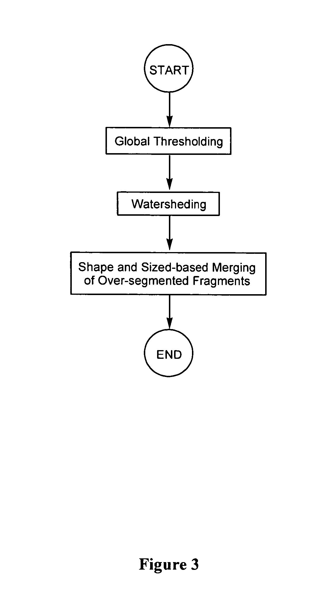
Segmentation
[0156] To test the segmentation algorithm disclosed herein (i.e., a global thresholding / watershed algorithm combined with shape and size-based merging technique), four images were selected from each cell sequence, generating a test set of 16 images containing a total of 3,071 nuclei. Two other segmentation techniques (namely, a simple watershed algorithm without fragment merging; and the watershed algorithm combined with connectivity-based merging described by Umesh Adiga and Chaudhuri (Pattern Recognition, 2001, 34: 1449-1458)) were also used for comparison purposes.
[0157]FIG. 15 shows examples of results obtained using these different segmentation techniques. Clearly, the inventive shape and size-based merging method can merge a lot more over-segmented nuclei than the other two methods.
[0158] Table 1 presents the segmentation results, which are compared with results obtained by manual analysis. The inventive method correctly segmented 97.8% of the nuclei. The waters...
example 2
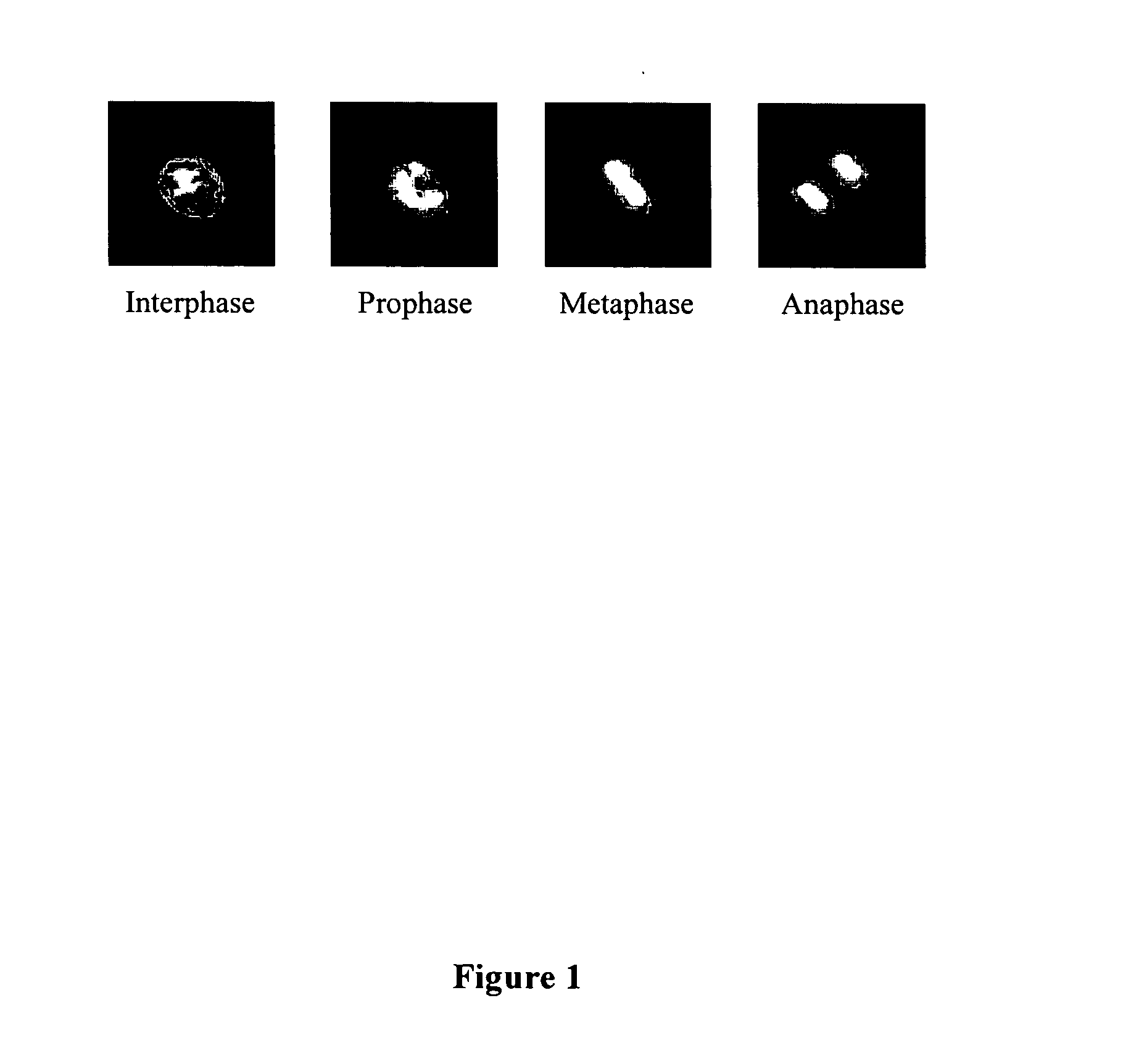
Cell Phase Identification
[0159] The training of the feature selection method was carried out using 100 nuclei for each cell cycle phase which resulted in a training set of 400 nuclei. The 400 cell nuclei were evenly divided into five disjointed subsets. Selection performance was evaluated by a five-fold cross validation in five individual tests with ⅘th of the initial data serving as the training set for the selection algorithm. The remaining ⅕th of the data served as the test set. In exhaustive experiments, a six nearest-neighbor (6-NN) rule delivered the most reliable results for the different selection strategies.
[0160]FIG. 16 shows the variation of the performance of the classifier (which is defined as the ratio between the number of nuclei correctly identified and the total number of nuclei) as a function of the size of the feature subset. The best performances were achieved with a subset size of seven features. Addition of the remaining 5 features caused a decrease in the se...
example 3
Cell Nuclei Tracking
[0164] To establish a metric for the performance of the tracking algorithm, three types of factors have been considered: [0165] (a) percentage of nuclei tracked (which is the number of nuclei tracked without termination through the entire sequence divided by the total number of nuclei at the beginning); [0166] (b) percentage of divisions detected (which is the ratio between the number of cell divisions for which the daughter cell nuclei were correctly assigned to their parent and the total number of cell divisions); and [0167] (c) false division number (which is the number of false divisions detected where two or more nuclei are associated with one nucleus in a previous frame, which did not undergo division).
[0168] Two tracking methods have been used to track the nuclei in all of the four sequences described above. The first method used was the location and size-based tracker disclosed herein and the second method was the centroid tracker (A. P. Goobic et al., ...
PUM
| Property | Measurement | Unit |
|---|---|---|
| Fraction | aaaaa | aaaaa |
| Fraction | aaaaa | aaaaa |
| Fraction | aaaaa | aaaaa |
Abstract
Description
Claims
Application Information