Particle analysis assay for biomolecular quantification

a biomolecular and particle analysis technology, applied in the field of particle analysis assay for biomolecular quantification, can solve the problems of reducing the sensitivity of fish assay, not being able to detect chromosome translocation, and not being able to determine the frequency of chromosome translocation

Inactive Publication Date: 2006-06-15
UNIV OF UTAH RES FOUND
View PDF10 Cites 29 Cited by
  • Summary
  • Abstract
  • Description
  • Claims
  • Application Information

AI Technical Summary

Benefits of technology

[0026] The method of the present invention increases by orders of magnitude the speed of detecting nucleotide sequence aberrations, such as chromosome translocations, over current detection methods, including FISH assays.

Problems solved by technology

Because chromosome translocations involve the movement of a nucleotide sequence within a sample, as opposed to the appearance or disappearance of the nucleotide sequence, it generally is not possible to detect a chromosome translocation merely by assaying for the presence or absence of a particular nucleotide sequence.
Because each FISH probe hybridizes to a specific chromosome type and not to the chromosome translocation itself, it is not possible to determine the frequency of chromosome translocations directly from the fluorescence signal emanating from a FISH probe.
The need to visually scan such individual cells effectively limits the number of cells that can be assayed, thereby reducing the sensitivity of the FISH assay, introducing the possibility of human error, and greatly increasing cost per analysis.
The requirement for re-attachment of the target sequence is particularly problematic and would add significantly to the complexity and cost of commercial separation kits using such methodology and reduce the precision of the assay because of variability in the detachment / reattachment step.
Further, this procedure is limited to two types of solid supports, but it would be useful to have more support options to facilitate multiple simultaneous analyses.
Moreover, the preferable methods for quantification described in U.S. Pat. No. 5,731,153 require either very expensive and uncommon equipment (e.g., measure 14C by accelerator mass spectrometry) or much less quantitative methods such as the detection of fluorescence labels on reporter nucleic acid probes.
Unfortunately, methods available for the quantification of chromosomal rearrangements are either very costly and inefficient, e.g., cytogenetic-type analyses (H. J. Evans et al., 35 Chromosoma 310-325 (1971); D. Pinkel et al., 83 Proc. Natl. Acad. Sci.
PCR is less labor intensive than cytogenetics but is of limited utility in direct DNA-based detection of most chromosomal translocations because the fusion points tend to be variable.

Method used


Image

  • Particle analysis assay for biomolecular quantification
  • Particle analysis assay for biomolecular quantification
  • Particle analysis assay for biomolecular quantification

Examples


example 1

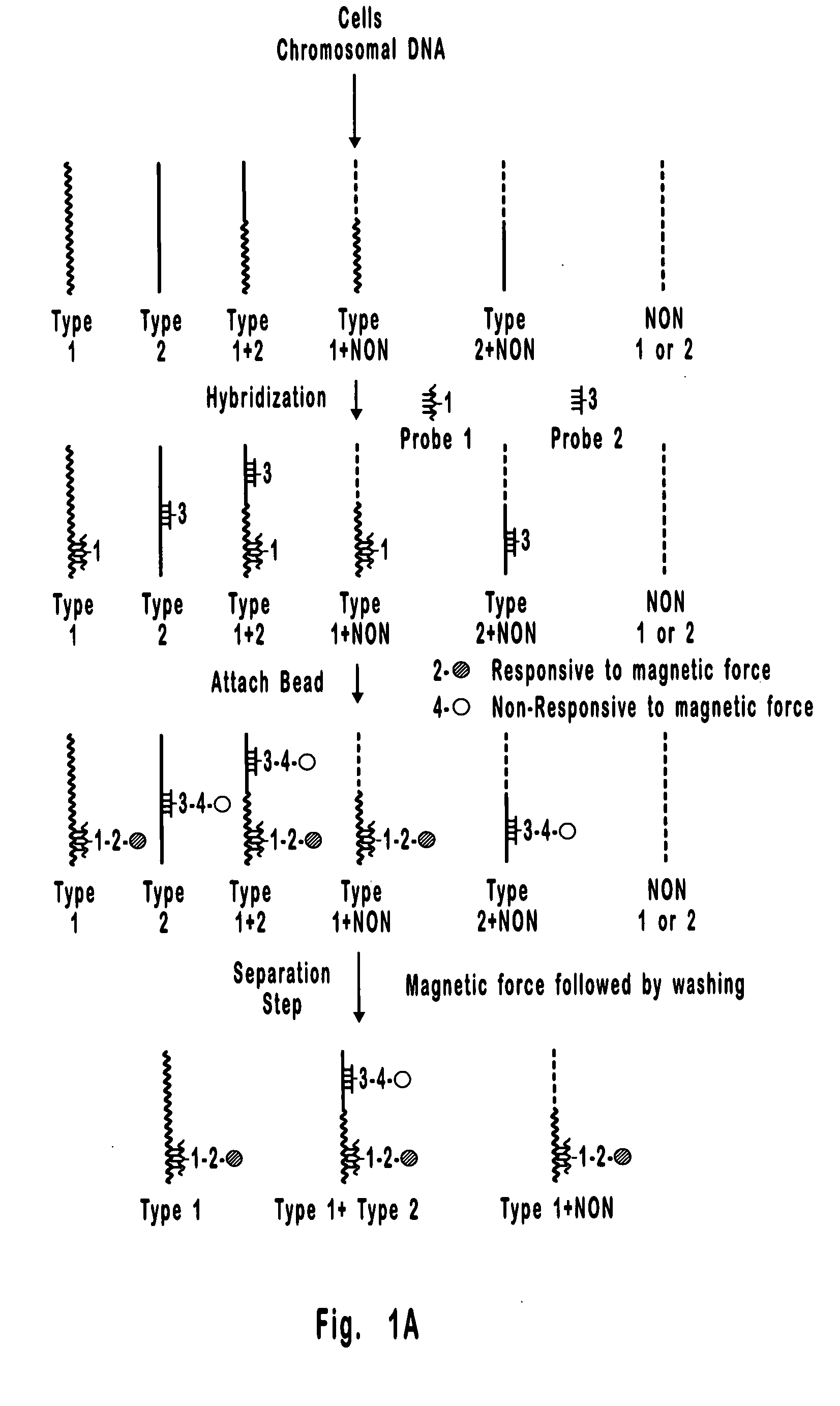
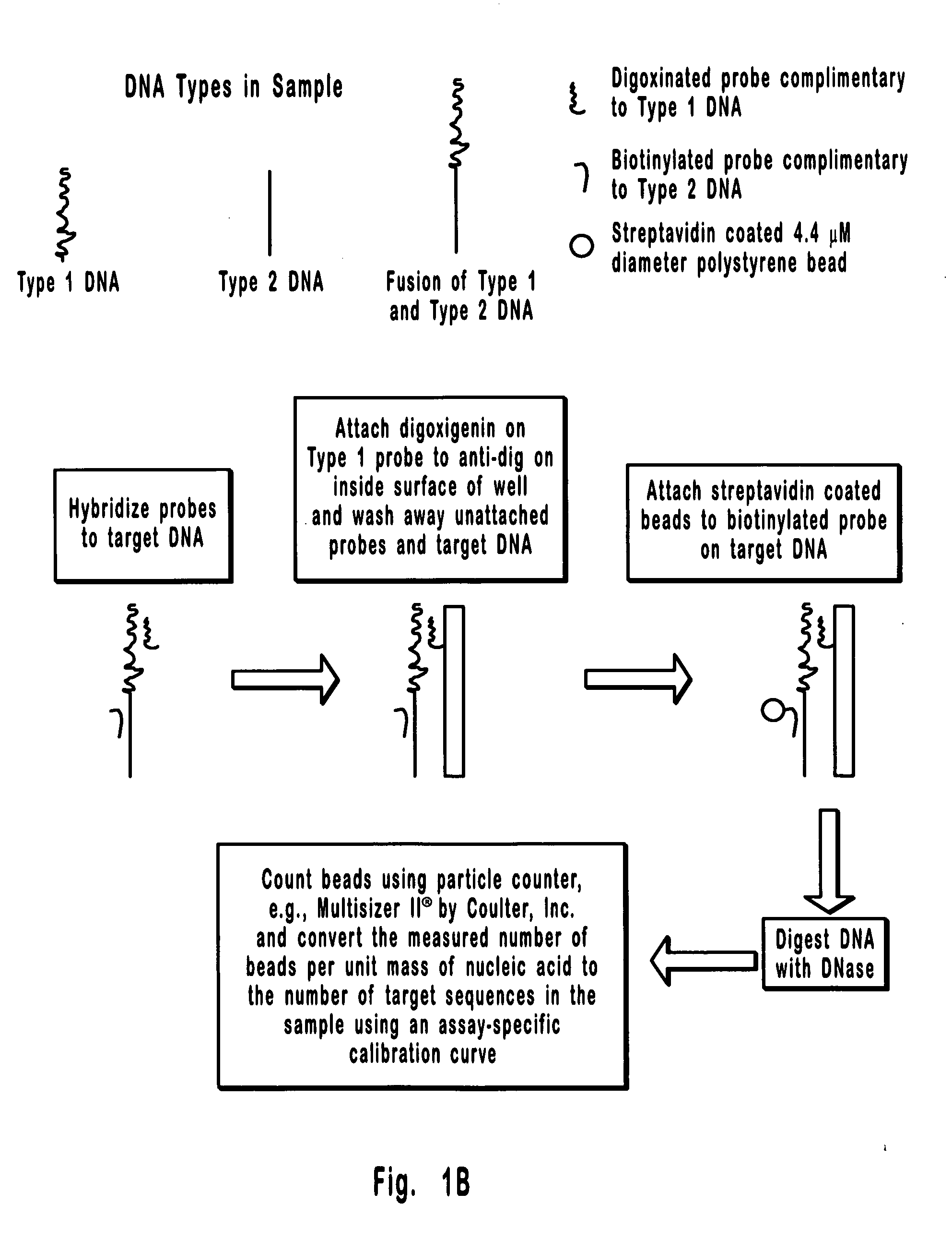
[0087] This is an example of a method that can be used to separate and quantify DNA with two unique and non-overlapping sequences. In this example, the sequences (identified here as Type 1 and Type 2 DNA) span 0.8 kb each and are about 17 kb apart on a contiguous 18.2 kb double stranded DNA molecule. Importantly, these sequences are too far apart for detection by PCR, which is limited to less than about 10 kb, and the 18.2 kb extended DNA molecule is too small for detection by FISH which is generally limited to the detection of condensed DNA such as that in metaphase chromosomes. Hence, the separation and detection demonstrated in the present example could not have been accomplished using available methods. It is also important to note that the DNA molecule selected for this example could just as well have been the result of a rearrangement between Type 1 and Type 2 DNAs, where, for example, Type 1 DNA is from one chromosome and Type 2 DNA is from another chromosome. Hence, the meth...

example 2

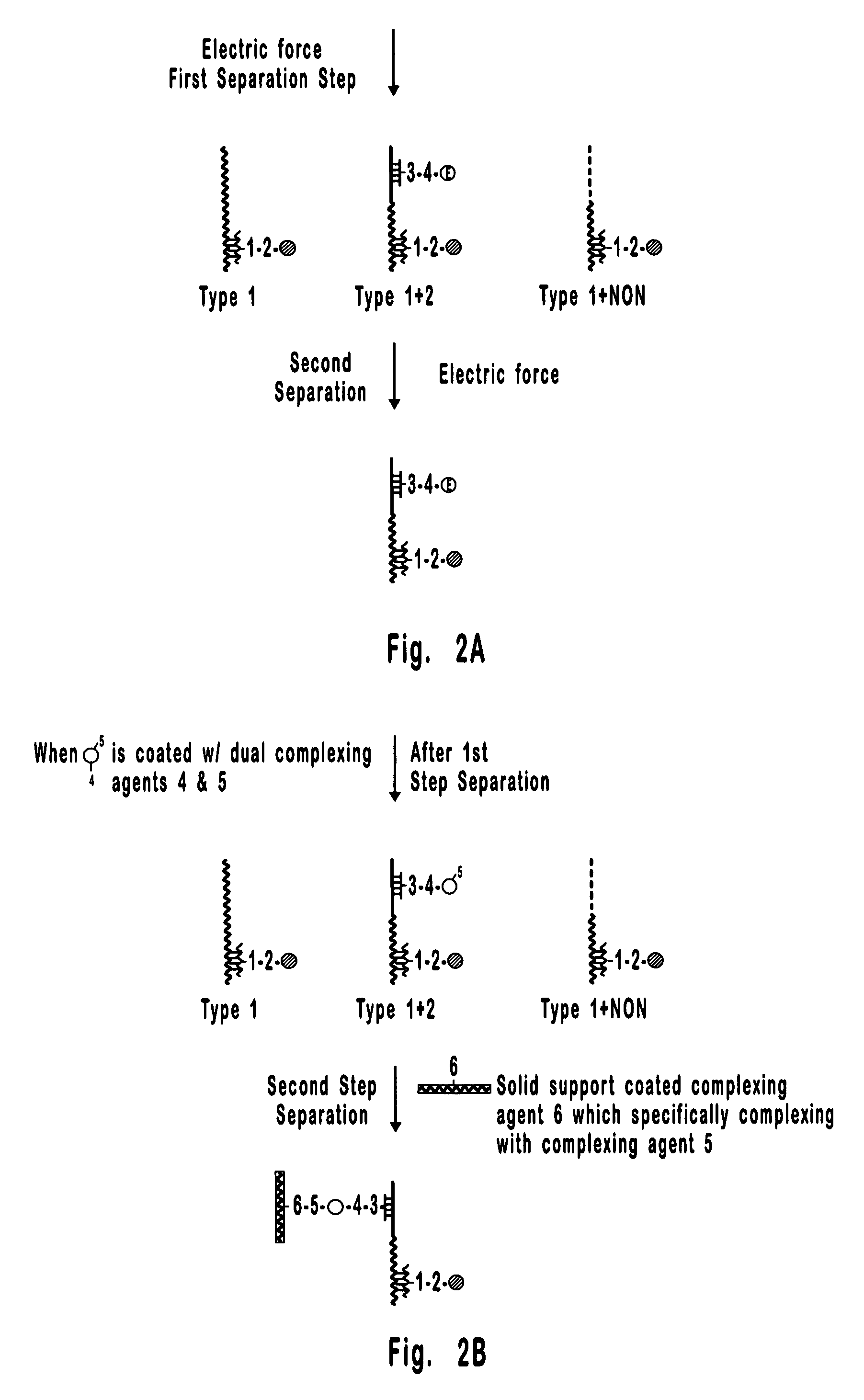
[0127] As discussed above, Stage 2 separation and quantification can also be accomplished by particle counting and size distribution analysis.

[0128] Filtration. For example, if Stage 1 separation is by magnetic force and Stage 2 is by filtration of beads, then the magnetically responsive beads would simply be selected to be larger than the non-magnetic beads. This would permit rapid separation of magnetically-responsive beads from non-magnetic beads. Because the only magnetically non-responsive beads remaining after Stage 1 separation are those complexed with magnetically-responsive beads via hybridization to the same contiguous nucleic acid molecule, the number of non-magnetic beads after Stage 1 would be proportional to the number of target nucleic acid molecules in the hybridization mix.

[0129] To facilitate separation of the beads by filtration, Stage I could be followed by detaching DNA from beads (e.g., via DNase treatment or cleavable linker) and filtering the beads through ...

example 3

[0142] This is an example of a method that can be used to separate and quantify nucleic acids (or chromosomes) with two unique and non-overlapping sequences, both on a contiguous nucleic acid molecule or chromosome. In this example, the two different sequences are identified as Type 1 and Type 2 nucleic acid sequences.

[0143] The method of the present disclosure includes, but is not limited to, the use of two kinds of microbeads. One kind of microbead is responsive to a magnetic field (e.g., Dynabeads Streptavidin, Product #112.05 from Dynal A / S, Oslo, Norway) and could be coated with biotinylated nucleic acid probes complementary to Type 1 nucleic acid sequences by attaching to the avidin on the surface of the magnetically-responsive beads. The other kind of microbead is not responsive to a magnetic field (e.g., streptavidin-coated polystyrene beads from Bangs Labs, Fishers, Ind.) and could be coated with biotinylated nucleic acid probes complementary to Type 2 nucleic acid sequenc...


PUM

PropertyMeasurementUnit
diameteraaaaaaaaaa
diameteraaaaaaaaaa
diameteraaaaaaaaaa

Abstract

A method is provided for carrying out multi-step separations of objects bearing at least two binding sites. In the first step, a first binder / bead composition is bound to objects that bear the first binding site, and then unbound objects, i.e. objects not bearing the first binding site, are separated from bound objects. In the second step, a second binder / bead composition is bound to the remaining objects that bear the second binding site, and then the objects that are bound to both beads are removed from those objects that are bound to only one bead. The beads can differ in magnetic responsiveness, charge, size, color, and the like, and these differences can be used to carry out the separation steps.

Description

CROSS-REFERENCE TO RELATED APPLICATIONS [0001] This application is a division of application Ser. No. 10 / 089,560, filed Apr. 1, 2002, which was the National Stage of International Application No. PCT / US00 / 27883, filed Oct. 10, 2000, which claims the benefit of U.S. Provisional Application No. 60 / 158,664, filed Oct. 8, 1999, which applications are incorporated herein by this reference.BACKGROUND OF THE INVENTION [0002] The present invention relates to methods for isolation, separation, and detection of selected objects. More particularly, the invention relates to methods for performing separations of objects, such as, without limitation, nucleic acids, proteins, cells, organelles, and the like. [0003] Separations of biological objects, such as proteins, chromosomes, nucleic acids, cells, organelles, and the like, and other types of objects are important in various detection, isolation, quantification, and diagnostic processes. Specificity and sensitivity are two important parameters ...

Claims


Application Information

Patent Timeline
Patent Type & Authority Applications(United States)
IPC IPC(8): C12Q1/68
CPCC12Q1/6816G01N33/54333C12Q2563/155C12Q2563/143C12Q2545/114
Inventor STRAUME, TORELIU, GANG
Owner UNIV OF UTAH RES FOUND
Why Eureka
Try Eureka