Autologous Growth Factors to Promote Tissue In-Growth in Vascular Device

a technology vascular devices, which is applied in the field of autologous growth factors to promote tissue ingrowth in vascular devices, can solve the problems of excessively high risk of conventional aneurysm repair, underreported aneurysm deaths, and increased so as to prevent stent graft migration and reduce the risk of stent graft migration , the effect of promoting healing

Inactive Publication Date: 2006-06-22
MEDTRONIC VASCULAR INC
View PDF14 Cites 7 Cited by
  • Summary
  • Abstract
  • Description
  • Claims
  • Application Information

AI Technical Summary

Benefits of technology

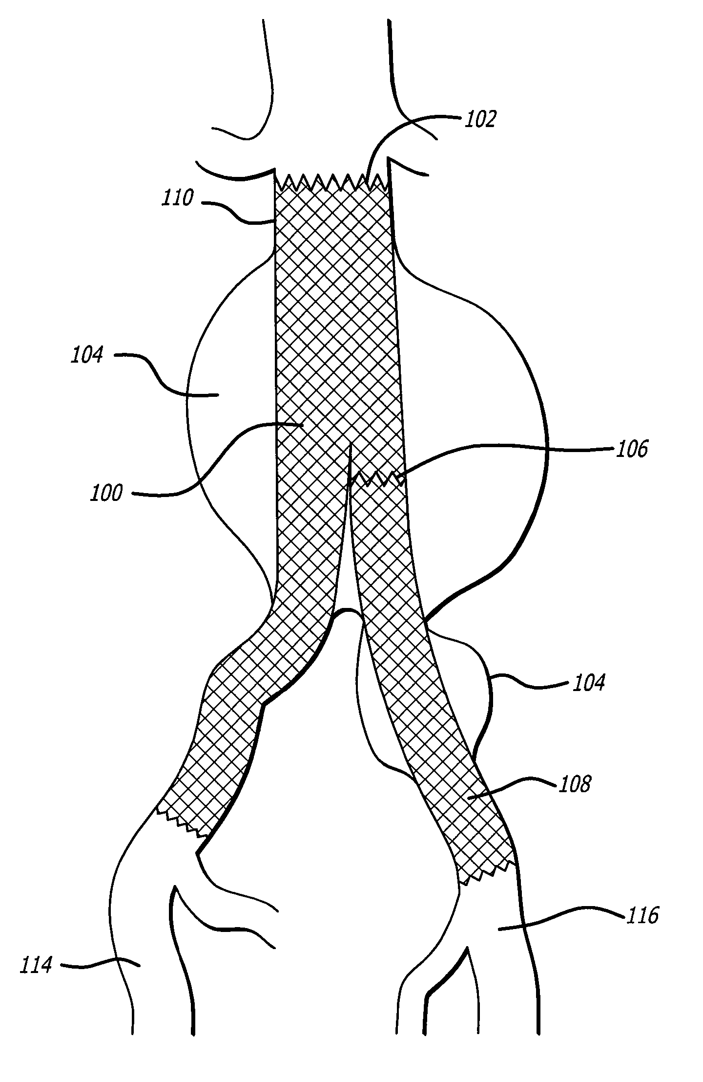
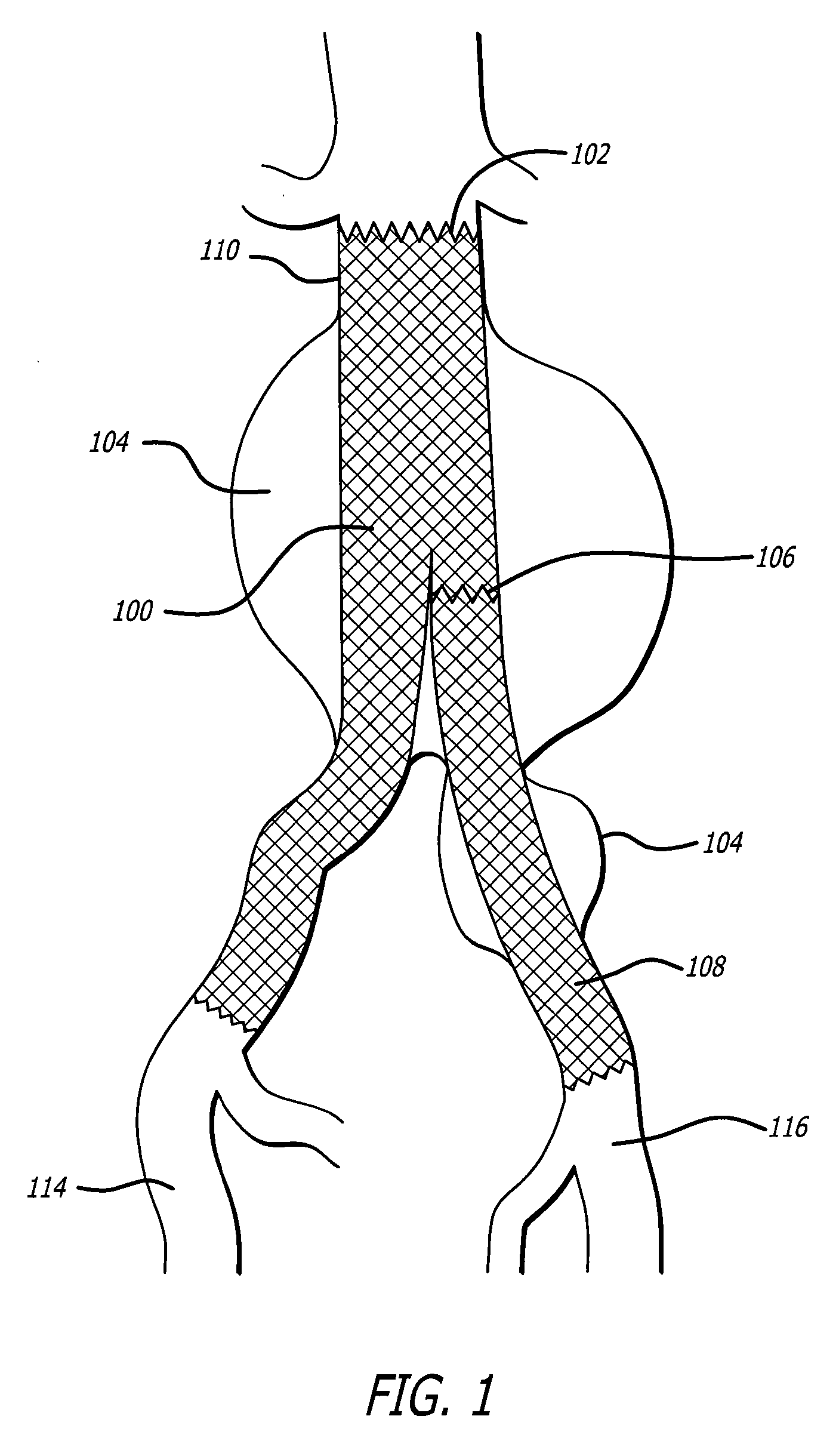
[0013] The risk of stent graft migration can be reduced by delivering to the treatment site, as a coating on the stent graft, endothelialization factors such as autologous growth factor compositions. This administration can be beneficial because, normally, the endothelial cells that make up the portion of the vessel to be treated are quiescent at the time of stent graft implantation and do not multiply. As a result, the stent graft rests against a quiescent endothelial cell layer. If autologous growth factor compositions are administered to the treatment site with the stent graft deployment, the normally quiescent endothelial cells lining the vessel wall, and in intimate contact with the stent graft, will be stimulated to proliferate. The same will occur with smooth muscle cells and fibroblasts found within the vessel wall. As these cells proliferate they can grow into and around the stent graft lining such that the stent graft becomes physically attached to the vessel lumen rather than merely resting against it. This endothelialization helps to prevent stent graft migration. These methods can promote healing, reduce endoleaks and minimize device migration by promoting endothelial tissue in-growth.
[0014] Based on the foregoing, embodiments according to the present invention provide stent grafts having autologous growth factor compositions coated thereon for the treatment of aneurysms, and associated methods for using and / or manufacturing the stent grafts. Additionally, stent grafts are disclosed which provide structural support for weakened arterial walls while the accompanying compositions promote tissue in-growth to reduce the chance of graft migration and endoleaks.

Problems solved by technology

These thinned and weakened sections of vessel walls can burst, causing an estimated 32,000 deaths in the United States each year.
Additionally, aneurysm deaths are suspected of being underreported because sudden unexplained deaths, about 450,000 in the United States alone, are often simply misdiagnosed as heart attacks or strokes while many of them may be due to aneurysms.
Additionally, patients whose multiple medical comorbidities make them excessively high risk for conventional aneurysm repair are candidates for stent grafting.
While stent grafts and stents (hereinafter collectively referred to as “vascular devices”) represent improvements over previously-used vessel treatment techniques, there are still risks associated with them.
The most common of these risks is migration of the vascular device due to hemodynamic forces within the vessel.
Stent migration can leave a treated area of a vessel more susceptible to reclosure.
Additionally, the asymmetrical hemodynamic forces can cause remodeling of an aneurysm sac which leads to increased risk of aneurysm rupture and increased endoleaks.
The magnetic fields used in this imaging process, when moving across the body, may cause insufficiently fixated metal-containing stents to migrate.
This natural endothelialization is not complete or consistent, however, and therefore does not in some cases prevent the stent graft migration and endoleak.

Method used


Image

  • Autologous Growth Factors to Promote Tissue In-Growth in Vascular Device
  • Autologous Growth Factors to Promote Tissue In-Growth in Vascular Device
  • Autologous Growth Factors to Promote Tissue In-Growth in Vascular Device

Examples


example 1

Properties of Platelet Rich Plasma

[0065] Aliquots of human peripheral blood (30-60 mL) are passed through the Magellan™ Autologous Platelet Separator System (the Magellan™ system, Medtronic, Inc., Minneapolis, Minn.) and the concentrated, platelet-rich plasma fraction (PRP) assayed for platelets (PLT), white blood cells (WBC) and hematocrit (Hct) (Table 1). The Magellan™ system concentrated platelets and white blood cells six-fold and three-fold respectively.

TABLE 1Blood cell yields after passing through the Magellan ™ system.Mean ± SDn = 19Initial BloodPRPYieldPLT (×1000 / μL)220.03 ± 48.58 1344.89 ± 302.00 6.14 ± 0.73WBC5.43 ± 1.4317.04 ± 7.01 3.12 ± 0.90(×1000μ / L)Hct (%)38.47 ± 2.95 6.81 ± 1.59

Cell Yield = cell count in PRP / cell count in initial blood = [times baseline]

[0066] Additionally, PRP was assayed for levels of the endogenous growth factors platelet-derived growth factor (PDGF), transforming growth factor-beta (TGF-β), basic fibroblast growth factor (bFGF), vascular endo...

example 2

Autologous Platelet Gel Generation

[0067] Autologous Platelet Gel (APG) is generated from the PRP fraction produced in the Magellan™ system by adding thrombin and calcium to activate the fibrinogen present in the PRP as well as causing the platelets to release additional stores of growth factors. For each approximately 7-8 mL of PRP, approximately 5000 units of thrombin in 5 mL 10% calcium chloride are required for activation. The APG is formed immediately upon mixing of the activator solution with the PRP. The concentration of thrombin can be varied from approximately 1-1,000 U / mL, depending on the speed required for setting to occur. The lower concentrations of thrombin will provide slower gelling times.

example 3

Effects of Platelet Releasates on Cell Proliferation

[0068] A series of in vitro experiments were conducted evaluating the effect of released factors from platelets on the proliferation of the human microvascular endothelial cells and human coronary artery smooth muscle cells. Primary cell cultures of both cell types were established according to protocols well known to those skilled in the art of cell culture. Autologous platelet gel was used as the source of platelet releasates. For each cell type, five culture conditions were evaluated; basal medium (BM)+APG; BM+platelet-free plasma (PFP); growth medium (GM); BM alone; and BM+thrombin. Growth medium is the standard culture medium for the cell type and contains optimal growth factors and supplements.

[0069] Autologous platelet gel had a significant growth effect on human microvascular endothelial cells after four days of culture (FIG. 3) and on human coronary artery smooth muscle cells after five days of culture (FIG. 4).


PUM


Abstract

Methods and devices for ameliorating stent graft migration and endoleak using treatment site-specific cell growth promoting compositions in combination with stent grafts are disclosed. Also disclosed is coating of autologous growth factor compositions onto stent grafts prior to stent graft implantation. Additional embodiments include stent grafts having autologous growth factor composition coatings useful for treating aneurysms.

Description

RELATED APPLICATIONS [0001] This application is a continuation-in-part of U.S. patent application Ser. No. 10 / 977,545, filed Oct. 28, 2004, which is herein incorporated by reference in its entirety.FIELD OF THE INVENTION [0002] Methods and devices for preventing vascular device migration using autologous growth factor composition-coated vascular devices are disclosed. Specifically, methods for producing autologous growth factor compositions and coating vascular devices with the compositions before device implantation are provided. BACKGROUND OF THE INVENTION [0003] A variety of implantable vascular devices, including stent grafts and stents, have been developed to treat abnormalities of the vascular system. Stent grafts are used to treat aneurysms of the vascular system and have also emerged as a new treatment for a related condition, acute blunt aortic injury, where trauma causes damage to an artery. Stents are used to treat areas of vessel narrowing or atherosclerosis. [0004] Aneu...

Claims


Application Information

Patent Timeline
Patent Type & Authority Applications(United States)
IPC IPC(8): A61F2/06
CPCA61L27/3616A61L27/3641A61L2300/414A61L27/54A61L27/3645
Inventor CHU, JACKDOIG, SCOTTFERNANDES, BRIANHUANG, TREVORWILCOX, JOSIAH
Owner MEDTRONIC VASCULAR INC
Why Eureka
Try Eureka