

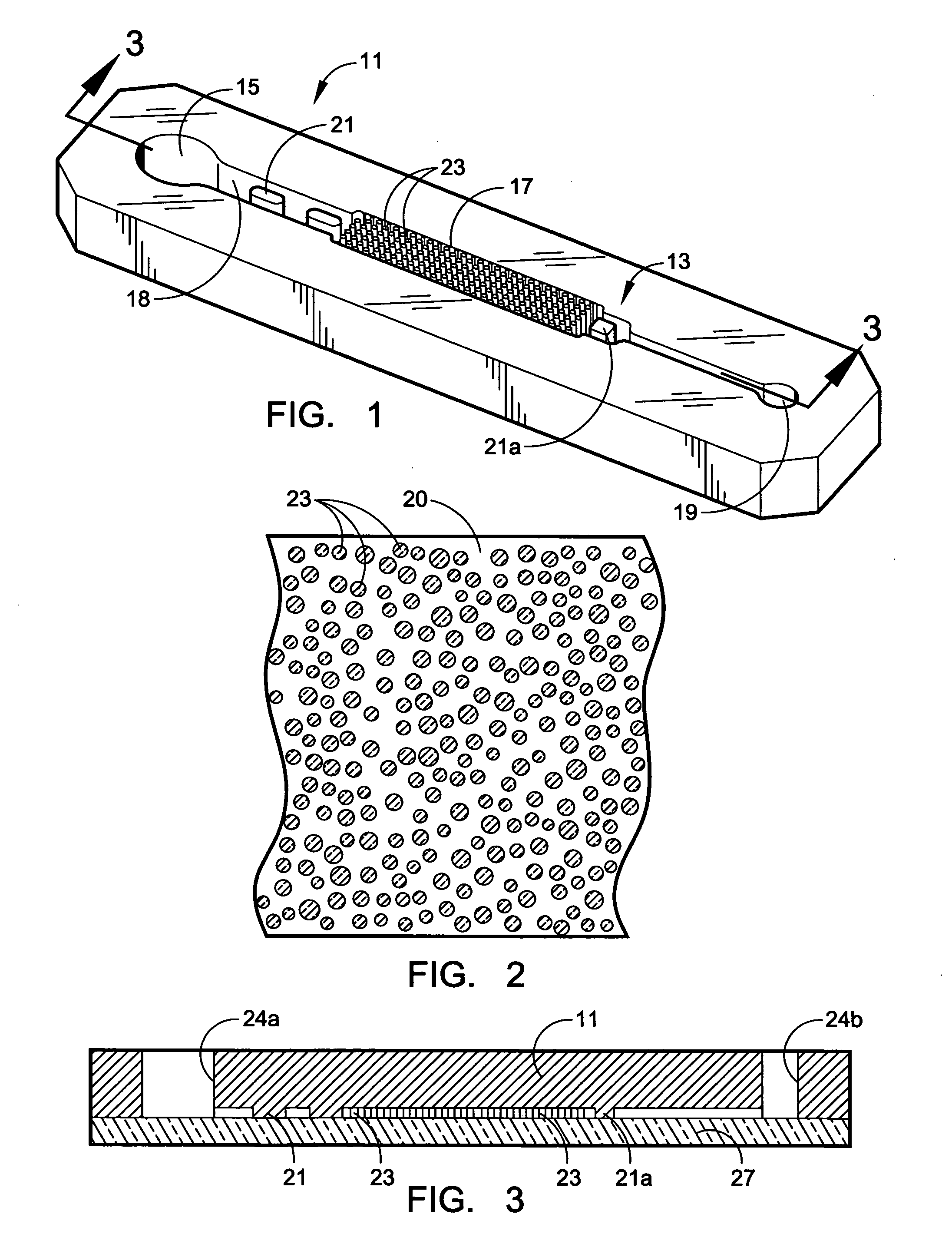
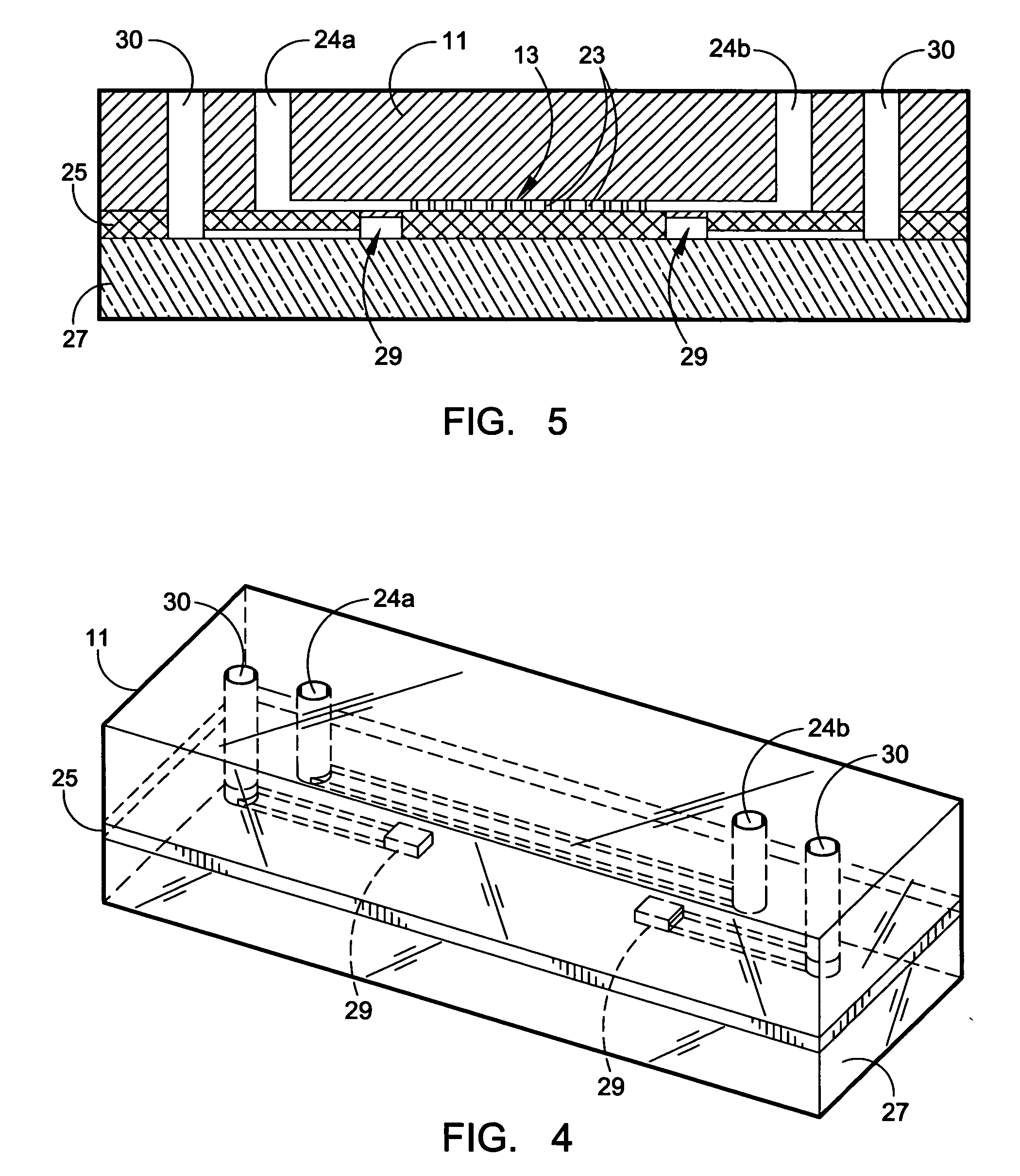
Recovery of rare cells using a microchannel apparatus with patterned posts
a microchannel apparatus and rare cell technology, applied in the field of rare cell recovery using a microchannel apparatus with patterned posts, can solve the problems of method risks, difficult non-specific binding, and less efficient collection of target cells
- Summary
- Abstract
- Description
- Claims
- Application Information
AI Technical Summary
Benefits of technology
Problems solved by technology
Method used
Image


Examples
example 1
[0065] A microflow apparatus for separating biomolecules is constructed using a prototype substrate as in Example 1. The substrate is formed from PDMS and is bonded to a flat glass plate to close the flow channel. The interior surfaces throughout the collection region are derivatized by incubating for 30 minutes at room temperature with a 10 volume % solution of Dow Corning Z-6020. After washing with ethanol, they are treated with nonfat milk at room temperature for about one hour to produce a thin casein coating. Following washing with 10% ethanol in water, a treatment is effected using a coating. Following washing with 10% ethanol in water, a treatment is effected using a hydrogel based on isocyanate-capped PEG triols, average MW of 6000. The formulation used consists of about 3% polymer. A hydrogel prepolymer is made using 1 part by weight polymer to 6 parts of organic solvent, i.e. Acetonitrile and DMF, was mixed with an 1 mg / ml antibody solution in 100 mM Sodium Borate pH 8.0 c...
example 2
[0071] A microflow apparatus for separating biomolecules is constructed using a prototype substrate as in Example 1. The substrate is formed from PDMS and is bonded to a flat glass plate to close the flow channel. The interior surfaces throughout the collection region are derivatized by incubating for 30 minutes at room temperature with a 10 volume % solution of Dow Corning Z-6020. After washing with ethanol, they are treated with nonfat milk at room temperature for about one hour to produce a thin casein coating. Following washing with 10% ethanol in water, a treatment is effected using a hydrogel based on isocyanate-capped PEG triols, average MW of 6000. The formulation that is used consists of about 3% polymer. A hydrogel prepolymer is made using 1 part by weight polymer to 6 parts of organic solvent, i.e. acetonitrile and DMF, and is mixed with an 1 mg / ml antibody solution in 100 mM sodium borate, pH 8.0, containing BSA. The resultant specific formulation comprises 100 mg prepol...
example 3
[0075] Another microflow apparatus for separating biomolecules is constructed using a prototype substrate as in Example 1. The substrate is formed from PDMS and is bonded to a flat glass plate to close the flow channel. The interior surfaces throughout the collection region are derivatized by incubating for 30 minutes at room temperature with a 10 volume % solution of Dow Corning Z-6020. After washing with ethanol, they are treated with nonfat milk at room temperature for about one hour to produce a thin casein coating.
[0076] Following washing with 10% ethanol in water, a treatment is effected using 10 μl of 2.5 mM NHS-polyglycine (ave. MW about 4500) in 0.2 MOPS / 0.5M NaCl, pH 7.0, by incubating at RT for 2 hours with gentle pumping of the solution back and forth in the channel to provide agitation. The microchannel is washed three times with 500 μl of pH 7.0 MOPS buffer to obtain maleimido-polyGly-coated channels.
[0077] Antibodies Trop-1 and Trop-2, which are specific to ligands ...
PUM
| Property | Measurement | Unit |
|---|---|---|
| thick | aaaaa | aaaaa |
| molecular weight | aaaaa | aaaaa |
| molecular weight | aaaaa | aaaaa |
Abstract
Description
Claims
Application Information