

Animal cell confluence detection method and apparatus
a technology of confluence detection and apparatus, applied in the field of apparatus and methods for detecting confluence in animal cells, can solve the problems of damage to experiments, high time-consuming manual visual inspection of confluence, non-auditability and non-repeatability
- Summary
- Abstract
- Description
- Claims
- Application Information
AI Technical Summary
Benefits of technology
Problems solved by technology
Method used
Image

Examples
Embodiment Construction
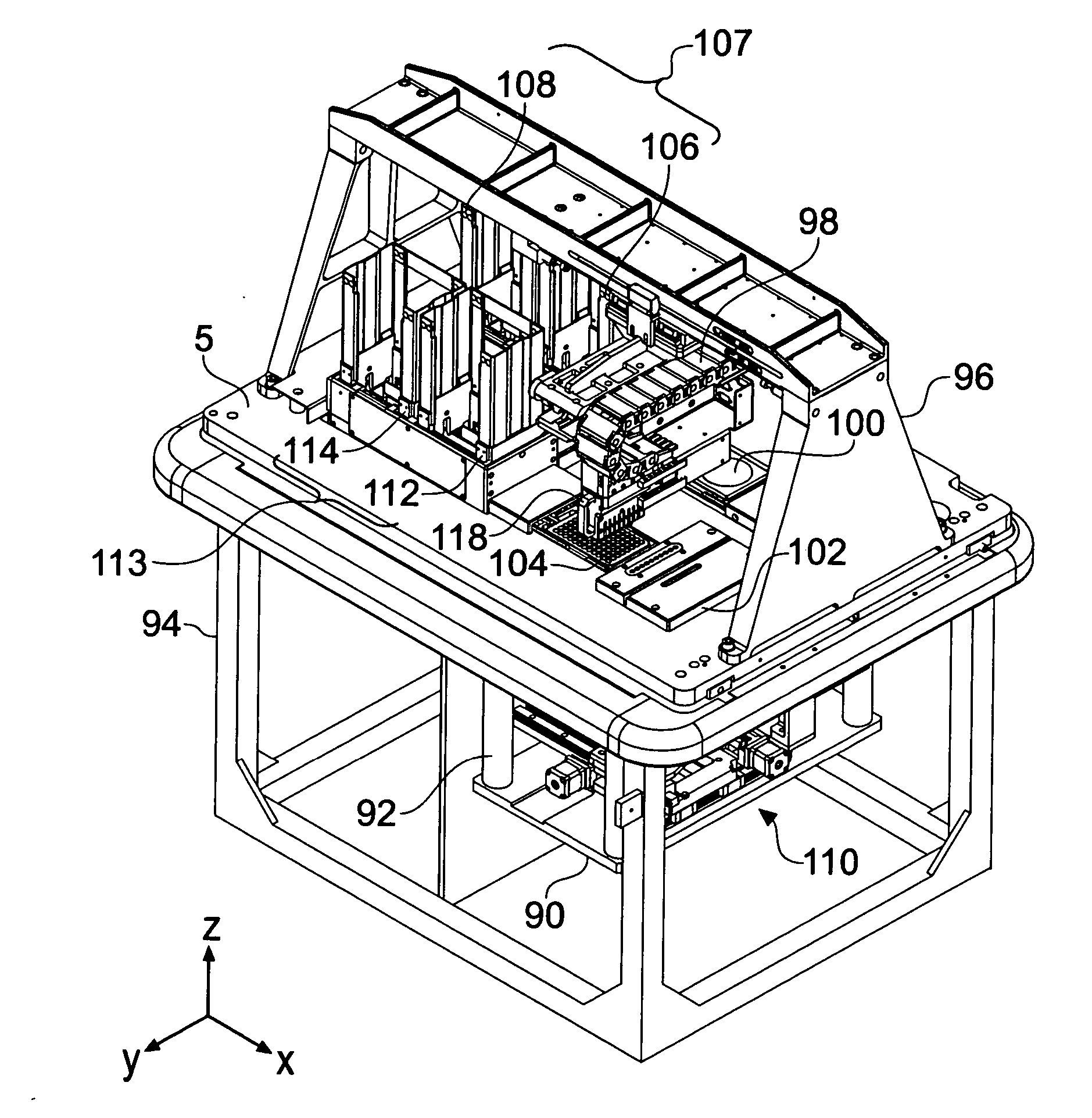
[0043]FIG. 1 is a perspective view of an apparatus embodying the invention.
[0044] The apparatus may be considered to be a picking robot with integrated confluence detection optics. The apparatus can be subdivided notionally into two half spaces existing above and below a main bed 5 which is supported by a frame 94.
[0045] Above the main bed 5, the apparatus appears as similar to a conventional picking robot. A cell picking head 118 is provided that comprises a plurality of hollow pins for aspirating animal cells. The cell picking head 118 is movable over the main bed 5 by a head position system made up of x- y- and z-linear positioners 98 connected in series and suspended from a gantry 96. A wash / dry station 102 is also provided on the main bed 5 for cleansing the pins. The whole upper half space of the apparatus will typically be enclosed in a housing (not shown) including a hinged door extending over one side and part of the top of the apparatus.
[0046] Below the main bed 5, an o...
PUM
 Abstract
Description
Claims
Application Information