Systems and methods for detection of wound fluid blood and application of phototherapy in conjunction with reduced pressure wound treatment system

Active Publication Date: 2006-08-03
KCI LICENSING INC
View PDF99 Cites 194 Cited by
  • Summary
  • Abstract
  • Description
  • Claims
  • Application Information

AI Technical Summary

Benefits of technology

[0025] In fulfillment of these and other objectives, the present invention provides wound fluid blood detection systems and methods operable in conjunction with reduced pressure wound treatment (RPWT) systems, as well as additional ancillary therapy and monitoring systems applied with RPWT systems. The blood detection monitor operates by optically characterizing the content of wound fluids to the extent of identifying a percentage blood content in the fluids. The optical identification relies upon the transmission of select wavelengths of light across a volume of wound fluid to a photo detector system capable of quantifying the absorption characteristics of the fluid at the select

Problems solved by technology

Unfortunately, this process is hampered when a wound is large or has become infected.
Without sufficient blood flow, the epithelial and subcutaneous tissues surrounding the wound not only receive diminished oxygen and nutrients, but are also less able to successfully fight bacterial infection and thus are less able to naturally close the wound.
Although still widely practiced and often effective, such mechanical closure techniques suffer a major disadvantage in that they produce tension on the skin tissue adjacent the wound.
In particular, the tensile force required in order to achieve closure using sutures or staples causes very high localized stresses at the suture or staple insertion point.
These stresses commonly result in the rupture of the tissue at the insertion points, which can eventually cause wound dehiscence and additional tissue loss.
Additionally, some wounds harden and inflame to such a degree due to infection that closure by stapling or sutu

Method used


Image

  • Systems and methods for detection of wound fluid blood and application of phototherapy in conjunction with reduced pressure wound treatment system
  • Systems and methods for detection of wound fluid blood and application of phototherapy in conjunction with reduced pressure wound treatment system
  • Systems and methods for detection of wound fluid blood and application of phototherapy in conjunction with reduced pressure wound treatment system

Examples


Example

[0042] Although those of ordinary skill in the art will readily recognize many alternative embodiments, especially in light of the illustrations provided herein, this detailed description is exemplary of the preferred embodiment of the present invention, the scope of which is limited only by the claims which may be drawn hereto.

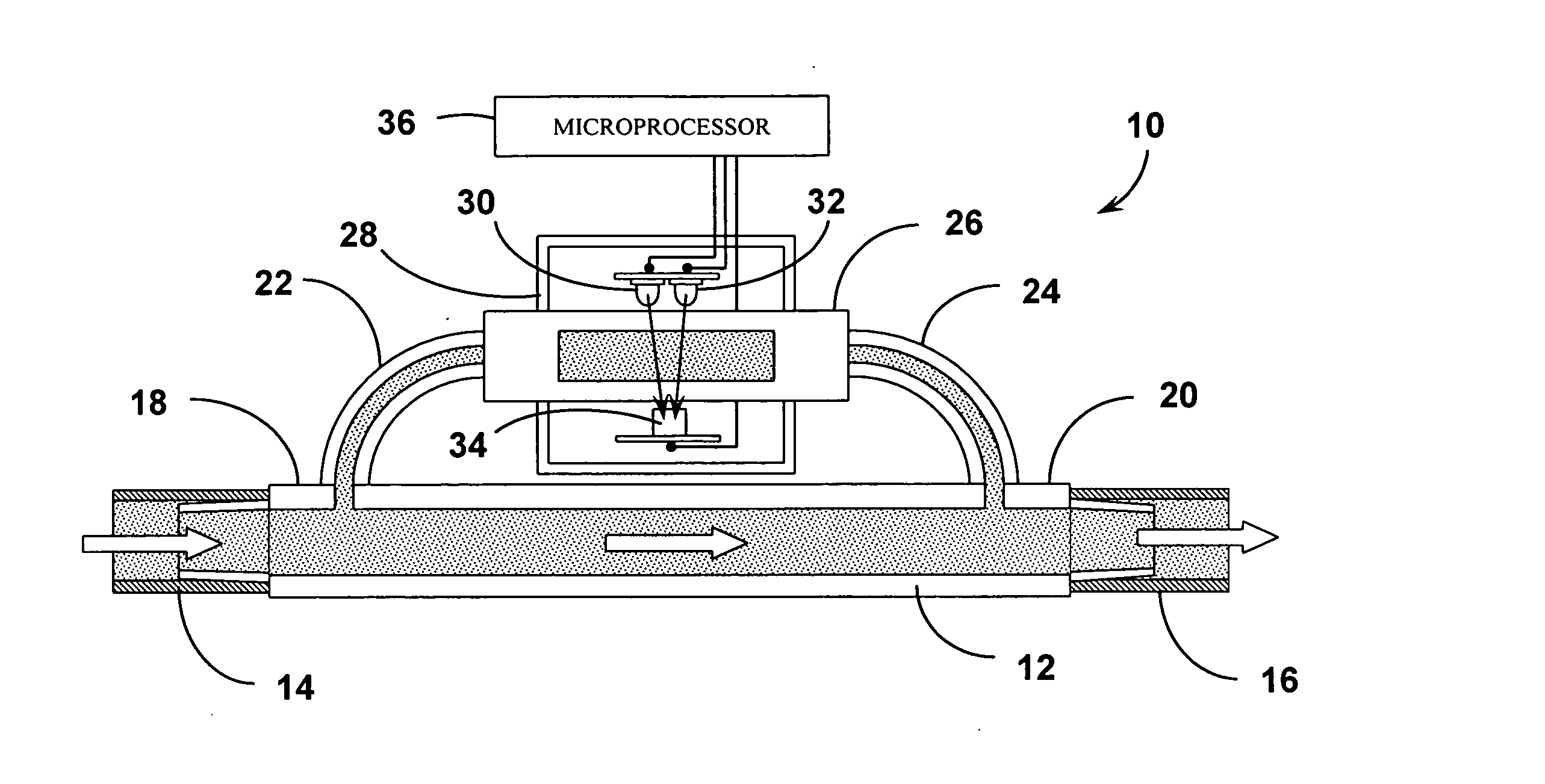
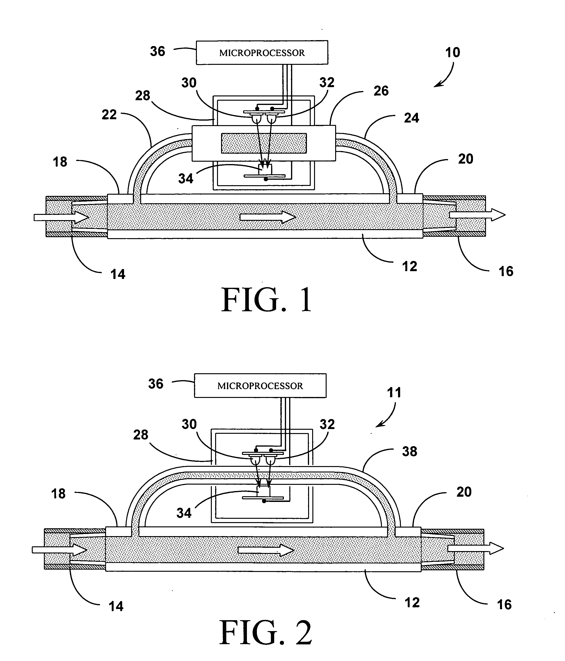
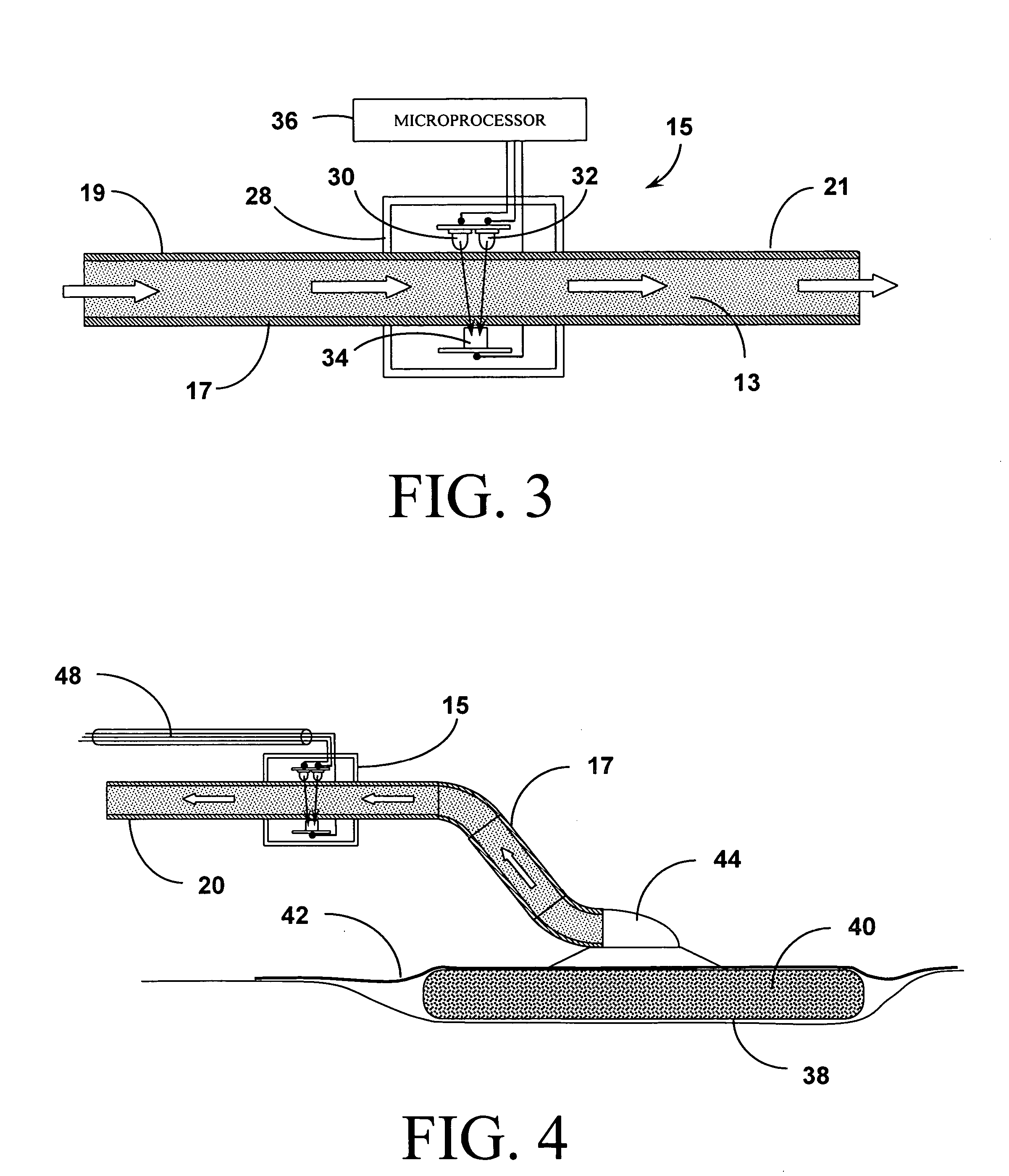
[0043] The systems and methods of the present invention as shown in the attached figures employ photometric or optical methods for detecting the presence (and ultimately, the concentration) of blood in wound fluid being drawn away from the wound by Reduced Pressure Wound Treatment (RPWT) devices and systems. In general, LEDs in the 540 / 560 / 580 / 620 / 640 / 660 nm and 800 nm ranges are used as the emitters and a photo detector sensitive to the same range of wavelengths is used as the receptor. These solid state optical components are positioned across a flow stream of the wound fluid and measurements are taken of the absorption of the illuminating light in a manne...


PUM


Abstract

Wound fluid blood detection systems and methods are described that are operable in conjunction with reduced pressure wound treatment (RPWT) systems, as well as ancillary therapy and monitoring systems applied concurrently with RPWT systems. The blood detection monitor operates by optically characterizing the content of wound fluids to the extent of identifying percentage blood content. This identification relies upon the transmission of select wavelengths of light across a volume of wound fluid to a photo detector (connected to signal processing instrumentation) capable of quantifying the absorption characteristics of the fluid. The detection components may be implemented in conjunction with either a fluid flow conduit (i.e. the reduced pressure tubing directing fluid away from the wound dressing) or more directly in association with the materials that comprise the wound dressing positioned within the wound bed itself. In addition, the present invention is configured to operate in conjunction with blood gas monitoring systems operating with the RPWT. Finally, the components of the systems that serve to illuminate the wound bed, lend themselves to use in conjunction with illumination in discreet electromagnetic wave wavelengths suitable for the application of phototherapy.

Description

CROSS-REFERENCE TO RELATED APPLICATIONS [0001] The present application is a continuation-in-part of U.S. patent application Ser. No. 10 / 867,990, filed on Jun. 15, 2004, which is a continuation of U.S. patent application Ser. No. 10 / 085,321, filed on Feb. 28, 2002, now U.S. Pat. No. 6,856,821, issued on Feb. 15, 2005, which is a continuation-in-part of U.S. patent application Ser. No. 09 / 579,755, filed on May 26, 2000, now abandoned; the present application is also a continuation-in-part of U.S. patent application Ser. No. 09 / 544,399, filed on Apr. 6, 2000, which claims the benefit of the filing of U.S. Provisional Patent Application Ser. No. 60 / 127,936, filed on Apr. 6, 1999; and the present application is also a continuation-in-part of U.S. patent application Ser. No. 10 / 090,358, filed on Mar. 4, 2002, which claims the benefit of the filing of U.S. Provisional Patent Application Ser. No. 60 / 273,587, filed on Mar. 5, 2001.BACKGROUND OF THE INVENTION [0002] 1. Field of the Invention ...

Claims


Application Information

Patent Timeline
IPC IPC(8): A61B5/00A61N5/06
CPCA61B5/0059A61B5/14542A61B5/1468A61B5/445A61B5/6834A61M1/0088A61N2005/0651A61M1/0025A61M1/73A61M1/90A61M1/95
Inventor GANAPATHY, PREMNARAYANJOHNSON, ROYCEROBINSON, TIMLOCKE, CHRISTOPHER B.
Owner KCI LICENSING INC
Why Eureka
Try Eureka