Ultrasonograph
- Summary
- Abstract
- Description
- Claims
- Application Information
AI Technical Summary
Benefits of technology
Problems solved by technology
Method used
Image
Examples
embodiment 1
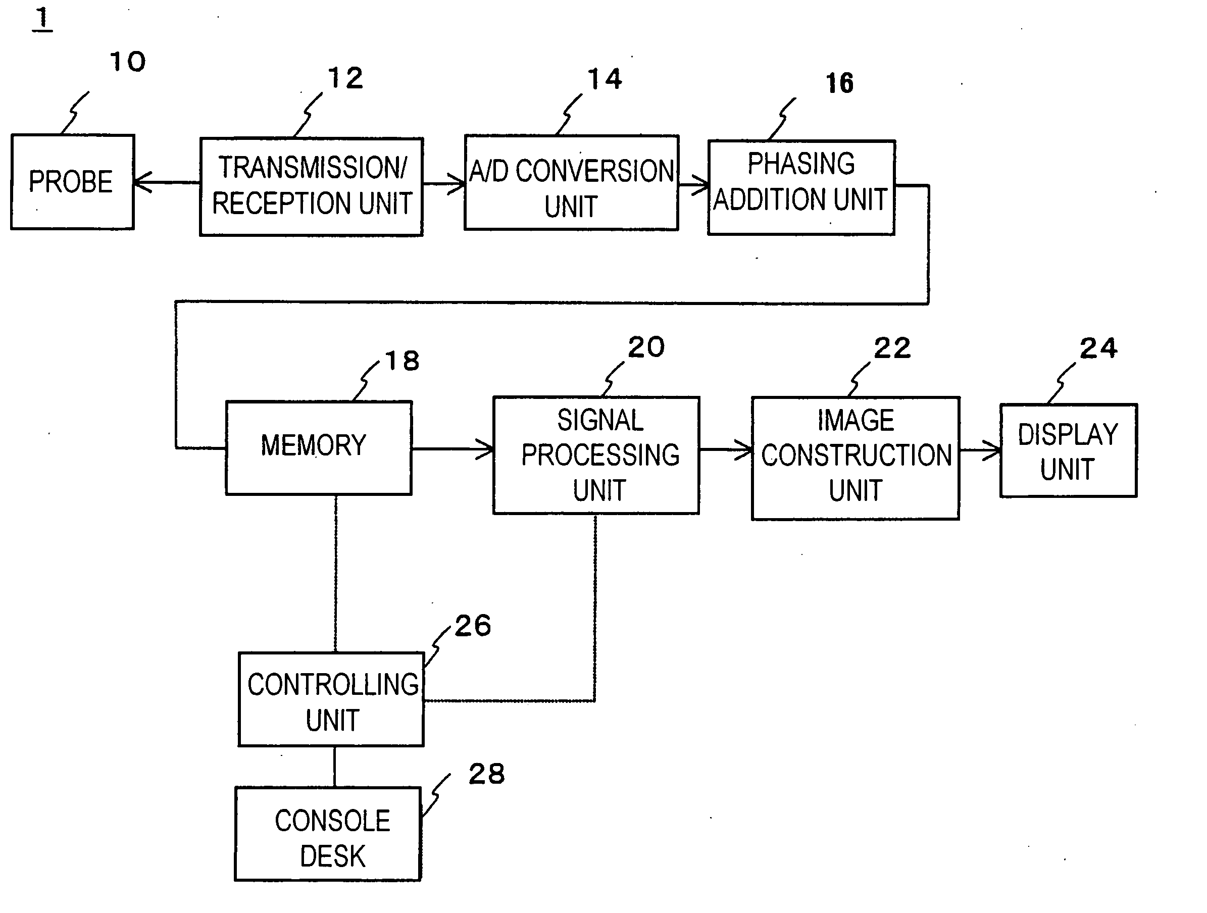
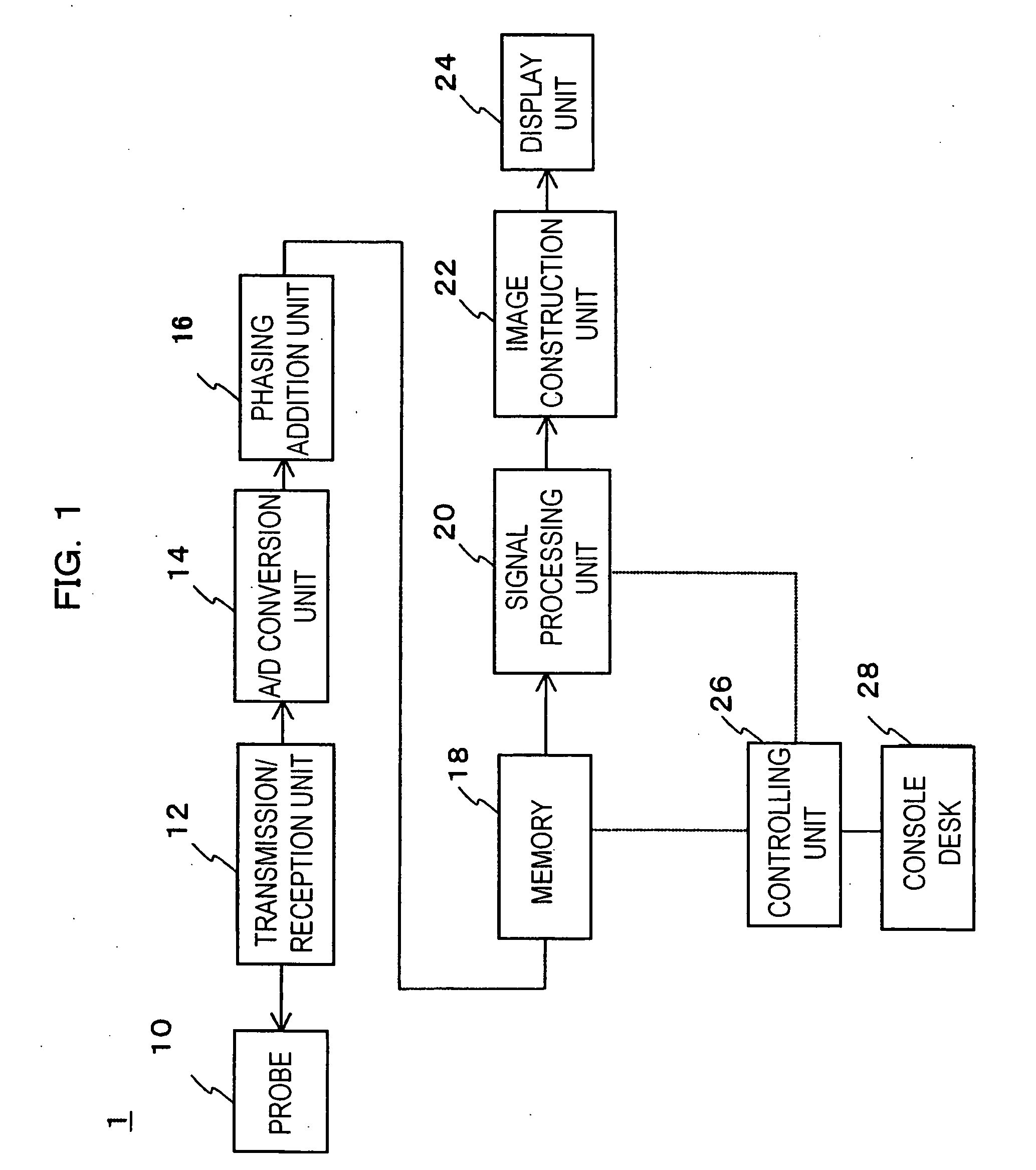
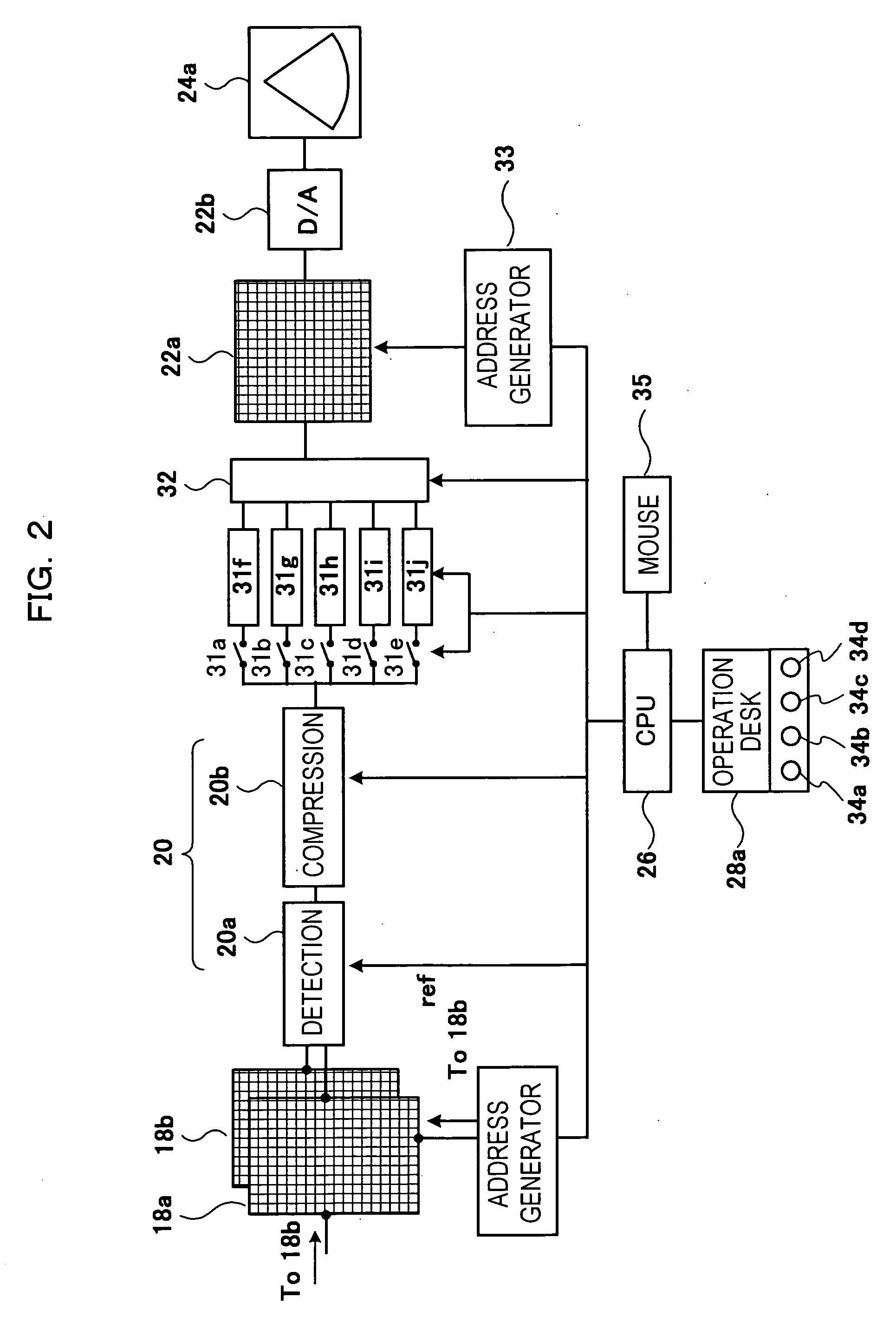
[0041] The first embodiment of an ultrasonic diagnostic apparatus relating to the present invention will now be described referring to FIG. 1 to FIG. 8. First, the configuration of the ultrasonic diagnostic apparatus relating to the present invention will be described using FIG. 1 and FIG. 2.
[0042] Ultrasonic diagnostic apparatus 1 is configured of probe 10, transmission / reception unit 12 that has a transmission unit and a reception unit, AD conversion unit 14, phasing addition unit 16, memory 18, signal processing unit 20, image construction unit 22, display unit 24 that has a monitor, controlling unit (CPU) 26, console desk 28, and so forth.
[0043] Next, the operation of the above-mentioned ultrasonic diagnostic apparatus 1 will be explained. First, a doctor applies probe 10 on the body surface of an object to be examined. Next, the doctor inputs an order to start ultrasound examination using console desk 28. Corresponding to a starting order, an order to output driving pulse is ...
embodiment 2
[0083] The second embodiment relating to the present invention will now be described referring to FIG. 11 and FIG. 12. The difference between the present embodiment and the first embodiment is that the intended ultrasound images are M-mode images, not B-mode images. In this embodiment, explanation will be omitted for the equivalent with embodiment 1 in function as well as configuration, with encoding remaining the same. FIG. 11 shows the M-mode images indicating the morphological variation of a heart wall in one heartbeat. FIG. 12 shows a display example of the plurality of M-mode images with different sensitivity range being arranged and displayed on the same screen simultaneously.
[0084] As shown in FIG. 11, M-mode images indicate temporal morphological variation toward ultrasound beam direction of, for example, heart wall 50 and 51 that form ventriculus sinister 52, and electrocardiographic (ECG) waveform 53 measured by an electrocadiograph (not shown in the diagram) is temporall...
PUM
 Abstract
Description
Claims
Application Information
 - R&D
- Intellectual Property
- Life Sciences
- Materials
- Tech Scout
- Unparalleled Data Quality
- Higher Quality Content
- 60% Fewer Hallucinations
Browse by: Latest US Patents, China's latest patents, Technical Efficacy Thesaurus, Application Domain, Technology Topic, Popular Technical Reports.
© 2025 PatSnap. All rights reserved.Legal|Privacy policy|Modern Slavery Act Transparency Statement|Sitemap|About US| Contact US: help@patsnap.com


