Apoptosis-associated protein and use thereof

a technology of apoptosis and a protein, applied in the field of functional fragments of proteins, can solve the problems of abnormal proliferation of cells, various diseases, and possible aggravating factors of various diseases, and achieve the effects of suppressing apoptosis or inflammatory cytokine production, and reducing the expression or activity of a protein

Inactive Publication Date: 2006-10-26
TAKEDA PHARMA CO LTD
View PDF2 Cites 1 Cited by
  • Summary
  • Abstract
  • Description
  • Claims
  • Application Information

AI Technical Summary

Problems solved by technology

Such molecular abnormalities and the collapse of the control mechanism damage the physiological functions of apoptosis, and result in causal factors or aggravation factors for various diseases.
For example, excessively suppressed apoptosis leads to abnormal proliferation of cells to be removed originally, which in turn induces tumoral diseases, autoimmune diseases and the like.
Conversely, abnormally promoted apoptosis leads to the death of cells that must exist originally, which in turn causes neurodegenerative diseases and the like.

Method used


Image

  • Apoptosis-associated protein and use thereof
  • Apoptosis-associated protein and use thereof
  • Apoptosis-associated protein and use thereof

Examples


example 1

Cloning of ABP1 cDNA

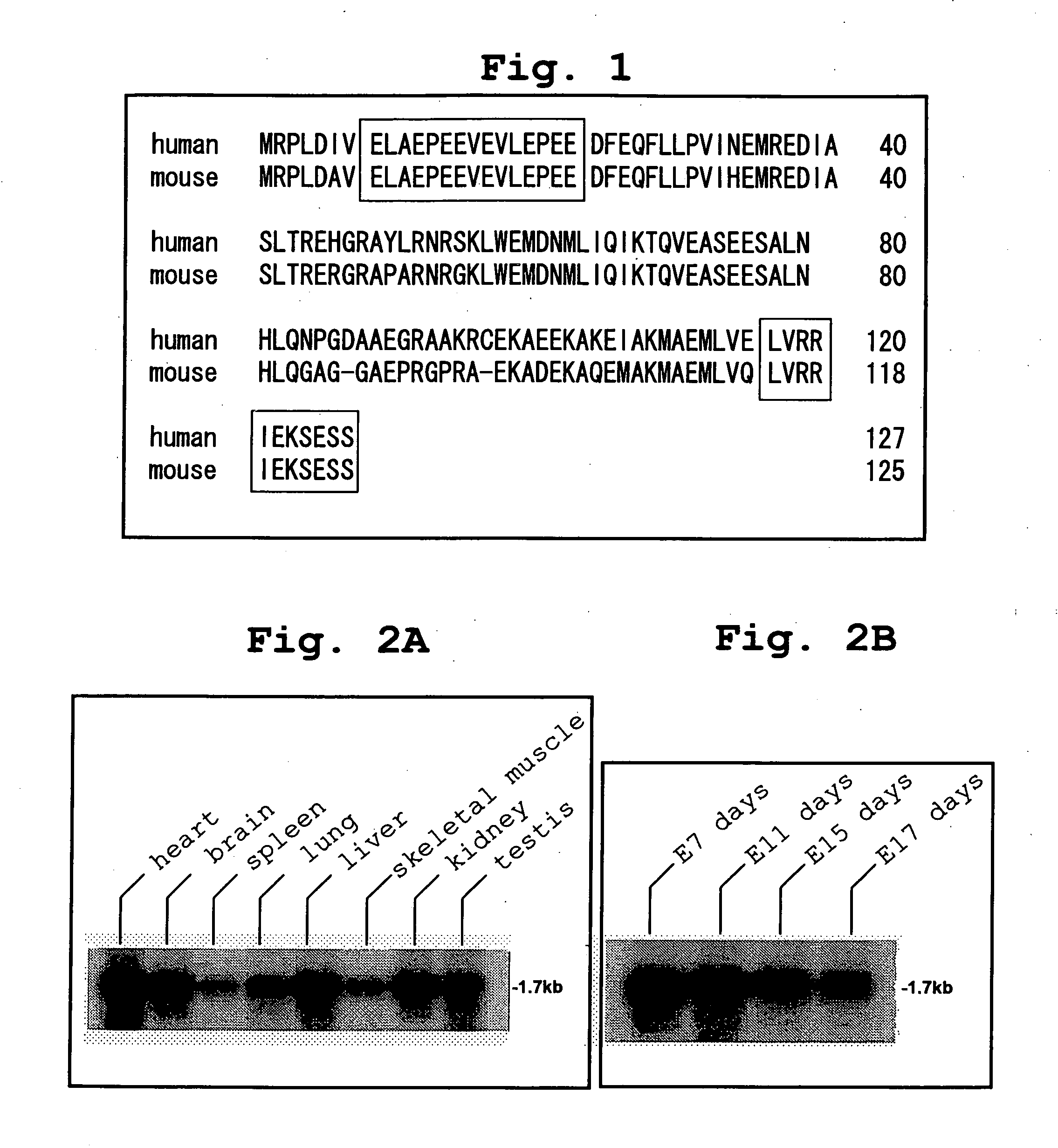
[0396] To identify the ASK1-binding protein, screening was conducted by the yeast two-hybrid method using Brent's system [Zervos et al., Cell, Vol. 72, p. 223-232, 1993; Gyuris et al., Cell, Vol. 75, p. 791-803, 1993] with ASK1-KR, prepared by replacing lysine (K) at the ATP-binding site (position 709) in the kinase domain of human ASK1 with arginine (R), as a bait and an expression library of fetal human brain origin as a prey. As a result, about 1200 positive clones were obtained. Database search of one of them based on its base sequence identified it as a portion of the protein coding region of the gene designated as “PGR1” (a portion corresponding to amino acid positions 35 to 127). The protein encoded by this gene consists of 127 amino acids, but information on the database was only relevant to the structure of the gene, and did not contain a report on its function. Hence, the full-length cDNA that encodes the protein of this gene was cloned by the PCR meth...

example 2

Expression Distribution of ABP1 mRNA

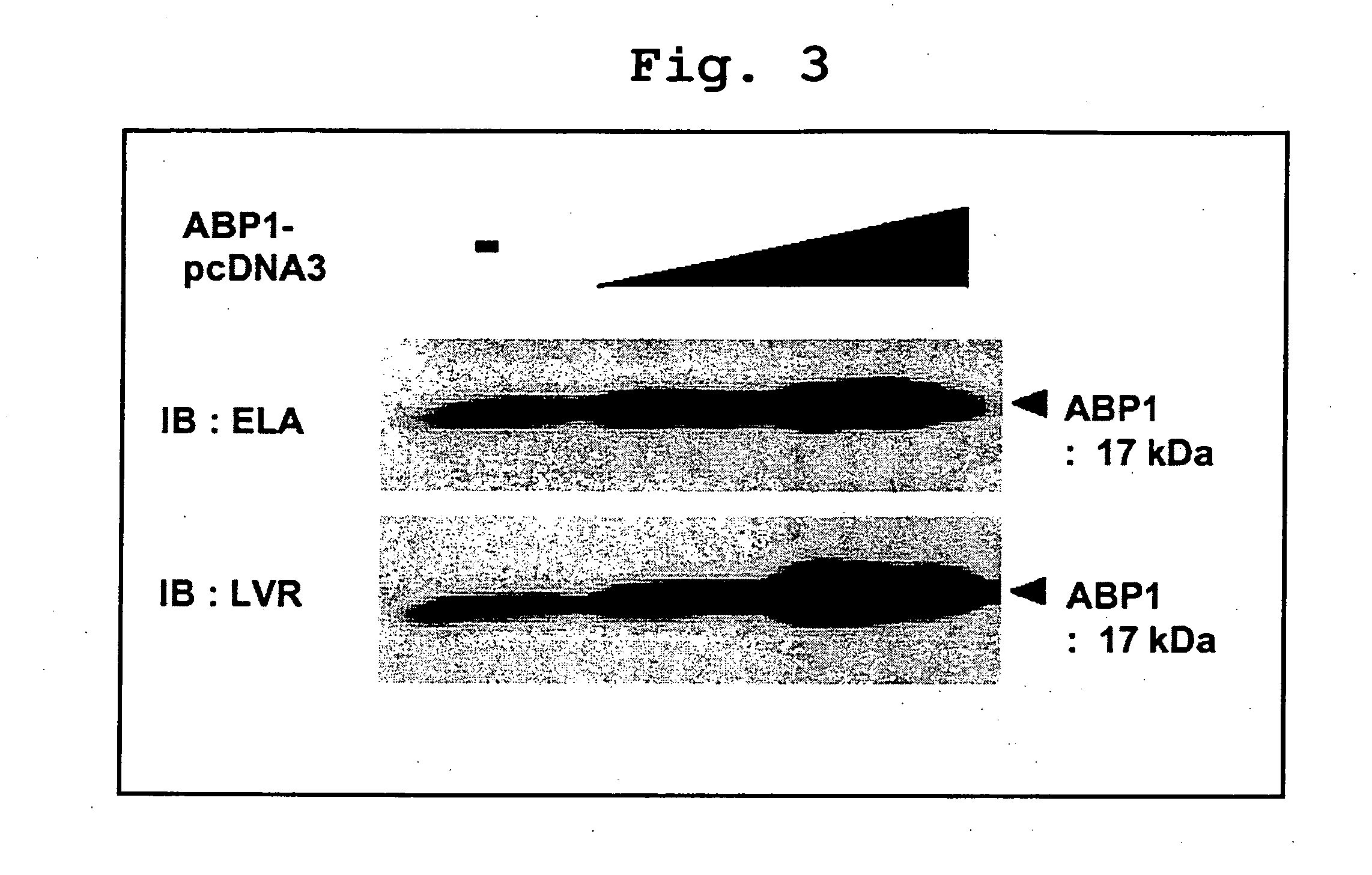
[0397] Using Mouse (#7762-1) and Mouse Embryo (#7763-1) in the Multiple Tissue Northern Blot (CLONTECH) with the full-length human ABP1 cDNA as a probe, Northern blot was conducted as directed in the instruction manual of the above-described product. As a result, the length of the ABP1 mRNA was determined to be about 1.7 kb, and the mRNA was ubiquitously expressed throughout all tissues (FIG. 2A). Fetal mouse blot also revealed the expression of ABP1 mRNA from relatively early time of E7 days, with no major changes during the course of embryogenesis (FIG. 2B).

example 3

Production of ABP1 Protein in Various Animal Cells

[0398] Next, a rabbit polyclonal antibody against ABP1 was prepared. Two kinds of peptide antibodies were prepared with ABP1-specific peptide (SEQ ID NO:5 and SEQ ID NO:6) as antigens on the basis of amino acid sequence information from the base sequence of the ABP1 cDNA, and were designated as ELA antibody and LVR antibody, respectively. The two antibodies were both used after being purified on the basis of affinity for antigen peptide.

[0399] HeLa cells and HEK293 cells were cultivated in the presence of 5% CO2 using Dulbecco's modified Eagle medium.

[0400] (DMEM; SIGMA) containing a high concentration of glucose (4.5 mg / ml) as a culture medium. The culture medium was supplemented with 10% fetal bovine serum (FBS) and 100 Units / ml penicillin. Porcine aortic endothelial (PAE) cells were cultivated using an F12 culture medium (Invitrogen) supplemented with 10% FBS, 10 mM HEPES, and 100 Units / ml penicillin.

[0401] Each cell was lysed...


PUM


Abstract

The present invention provides a partial peptide comprising the functional domain of a protein comprising an amino acid sequence which is the same or substantially the same as an amino acid sequence represented by SEQ ID NO:2 or SEQ ID NO: 4; a screening method and a kit therefor for a substance which regulates ASK1 activation, or a prophylactic or therapeutic substance for diseases associated with apoptosis or inflammation, which comprises using the protein or the partial peptide or a cell which produces the same, and optionally ASK1 or a partial peptide thereof or a cell which produces the same; and an agent for regulating apoptosis or inflammatory cytokine production, or a prophylactic or therapeutic agent for diseases associated with apoptosis or inflammation, which comprises a substance for regulating an activity of a protein comprising an amino acid sequence which is the same or substantially the same as an amino acid sequence represented by SEQ ID NO:2 or SEQ ID NO: 4.

Description

FIELD OF THE INVENTION [0001] The present invention relates to a functional fragment of a protein that bind to ASK1 to activate, and various uses of the protein and the fragment, particularly pharmaceutical uses. BACKGROUND ART [0002] Apoptosis is responsible for the functions of removing cells that become no longer necessary during development and abnormal cells, of homeostasis, and of body defence reactions to remove injured cells, and its mechanisms have been elucidated gradually at the molecular level. Such molecular abnormalities and the collapse of the control mechanism damage the physiological functions of apoptosis, and result in causal factors or aggravation factors for various diseases. For example, excessively suppressed apoptosis leads to abnormal proliferation of cells to be removed originally, which in turn induces tumoral diseases, autoimmune diseases and the like. Conversely, abnormally promoted apoptosis leads to the death of cells that must exist originally, which ...

Claims


Application Information

Patent Timeline
Patent Type & Authority Applications(United States)
IPC IPC(8): C12Q1/68G01N33/574C07H21/04C12P21/06A61K39/395C07K14/82C07K16/30A61K38/00A61P3/00A61P5/00A61P7/00A61P9/00A61P11/00A61P19/08A61P25/00A61P29/00A61P31/12A61P35/00A61P37/02A61P37/06A61P43/00C07K14/47
CPCA61K38/00G01N2500/00C07K14/4747A61P11/00A61P19/08A61P25/00A61P29/00A61P3/00A61P31/12A61P35/00A61P37/02A61P37/06A61P43/00A61P5/00A61P7/00A61P9/00
Inventor ICHIJO, HIDENORI
Owner TAKEDA PHARMA CO LTD
Why Eureka
Try Eureka