Fibrin cell supports and method of use thereof

a technology of fibrin cell and cell culture, which is applied in the direction of skeletal/connective tissue cells, prosthesis, peptide/protein ingredients, etc., can solve the problems of unable to restore the function of the cutaneous barrier of the skin, the reconstituted tissue is not in order, and the number of technical problems remains large, so as to reduce the probability of mechanical damage

Inactive Publication Date: 2006-10-26
DFB PHARMA
View PDF4 Cites 30 Cited by
  • Summary
  • Abstract
  • Description
  • Claims
  • Application Information

AI Technical Summary

Benefits of technology

[0014] In another aspect, the present invention provides a method for decreasing the probability of mechanical damage to the fibrin cell support during transport prior to transplantation.

Problems solved by technology

Artificial reconstitution of such a complex organ thus poses numerous problems.
However, this equivalent dermis is merely a temporary dressing as it cannot restore the skin's cutaneous barrier function.
However, recovery of the reconstituted tissue in order to make a graft therefrom still poses a number of technical problems.
These manipulations are both delicate and time consuming, which jeopardizes the quality of the graftable epithelium.

Method used


Image

  • Fibrin cell supports and method of use thereof
  • Fibrin cell supports and method of use thereof
  • Fibrin cell supports and method of use thereof

Examples


example 1

Preparation of a Fibrin Cell Support for Cell Cultures

[0039] A fibrin cell support for cell cultures is prepared by mixing a solution containing fibrinogen and a solution containing calcic thrombin.

[0040] Lyophilized fibrinogen (375-575 milligrams) is reconstituted with 5 ml of aprotinin (3,000 KIU / ml; kallikrein inhibitor units / ml) then combined with 5 ml of 2.2% NaCl containing 2 mM calcium chloride. Lyophilized thrombin (225 to 275 milligrams; approximately 2500 International Units) is diluted in 1.1% NaCl containing 1 mM calcium chloride to a final concentration of 0.225 to 0.275 milligrams or 2.5 IU. The solubilized fibrinogen and solubilized thrombin are mixed in a 1:1 ratio and dispensed into a cell culture dish or flask (2.5 mls of the fibrinogen-thrombin mixture per 100 cm2 of culture dish surface) to form a fibrin cell support. The fibrin cell support is then covered in cell culture medium.

example 2

Preparing a Keratinocyte Culture on the Fibrin Cell Support

[0041] Human keratinocytes originating from a skin biopsy are cultured in the presence of lethally-irradiated or mitomycin C-treated human fibroblasts, or lethally-irradiated or mitomycin C-treated murine feeder cells. (See Limat et al. (1986) J Invest Dermatol. 87(4):485-8; Green et al. (1979) Proc. Natl. Aced. Sci. 76:5665).

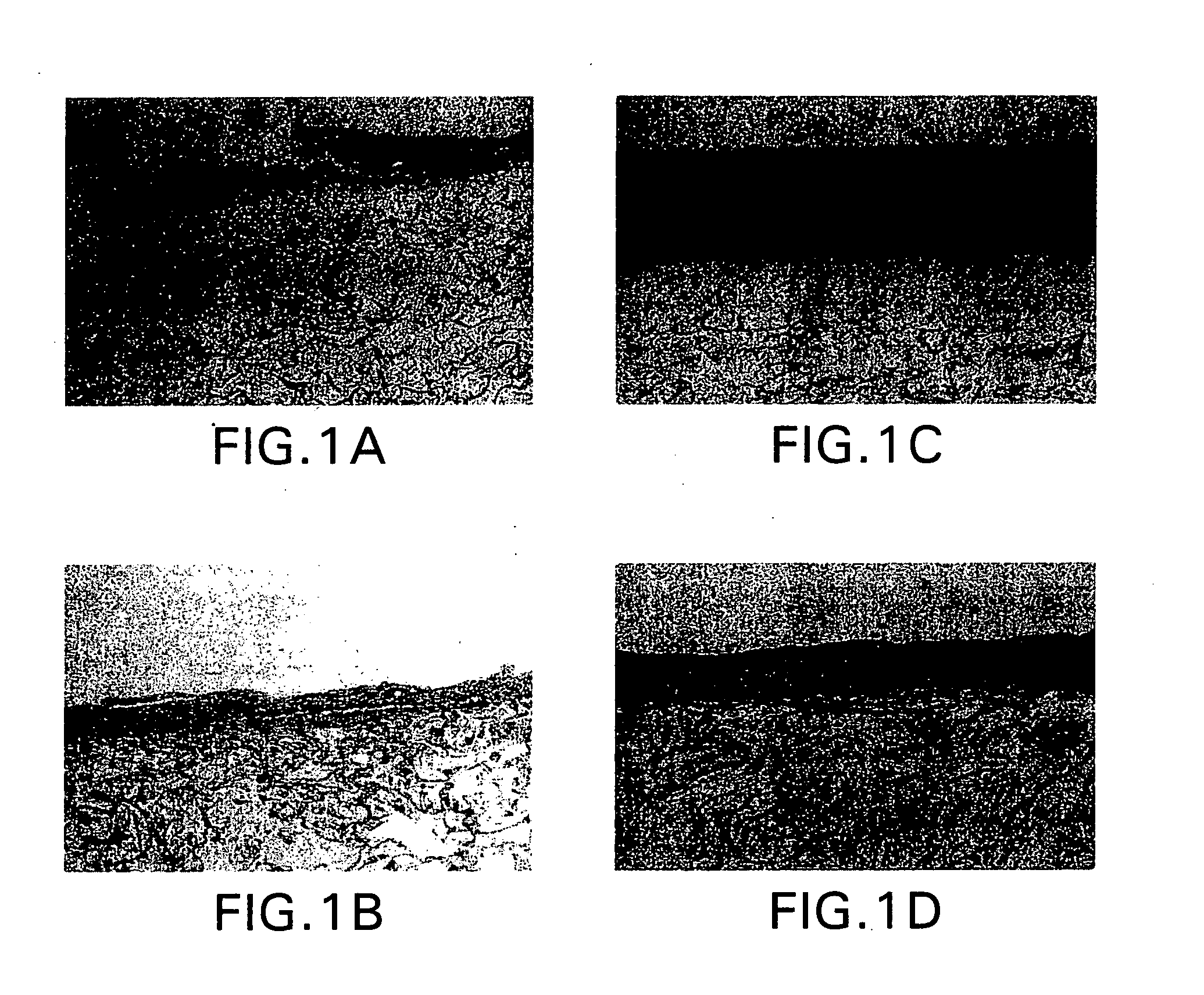
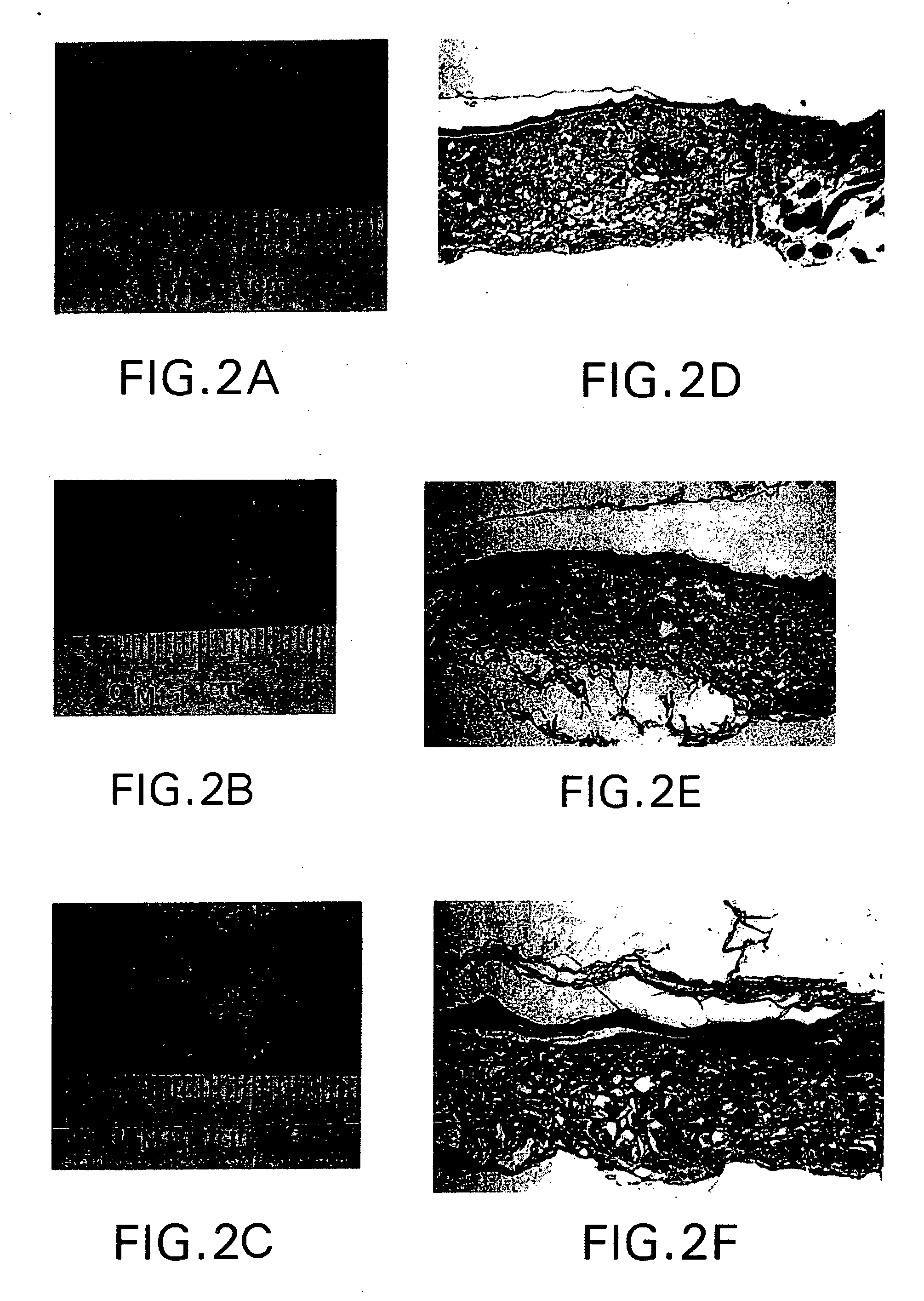
[0042] A layer of confluent keratinocytes is trypsinized, replaced in suspension in culture medium and seeded at subconfluent density (e.g., in a 1 / 10 dilution) on a tissue culture dish covered with the fibrin cell support prepared as described in Example 1. The keratinocytes are then allowed to reach confluence, at which point the resulting keratinocyte graft can be used in therapeutic methods. The fibrin cell support of this invention stands up well to handling and does not retract at the time of detachment, which makes it possible to recover 100% of the surface area of the cell layer of the culture...

example 3

Recovery of a Pre-Established Cell Layer Using the Fibrin Cell Support

[0045] Keratinocytes are inoculated according to Green's conventional method, in a Petri dish covered with a layer of lethally irradiated fibroblasts. (See Green et al. (1979) Proc. Natl. Aced. Sci. 76:5665). When keratinocytes are confluent and formed of several layers of cells, the culture medium is removed, an EDTA solution is added for 1 hour 30 minutes. This is followed by washing twice with PBS. The fibrin cell support prepared as described in Example 1 is then poured directly onto confluent keratinocytes.

[0046] Upon coagulation of the fibrin cell support, it can be detached mechanically and used as a graft, as demonstrated in Example 2.


PUM

PropertyMeasurementUnit
concentrationaaaaaaaaaa

Abstract

The present invention relates to fibrin cell supports for cell cultures formed by the mixture of plasma proteins including fibrinogen and thrombin. The fibrin cell supports are preferably used for preparing a culture of cells such as keratinocytes, recovering the culture in the form of a reconstituted tissue, and transporting same. The reconstituted tissue is particularly suitable for use as a skin graft.

Description

RELATED APPLICATIONS [0001] This application claims priority to U.S. Application Nos. 60 / 408,566, filed Sep. 6, 2002; and 60 / 433,715, filed Dec. 16, 2002, each of which are incorporated herein by reference in their entireties.FIELD OF THE INVENTION [0002] The present invention relates to a fibrin cell support for cell cultures, containing a coagulated mixture of plasma proteins including fibrinogen and thrombin; its use in the preparation of cell cultures; its transport and transplantation in the form of an isolated cell, colonies of cells, or a reconstituted epithelia; and it's use for therapeutic purposes. BACKGROUND OF THE INVENTION [0003] The reconstitution of a living skin similar to the human skin from a few cells obtained from a biopsy, or of a simplified skin performing the physiological functions of a normal skin, is being studied extensively with the aim of replacing skin damaged by disease (hemangiomas, keloids, hypertrophic scarring, bullous pemphigoid, viral or bacteial...

Claims


Application Information

Patent Timeline
Patent Type & Authority Applications(United States)
IPC IPC(8): C12N5/02C12N5/00A61K35/12A61K35/36C12N11/02A61L27/00A61L27/22A61L27/60C12N5/071
CPCA61K35/12A61K38/57A61K35/36A61K38/00C12N2533/56A61L27/225A61L27/3813A61L27/60C12N5/0068C12N5/0698C12N2502/094C12N2502/1323A61K2300/00
Inventor RONFARD, VINCENT
Owner DFB PHARMA
Why Eureka
Try Eureka