Cell sodding method and apparatus

Active Publication Date: 2006-11-16
KOLIGO THERAPEUTICS INC
View PDF11 Cites 47 Cited by
  • Summary
  • Abstract
  • Description
  • Claims
  • Application Information

AI Technical Summary

Benefits of technology

[0008] The present invention is based, in part, on the inventors' surprising discovery that a sustained low pressure gradient across a graft scaffold or substrate material for about 1 minute up to about 24 hours, preferably in a cell perfusion system, provides significantly more rapid and uniform cell adhesion and maturation on substrate materials than the transient high-pressure gradients employed by known

Problems solved by technology

Furthermore, application of a sustained low pressure gradient reduces stress, damage to the cells, and unnecessary activation of the cells during the adhesion process.
Employing sustained low magnitude

Method used


Image

  • Cell sodding method and apparatus
  • Cell sodding method and apparatus
  • Cell sodding method and apparatus

Examples


Example

EXAMPLE 1

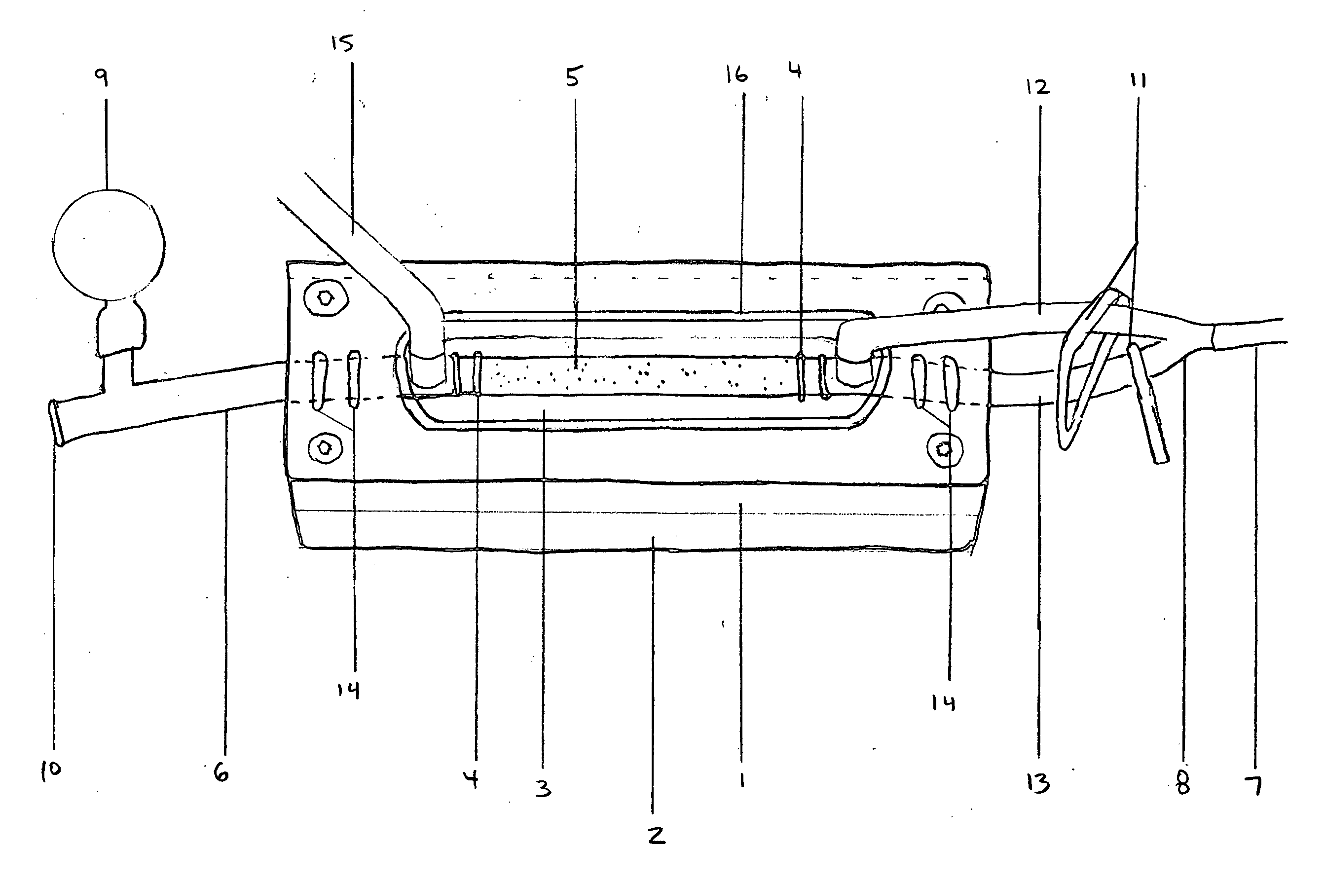
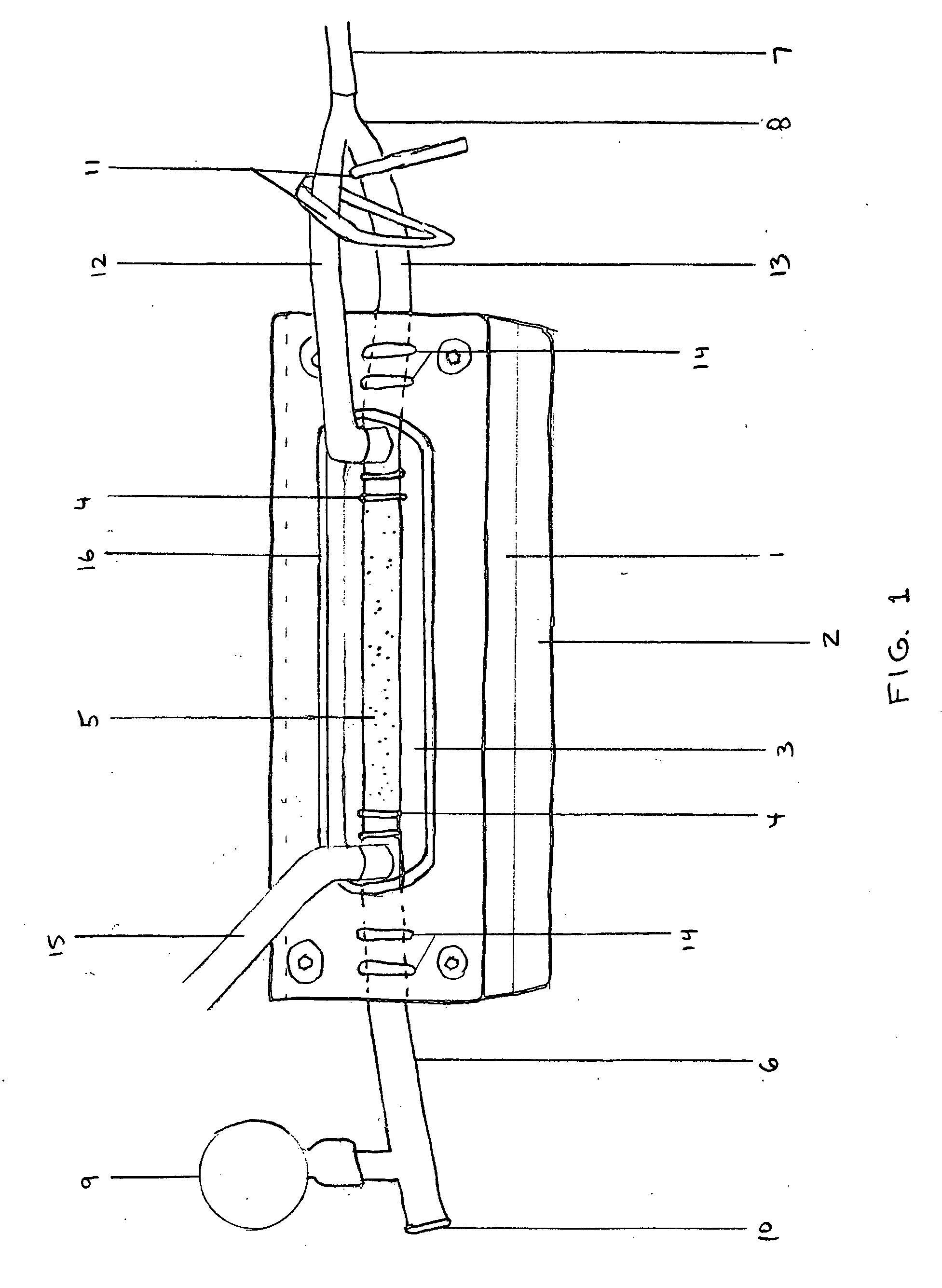
[0053] We used a sustained low pressure head (10-50 mmHg) to maintain a transmural pressure gradient for 1 min up to 24 hours to seed early passage microvascular endothelial cells isolated from adipose tissue onto commercially obtained (Impra) expanded Teflon (ePTFE) small diameter grafts (3-4 mm Inner Diameter). The microvascular endothelial cells were obtained according to the method of U.S. Pat. Nos. 4,820,626; 5,230,693; and 5,628,781. Grafts were primed with 70% ethanol which was then replaced with media (M199, Invitrogen) containing 20% FBS (Invitrogen) and antibiotics (Invitrogen) prior to being mounted in vascular biochambers. The cells were introduced into the biochamber at a density of 1×10⁶ cells / cm2.

[0054] In a series of experiments with transmural pressure, followed, in some cases, by lumenal perfusion, endothelial cells were shown to actively adhere to the ePTFE and elastin surfaces. A microscopic view of cross sections after 6 hours of perfusion showed expr...

Example

EXAMPLE 2

[0056] An ePTFE graft was cleared using 10 min 70% EtOH, then 10 min 100% EtOH, then 24 hours in MVEC media (M199E, 1 μg / mL Amphotericin B, 20 μg / mL Penicillin / Streptomycin, 15% heat-inactivated FBS). The graft was then sodded with 0.82×10⁶ MVEC cells / cm2 with a BOS gear-pump pressure head of about 20 mmHg for 4 hours. The sodded graft was then fixed in 10% neutral-buffered formalin and embedded in paraffin, then sectioned. After histoanalysis, one of two blocks was melted down, the ePTFE deparaffinized, and dehydrated in 100% EtOH. The resulting sodded graft was then critical point dried, mounted, and sputter coated for SEM, and imaged on an Hitachi S-800 field-emission SEM. FIG. 6 shows an SEM image of endothelial cell adhesion on an ePTFE graft substrate sodded with 0.82×10⁶ MVEC cells / cm2 for 4 hours (about 20 mmHg).

Example

EXAMPLE 3

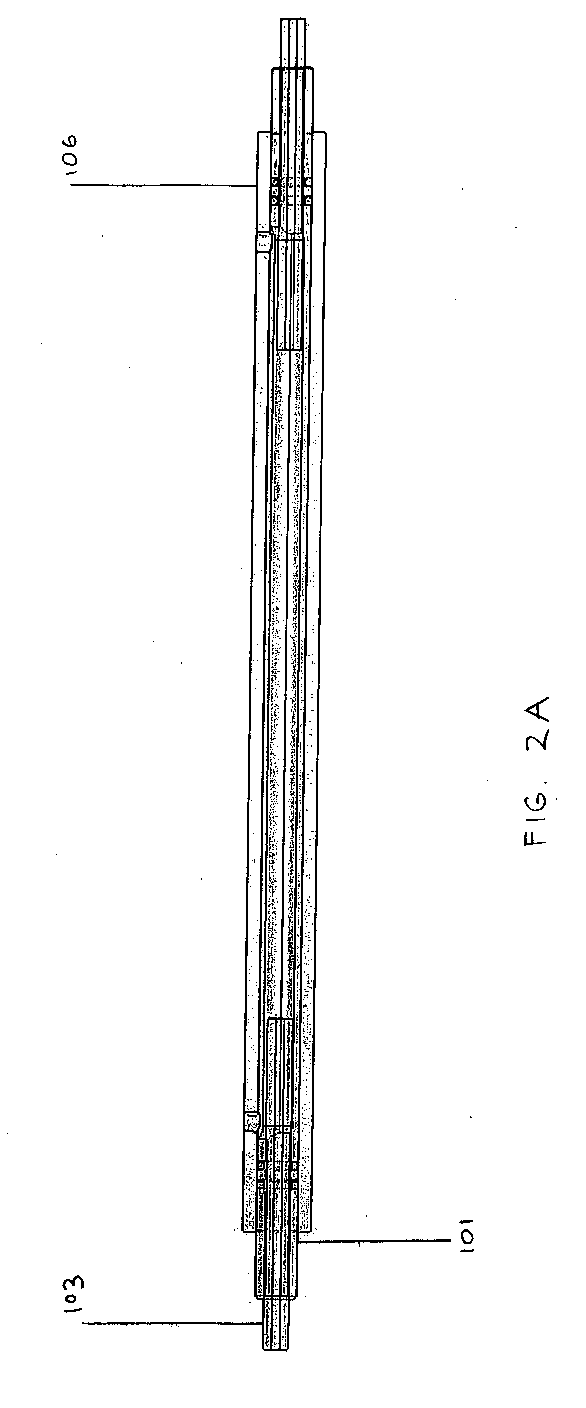
[0057] We denucleated a 4 mm internal diameter ePTFE graft substrate having a length of 10 cm using 10 min 70% ethanol, then 10 min 100% ethanol. The graft was then sodded in a biochamber as shown in FIG. 2 at 5×10⁵ cells / cm2by applying 10 minutes transmural flow (˜50 mmHg), followed by 20 minutes luminal flow (10 mL / min). The resulting MVEC-sodded graft was fixed, stained with BBI, and imaged. FIG. 7 shows the proximal and distal ends of a 10 cm long vascular ePTFE graft sodded with 5×10⁵ cells / cm2 for 10 minutes (about 50 mmHg) transmurally, followed by 20 minutes of luminal flow (10 mL / min), resulting in highly uniform cell adhesion at each end of the long graft.


PUM


Abstract

Tissue engineering methods and biochamber apparatus are provided for making tissue grafts for implantation into a patient. The methods include applying a sustained low magnitude pressure gradient transmurally across a permeable scaffold material using a media containing cells, preferably microvascular epithelial cells, to be deposited on the scaffold for the production of tissue grafts, preferably vascular grafts, to promote accelerated adhesion and maturation of cells on the scaffold material. Biochambers for preparing tubular tissue grafts are provided which contain connectors for holding a graft substrate, proximal and distal tubing for connection to an optional perfusion system, and structure for switching between transmural flow of a cell suspension across the graft substrate and translumenal flow through the lumen of the graft.

Description

[0001] This invention claims priority to U.S. Patent Application Ser. No. 60 / 638,199, filed on Dec. 23, 2004, which is hereby incorporated by reference herein.STATEMENT REGARDING FEDERALLY SPONSORED RESEARCH OR DEVELOPMENT [0002] This invention was made with Government support under #W81XWH-04-1-0503 and #W81XWH-05-1-0620 awarded by the United States Army Medical Research and Materiel Command.FIELD OF THE INVENTION [0003] The present invention relates to tissue engineering systems for the manufacture of tissue implants and grafts, including, but not limited to, prosthetic vascular and arteriovenous grafts. The present invention relates, in particular, to methods and apparatus for adhering cells to permeable scaffold materials by sustained low pressure cell sodding, which includes applying a sustained pressure gradient across a permeable material using media containing cells to be deposited on the material. The methods and the apparatus of the invention promote accelerated adhesion a...

Claims


Application Information

Patent Timeline
IPC IPC(8): C12N5/08A61F2/06C12N5/071
CPCC12M21/08C12M25/10C12M25/14C12N2533/30C12M29/10C12N5/0697C12M27/10
Inventor KOSNIK, PAUL E.ENGLAND, CHRISTOPHER T.DENNIS, ROBERT G.WILLIAMS, STUART K.
Owner KOLIGO THERAPEUTICS INC
Why Eureka
Try Eureka