

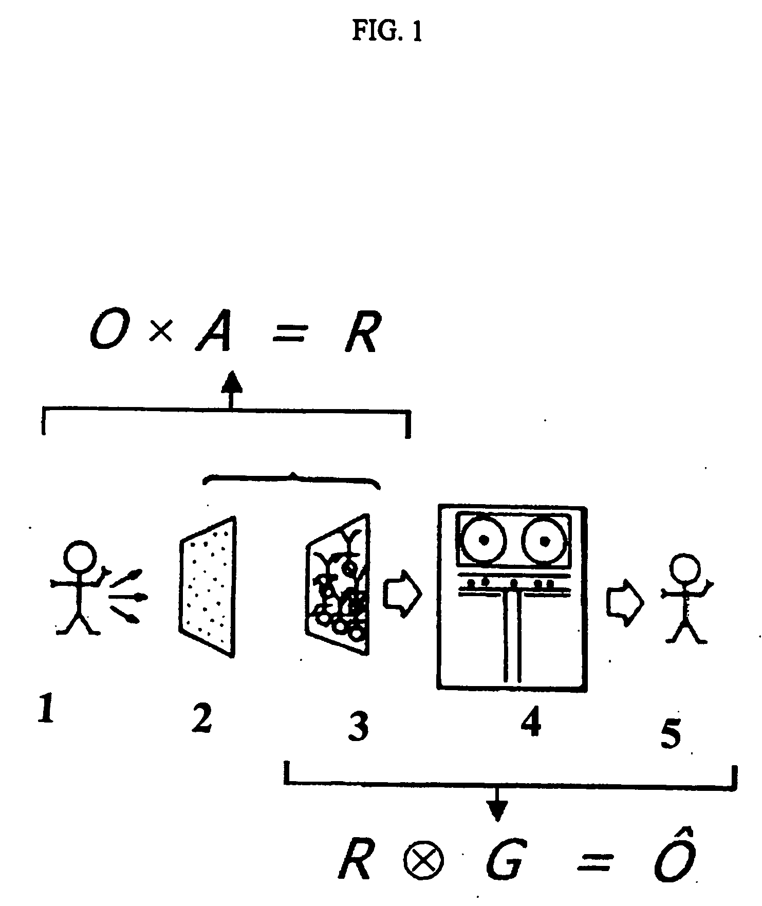
Soft x-ray imager with ten micrometer resolution
a soft x-ray imager and resolution technology, applied in the field of imaging of radiationemitting objects, can solve the problems of limited fov, limited number and magnification, and limited material thickness, so as to reduce penetration and increase detection efficiency
- Summary
- Abstract
- Description
- Claims
- Application Information
AI Technical Summary
Benefits of technology
Problems solved by technology
Method used
Image

Examples
Embodiment Construction
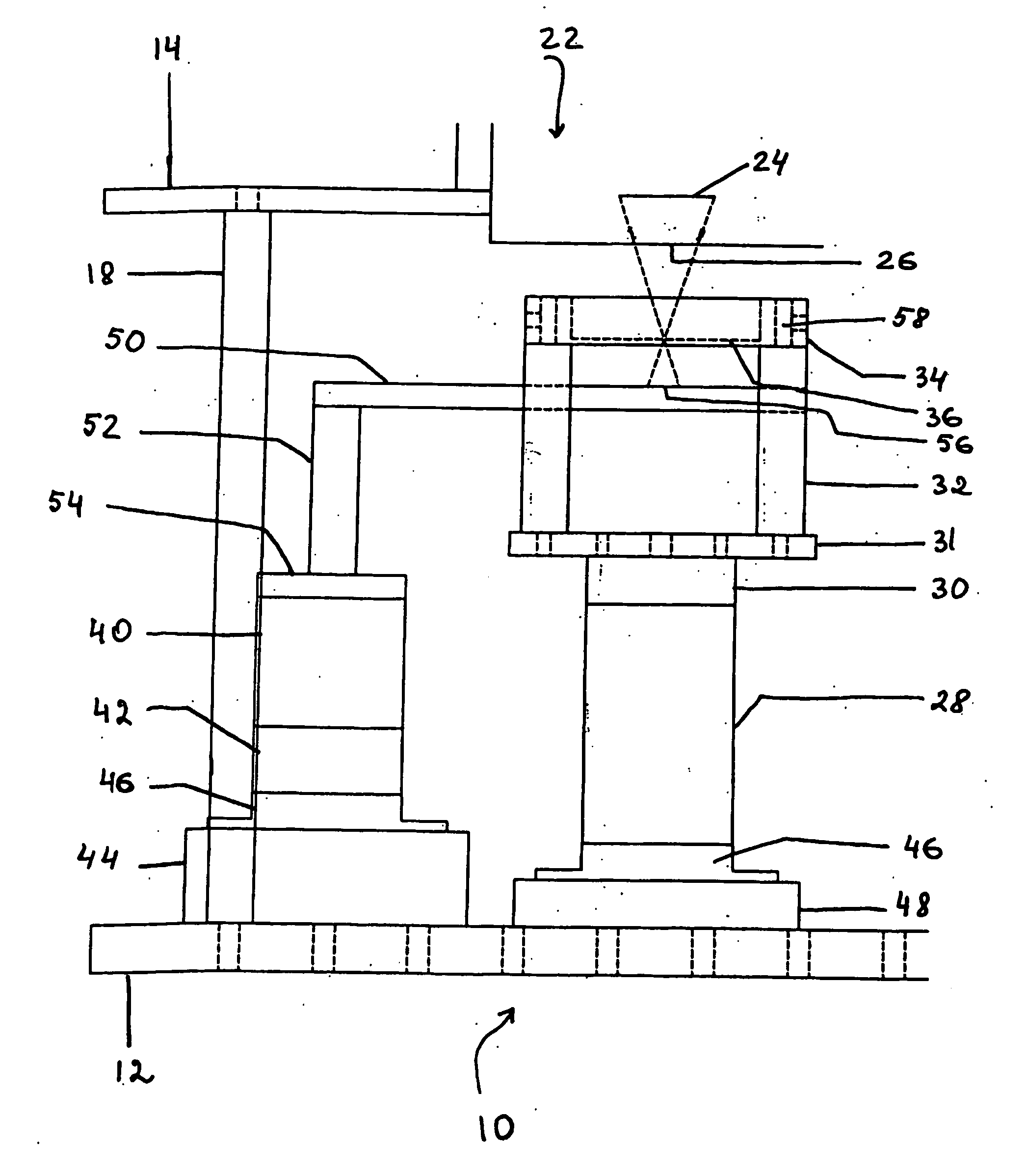
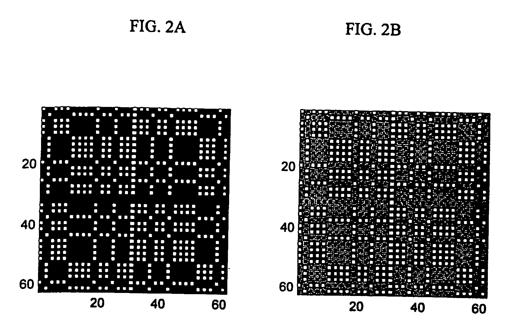
[0059] The invention was driven by the desire to develop a device capable of imaging objects on a scale of 280 microns or less. In the present invention, the synergy is achieved by the combination of low-energy photons (to obtain reduced penetration and high detection efficiency), a CCD detector (to obtain high-resolution), and coded aperture optics (to obtain high sensitivity and SNR). The low penetration of 3-10 keV photons in heavy materials opens the opportunity of designing high-resolution optics. The small solid angles involved cause low sensitivity but counts can be recovered with a desired coded aperture. A tool capable of imaging directly not large molecules, but single atoms, in a relatively short time, is appealing because of its potential for translating molecular imaging techniques to the space scale of a human somatic cell, thus making a powerful modality available to investigators in basic life science research.
[0060] The present invention can be used to study object...
PUM
 Abstract
Description
Claims
Application Information