
Automatic registration of images

a multi-spectral image and automatic registration technology, applied in the field of image processing, can solve the problems of compromising the blood flow of the eye, glaucoma patients' optic nerve fiber loss, and the inability to perform non-invasive oxygenation measurement of the human retina and optic nerve, and achieves more reliable and easy to obtain optical density ratios, the effect of more reliable measurements

Active Publication Date: 2006-12-07
OXYMAP EHF
View PDF4 Cites 43 Cited by
  • Summary
  • Abstract
  • Description
  • Claims
  • Application Information

AI Technical Summary

Benefits of technology

[0011] The present invention concerns methods, software, and a system that enables in real time the automatic analysis of spectrophotometric measurements of oxygen saturation in the retinal vasculature. The invention enables any number of spectral images to be examined in the same coordinate system, which means that optical density ratios are more reliably and easily obtained. The method is suitable for measuring large part of the fundus at once. The current invention, moreover, automatically finds vessels in the spectral images and calculates the optical density ratios automatically. This has never been done before and results in more reliable measurements than when measurement points are chosen by hand, and can be done automatically and extended by a novice operator. Terminology Used in the Present Context:
[0029] In another aspect the current invention relates to a method for detecting blood vessels in multi-spectral images, said method comprises the steps of: retrieving registered spectral images (14), for each spectral image, remove defective pixels (15), enhance image contrast (16), perform top-hat transform using structuring element larger than the largest vessel diameter (17), binarize each image (18), apply filter for smoothing the image (19), combine all the spectral images using binary AND operator (20), skeletonize the resulting image (21), prune the skeleton image (22), re-grow the pruned image (23), locate junction points (24), wherein the junction points are the intersection of two or more vessels.

Problems solved by technology

Some of the disease states that pose the greatest danger to vision are thought to involve abnormalities in the oxygen supply to the retina or the oxygen metabolism.
It has also been suggested that ocular blood flow is compromised in glaucoma, including normal tension glaucoma, and that the loss of optic nerve fibers in glaucoma patients may be due to ischemia.
Non-invasive measurement of oxygenation in the human retina and optic nerve has proved difficult but considerable progress has been made, particularly in recent years as digital technology has evolved.
Pigmentation may cause underestimation of ODs in vessels, in particular at 586 nm, because it causes stronger absorption of light.
One of the limitations of prior art retinal oximetry [13] includes difficulties in obtaining calibrated measures of actual oxygen saturation.
The problems with the prior art methods used in retinal oximetry is the need for an expert to perform the oximetry, the high variability of the results, and poor reproducibility.
The prior art methods are time consuming, and manual registration of the spectral images is inaccurate because of the error prone manual selection of sample points.

Method used


Image

  • Automatic registration of images
  • Automatic registration of images
  • Automatic registration of images

Examples


Embodiment Construction

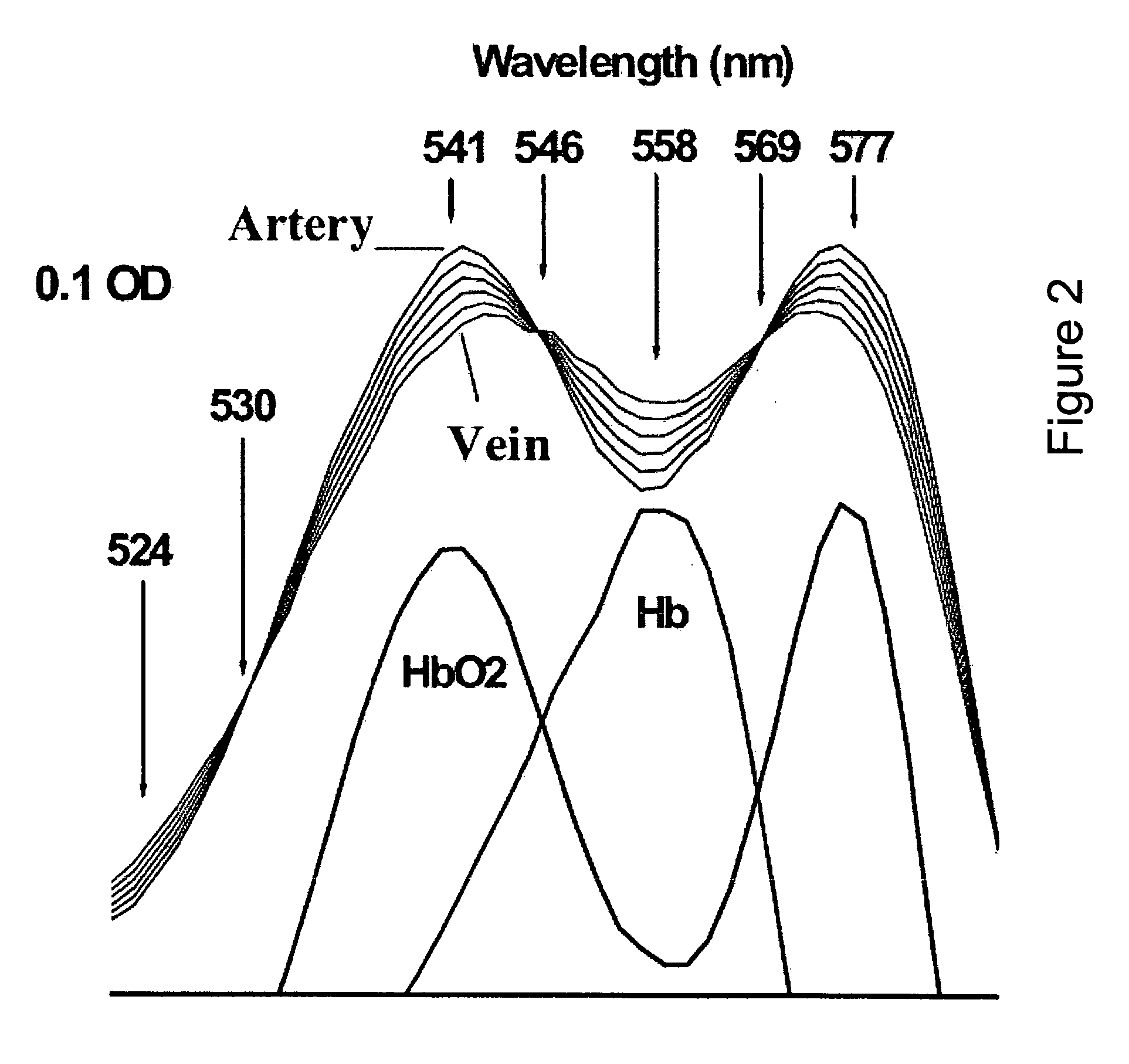
[0048] The retinal oxymetry process utilizes the fact that the amount of light absorbed by hemoglobin is dependent on the wavelength of the light and the oxygen saturation of the blood. To estimate the amount of light reflected, multi-spectral images containing sub-images at different wavelengths are acquired.

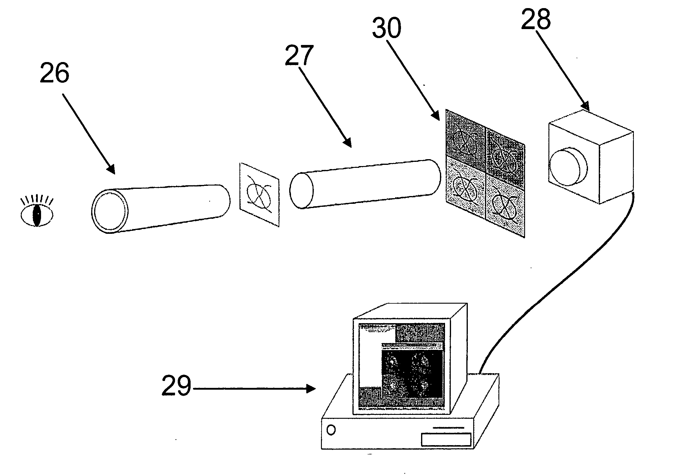
[0049] Therefore, the image from the digital camera is a multi-spectral image consisting of 2 or more sub-images. Each sub-image has been filtered with an optical narrow band-pass filter (with center wavelengths 542 nm, 558 nm, 586 nm, and 605 nm). The sub-images, here after referred to as spectral images, represent the same area of the retina. However, an accurate spectral image position and rotation on the multi-spectral image is not known because of the sensitivity of the beam splitter. For this reason, the spectral images may overlap and sometimes parts of the spectral images are outside of the multi-spectral image and therefore parts of the spectral image are missing.

[00...


PUM


Abstract

The present invention relates to a system for automatically evaluating oxygen saturation of the optic nerve and retina, said system comprising: image capturing system further comprising: a fundus camera (26), a four wavelengths beam splitter (27), a digital image capturing device (28), a computer system, image processing software performing in real-time the steps of: registering set of multi-spectral images (1) by, binarizing the multi-spectral image (7), find the all the border regions of each image by finding the region including the straight line that passes the most number of points in the region (8), use the orientation of the borders to evaluate the orientation of each spectral image (9), equalize the orientation of each spectral image by rotating the spectral image (10), edge detect each spectral image (11), estimate the translation between the spectral images based on the edges of adjacent images (12), transform the images to a stack of registered images (13), locating blood vessels (2) in each of the multi-spectral images by, retrieving registered spectral images (14), for each spectral image, remove defective pixels (15), enhance image contrast (16), perform top-hat transform using structuring element larger than the largest vessel diameter (17), binarize each image (18), apply filter for smoothing the image (19), combine all the spectral images using binary AND operator (20), skeletonize the resulting image (21), prune the skeleton image (22), re-grow the pruned image (23), locate junction points (24), evaluating the width of the blood vessels (3) by calculating the normal vector to the vessel direction to, estimate the direction and position of the vessel profile, evaluate positions of vessel walls based on the vessel profile obtained, evaluate vessel width based on the position of the vessel walls, selecting samples for calculating optical density, calculating the optical density ratio (4), evaluating oxygen saturation level (5), and presenting the results (6), wherein presenting the results may include presenting numerical and visual representation of the oxygen saturation.

Description

TECHNICAL FIELD [0001] The present invention relates to the field of image processing, more particularly to the automatic registration of multi-spectral images. The present invention, moreover, relates to the application of image processing in the field of ophthalmology for spectral, spatial and temporal analysis. BACKGROUND ART [0002] Some of the disease states that pose the greatest danger to vision are thought to involve abnormalities in the oxygen supply to the retina or the oxygen metabolism. These include diseases such as diabetic retinopathy, which is one of the most common causes of blindness in the world and retinopathy of prematurity, retinal vein and artery occlusions. It has also been suggested that ocular blood flow is compromised in glaucoma, including normal tension glaucoma, and that the loss of optic nerve fibers in glaucoma patients may be due to ischemia. [0003] The principle of spectrophotometric hemoglobin oximetry is old and the method has been developed into c...

Claims


Application Information

Patent Timeline
IPC IPC(8): A61B5/00
CPCA61B3/1241A61B5/7264A61B5/14555
Inventor HALLDORSSON, GISLI HREINNSTEFANSSON, EINARBENEDIKTSSON, JON ATLIKARLSSON, ROBERT ARNARHARDARSON, SVEINN HAKONEYSTEINSSON, THORBEACH, JAMES M.
Owner OXYMAP EHF
Learn More