Isolation of nucleic acid using colored buffers

a nucleic acid and color technology, applied in the direction of biochemistry apparatus and processes, sugar derivatives, organic chemistry, etc., can solve the problems of inferior nucleic acid products and poor execution, and achieve the optimization of the bacterial lysis and neutralization steps, the effect of reducing the cost of lysis and neutralization, and fast and reliable results

Inactive Publication Date: 2007-01-18
ZYMO RES CORP
View PDF8 Cites 26 Cited by
  • Summary
  • Abstract
  • Description
  • Claims
  • Application Information

AI Technical Summary

Benefits of technology

[0012] The present invention provides a fast, reliable, and efficient method for the isolation of pure plasmid DNA based on a modified alkaline lysis procedure. The present invention ensures optimization of the bacterial lysis and neutralization steps by visualization of color changes due to indicator dyes present in the individual buffers that can reflect the pH of the solution after mixing (FIG. 1). In a preferred embodiment the color change is based on indicator dyes whose color is related to the pH of the solution and allows the efficiency of lysis and neutralization, and clearing of the lysate, to be assessed visually. In other embodiments the indicator is different in each of the buffers and may not undergo a response to pH. The presence of one or more indicator dyes also guards against accidental errors in sample handling related to pipeting of the resuspension buffer, lysis buffer, and neutralization buffer, or any buffer component. It also allows one to determine if the removal of the media supernatant is complete. Furthermore, since it is easy to gauge the degree of lysis and neutralization by simply examining the color change

Problems solved by technology

It is common for poor execution of these basic pro

Method used


Image

  • Isolation of nucleic acid using colored buffers
  • Isolation of nucleic acid using colored buffers
  • Isolation of nucleic acid using colored buffers

Examples


example 1

Preferred Embodiment

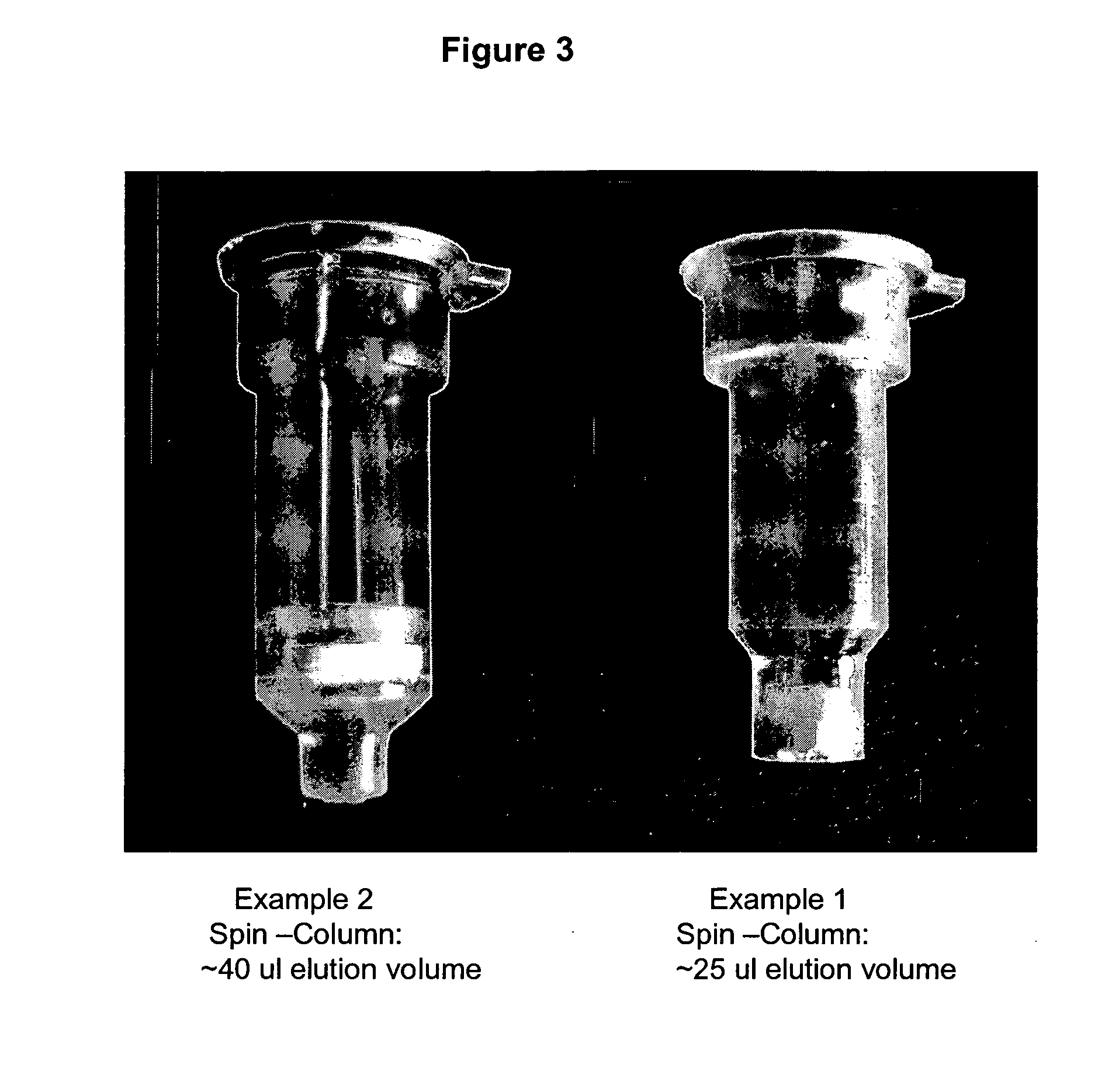
Spin-Column (Modified Low Volume Elution)

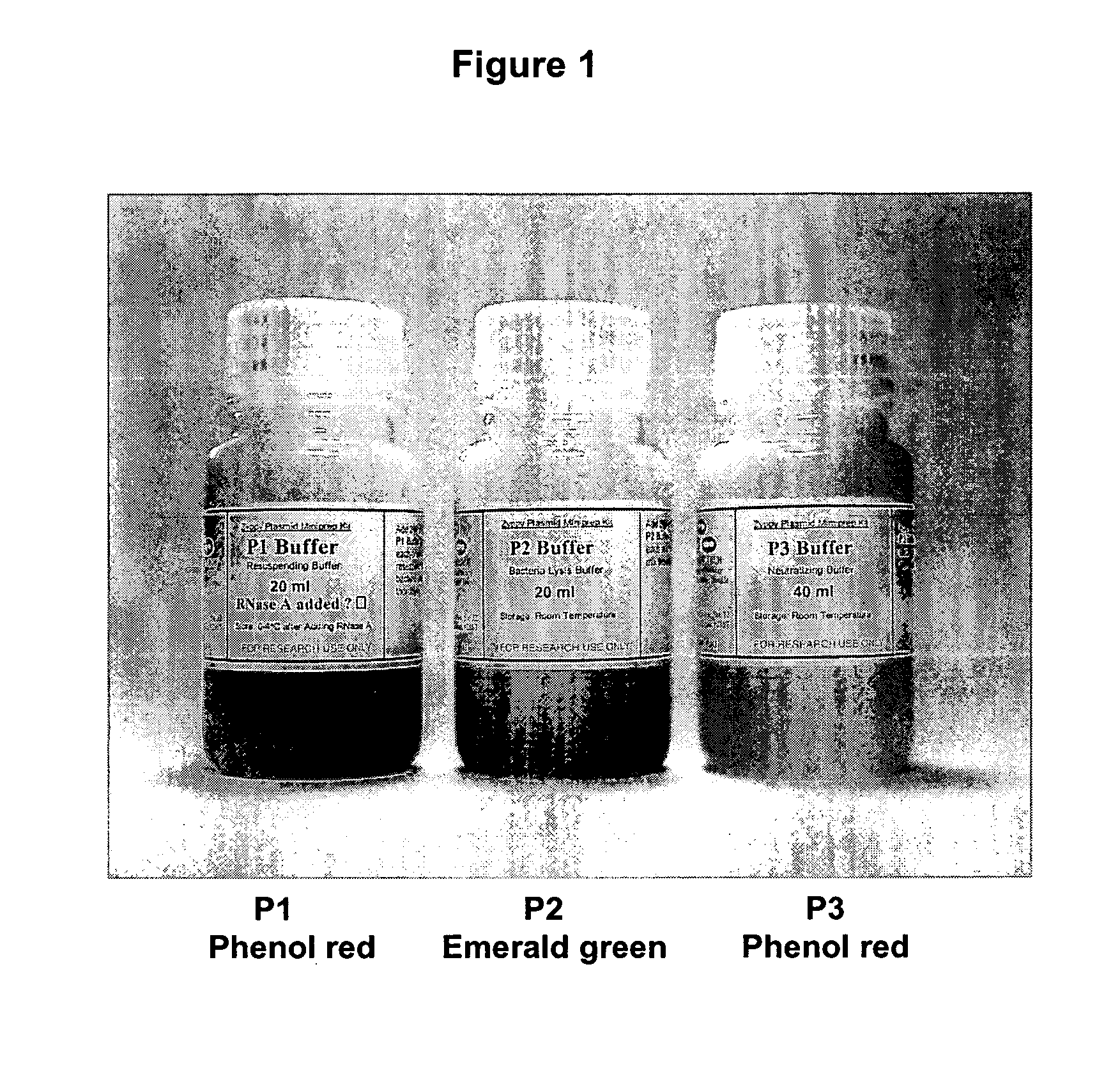
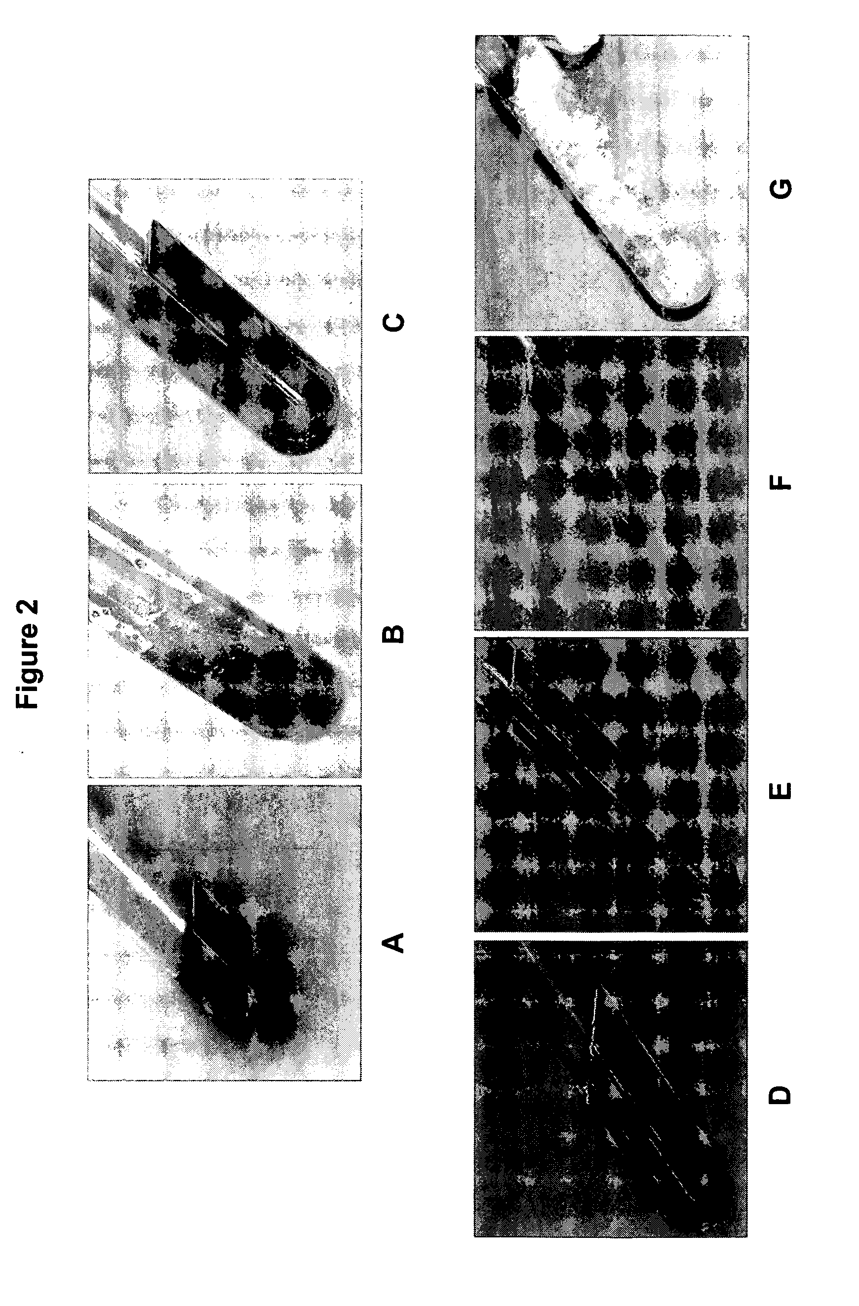
[0049] 1. Pellet 0.5-5 ml of overnight culture in a 1.5 ml microfuge tube by spinning for 15-20 seconds. [0050] 2. Discard the supernatant. [0051] 3. Add 200 ul of P1 buffer (red), containing RNAse A (100 ug / ml). Resuspend completely with gentle vortexing or by pipette. Solution is pink or light red in color to the naked eye. [0052] 4. Add 200 ul of P2 buffer (blue). Mix by inverting and swirling the microfuge tube 4-6 about times. The solution becomes clear and a deeper red in color. If lysis is incomplete the clearing and darkening to a red of the color is qualitatively less pronounced to the naked eye. [0053] 5. Add 400 ul pf P3 buffer (yellow) and mix thoroughly, but gently. Do not vortex this step hard. A white precipitate will form which consists of K-SDS and cellular debris. The buffer becomes yellow and cleared debris suspension. [0054] 6. Spin the microfuge tube for 3 minutes at maximum speed. [0055] 7. Load the...

example 2

Preferred Embodiment

Spin-Column (Standard: Microfuge or Vacuum Elution)

[0060] 1. Pellet 0.5-5 ml of overnight culture in a 1.5 ml microfuge tube by spinning for 15-20 seconds. [0061] 2. Discard the supernatant. [0062] 3. Add 200 ul of P1 buffer (red). Resuspend completely with gentle vortexing or by pipette. Solution becomes pink or light red in color to the naked eye. [0063] 4. Add 200 ul of P2 buffer (blue). Mix by inverting and swirling the microfuge tube 4-6 about times. The solution becomes clear and a deeper red in color. If lysis is incomplete the clearing and darkening to a red of the color is qualitatively less pronounced to the naked eye. [0064] 5. Add 400 ul pf P3 buffer (yellow) and mix thoroughly, but gently. Do not vortex this step hard. A white precipitate will form which consists of K-SDS and cellular debris. Shaking the tube inverted several times increases the efficiency of precipitate formation. [0065] 6. Spin microfuge tube for 3 minutes. [0066] 7. Load the sup...

example 3

Short Procedure

Alternative Embodiment

[0071] 1. Pellet 0.5-5 ml of overnight culture. Discard supernatant. [0072] 2. Add 200 ul of P1 buffer (red) and resuspend pellet, becomes light red or pink. [0073] 3. Add 200 ul of P2 buffer (blue) and mix, becomes deeper red and clear to the naked eye. [0074] 4. Add 400 ul of P3 buffer (yellow) and mix thoroughly, white cellular precipitate forms. [0075] 5. Spin full speed for 3 minutes. [0076] 6. Load supernatant to the spin-column. [0077] 7. Spin the column with the collection tube for 30 sec. [0078] 8. Discard the flow-through. [0079] 9. Add 600 ul of Wash buffer and spin for 30 sec. [0080] 10. Add 40 ul (25 ul) of Elution Buffer or water, put the column into a new microfuge tube, and spin for 10 sec.


PUM

PropertyMeasurementUnit
Coloraaaaaaaaaa
Responsivityaaaaaaaaaa

Abstract

The present invention describes isolation of plasmid DNA or nucleic acid material from Escherichia coli (E. coli) or other species of microorganisms, or from other sources. The addition of indicator dyes to the alkaline lysis based purification buffers allows for increased ease of visual monitoring of the mixing and lysis steps of the procedure. The yield and quality of plasmid DNA is improved and the chance of error is reduced. The practice may be applied to other nucleic acid purification procedures.

Description

CROSS REFERENCE TO RELATED APPLICATIONS [0001] This application claims priority to U.S. provisional application 60 / 699,957.BACKGROUND OF THE INVENTION [0002] 1. Field of the Invention [0003] The present invention relates to isolating pure plasmid DNA from E. coli and other organisms. The addition of indicator dyes to the alkaline lysis based buffers allows for easy and rapid visual monitoring of the resuspension, lysis, and neutralization steps. The ability to assess the process increases plasmid yield and also reduces errors. The present invention is useful in other nucleic acid purification applications. [0004] 2. Description of the Related Art [0005] The present invention relates to the purification of plasmid DNA and other nucleic acids from source materials. The purification of plasmid DNA from bacterial lysates is a standard molecular biological technique that is critical for downstream recombinant DNA manipulations. It is critical and consumes significant resources of academi...

Claims


Application Information

Patent Timeline
IPC IPC(8): C12Q1/68C07H21/04
CPCC07H21/04
Inventor JIA, XIYUJIA, LINDA
Owner ZYMO RES CORP
Why Eureka
Try Eureka