Medical and dental implant devices for controlled drug delivery

a controlled drug and implant device technology, applied in dental surgery, dental implants, colonoscopy devices, etc., can solve the problems of loss of bone tissue at the interface with the prosthetic device, the loosening of the joint/prosthetic, and the end of the prosthetic implan

Inactive Publication Date: 2007-01-18
MICROCHIPS INC
View PDF99 Cites 268 Cited by
  • Summary
  • Abstract
  • Description
  • Claims
  • Application Information

AI Technical Summary

Benefits of technology

[0015] In another aspect, a method for treating osteonecrosis is provided which includes the steps of removing necrotic bone tissue from a bone and creating one or more channels or voids in said bone; and inserting at least one drug delivery device into the one or more channels or voids, wherein the drug delivery device comprises a body portion having a plurality of discrete reservoirs containing at least one release system comprising one or more therapeutic or prophylactic agents for release in vivo. In one embodiment, two or more drug delivery devices are inserted into two or more channels formed in the bone. The channels may be separate and may be parallel. In another embodiment, the method further includes utilizing a fluid delivery means to wet the drug delivery device which is disposed in the one or more channels or voids. For example, the fluid delivery means may be a re-routed or grafted blood vessel, or it may be include a fluid source, pump, and at least one catheter, wherein the distal end of the catheter is inserted into at least one of the channels or voids containing the drug delivery device and delivers fluid from the fluid source via the pump. The fluid reservoir and pump may be integrated into a single device. The fluid source may be saline, blood, or a blood component, and may include hyaluronic acid. In one embodiment of the method, the step of removing necrotic bone tissue from a bone and creating one or more channels or voids in said bone may involve a light bulb or trapdoor surgical procedure.
[0016] In another aspect, a joint resurfacing device is provided that includes a body portion having a joint tissue interfacing surface and an opposing side; a plurality of discrete reservoirs located joint tissue interfacing surface; at least one release system disposed in one or more of the plurality of reservoirs containing at least one release system comprising one or more therapeutic or prophylactic agents for release in vivo; and an anchor portion extending from the opposing side away from the joint tissue interfacing surface, wherein the anchoring portion is adapted to secure the joint resurfacing device to a bone in need of resurfacing. The one or more therapeutic or prophylactic agents may be a bone growth promoter or may be selected from bone morphogenic proteins, angiogenesis promoters, analgesics, anesthetics, antibiotics, and combinations thereof. In one embodiment, the joint tissue interfacing surface comprises a rounded cap. In one embodiment, the reservoirs have chamfered openings in the surface of the joint tissue interfacing surface. The anchor portion may include on one or more screws.
[0017] In still another aspect, an implantable infection control device is provided, which includes a plurality of beads tethered together to form a chain, wherein the beads comprise a plurality of discrete reservoirs which are loaded with a release system comprising at least one anti-infective drug formulation for controlled release in vivo. The beads may be cylindrical, spherical, or elliptical shaped, and typically are made of a biocompatible material selected from polytetrafluoroethylenes, polyesters, polymethylmethacrylates, silicones, metals, glasses, ceramics, bone cements, and combinations t

Problems solved by technology

Risks that may follow the replacement surgery include infection and, in the long term with some types of devices, loss of bone tissue at the interface with the prosthetic device as the bone remodels and consequent loosening of the joint/prosthetic.
Another drawback of joint replacement is that the prosthetic implant eventually will wear out, for example, ten to twenty years following implantation.
This is problematic where the patient receiving the joint replacement is relatively young and might be expected to live well beyond the useful life of the joint prosthesis.
Replacement of the prosthesis may not be possible in some instances, and would nevertheless require another invasive surgery.
A significant challenge to the widespread use of dental implants is the often extended time to osseointegration or the growth action of bone tissue, as it assimilates surgically implanted devices or prostheses to be used as either replacement parts (e.g., hip) or as anchors (e.g., endosseous dental implants).
Coatings, however, substantially limit the selection of the coating materials and the drugs, as well as substantially limit the control over the release kinetics and spatial release patterns.
Unfortunately, coatings having improved mechanical stability and adhesion may tend to have decreased utility as a controlled drug delivery vehicle.
For example, when a coating material is selected that is robust enough not to crack or delaminate from an underlying substrate, it usually performs poorly as a drug delivery vehicle, in that drug does not release as well or as efficiently as would be desired.
Surface coatings, however, are vulnerable to mechanical failure and suffer

Method used


Image

  • Medical and dental implant devices for controlled drug delivery
  • Medical and dental implant devices for controlled drug delivery
  • Medical and dental implant devices for controlled drug delivery

Examples


Embodiment Construction

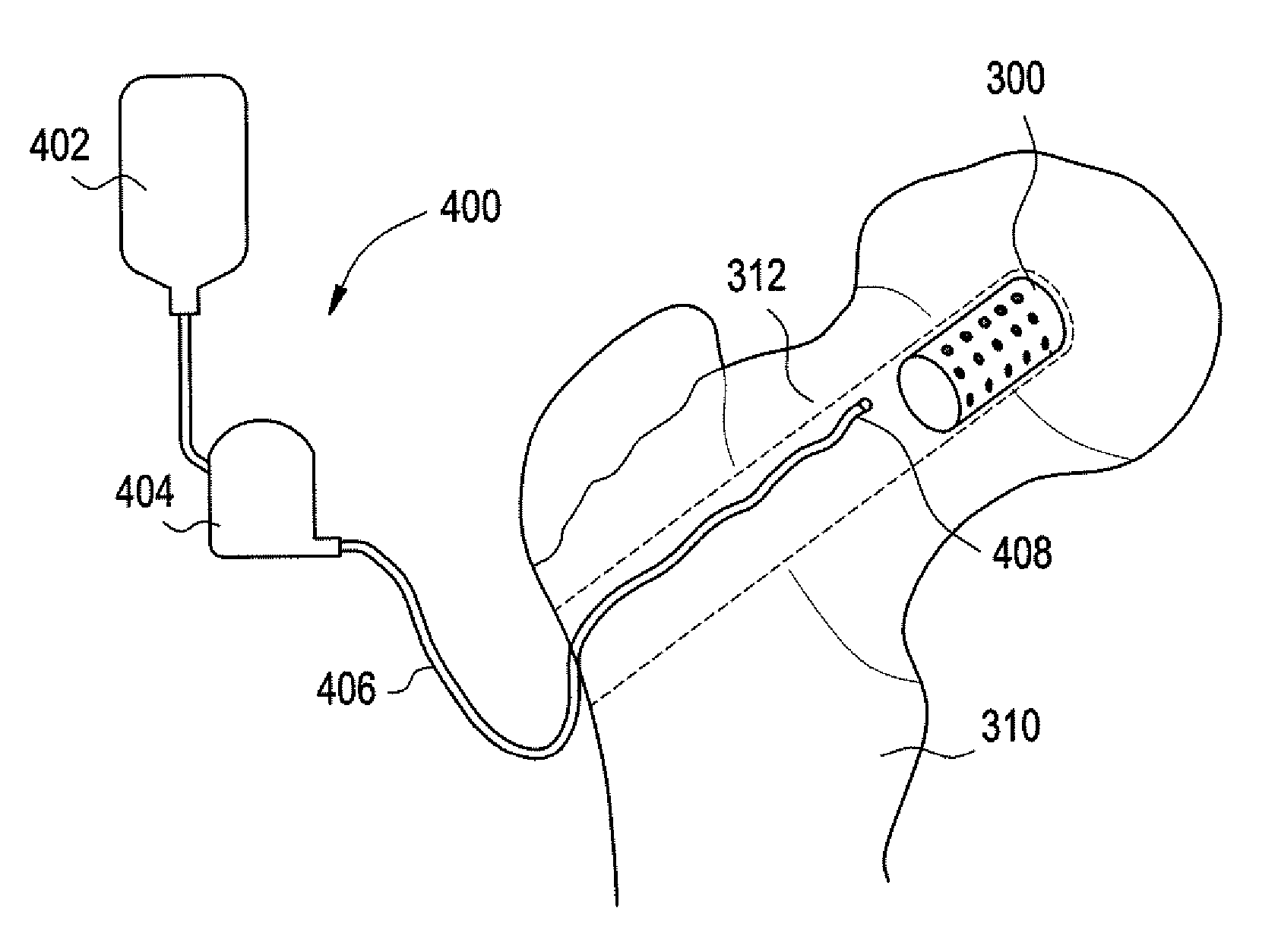
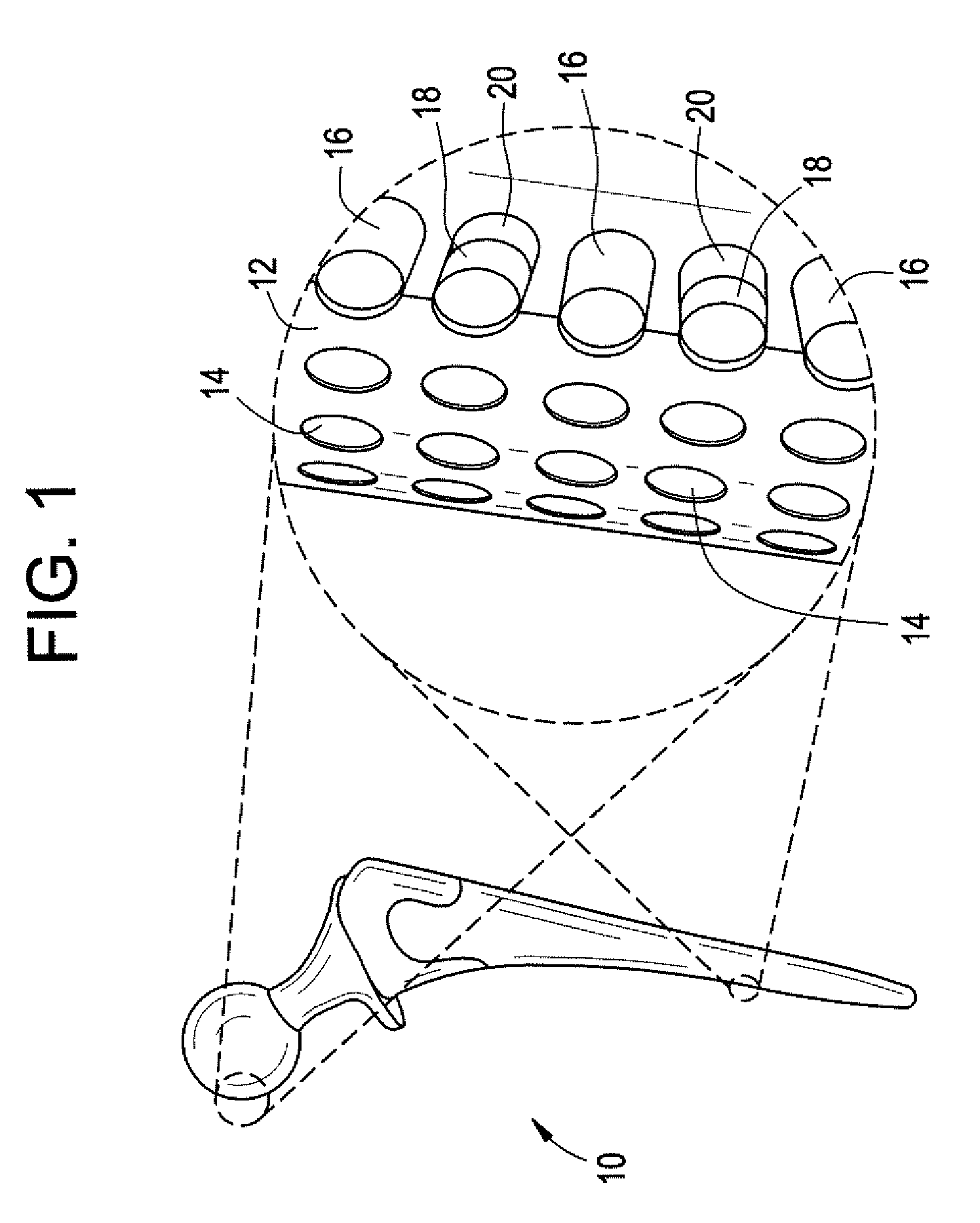
[0037] Implantable devices and methods of use have been developed to provide controlled delivery of therapeutic and prophylactic agents in the treatment and health of orthopedic, joint, spinal, and dental tissues. As used herein, the term “orthopedic” includes and is synonymous with the term “orthopaedic.” In one particular aspect, the devices and methods are used in the treatment of avascular necrosis, providing improved controlled delivery of bone growth promoters and other drugs directly where needed. As used herein, the terms “avascular necrosis” and osteonecrosis” are synonymous and may be used interchangeably.

[0038] In another aspect, the device is a prosthetic device. As used herein, the term “prosthetic” refers to medical and dental devices that are primarily used to secure together separate tissue portions or to provide a load bearing function. It is considered prosthetic in the sense that it is serving as a structural complement or substitute (permanently or temporarily) ...


PUM

PropertyMeasurementUnit
Temperatureaaaaaaaaaa
Pressureaaaaaaaaaa
Structureaaaaaaaaaa

Abstract

Implantable devices and methods for use in the treatment of osteonecrosisare provided. The device includes at least one implant device body adapted for insertion into one or more channels or voids in bone tissue; a plurality of discrete reservoirs, which may preferably be microreservoirs, located in the surface of the at least one implant device body; and at least one release system disposed in one or more of the plurality of reservoirs, wherein the release system includes at least one drug selected from the group consisting of bone growth promoters, angiogenesis promoters, analgesics, anesthetics, antibiotics, and combinations thereof. The device body may be formed of a bone graft material, a polymer, a metal, a ceramic, or a combination thereof. The device body may be a monolithic structure, such as one having a cylindrical shape, or it may be in the form of multiple units, such as a plurality of beads.

Description

CROSS-REFERENCE TO RELATED APPLICATIONS [0001] This application claims benefit under 35 U.S.C. §119(e) of U.S. Provisional Application No. 60 / 763,336, filed Jan. 30, 2006, U.S. Provisional Application No. 60 / 727,323, filed Oct. 17, 2005, and U.S. Provisional Application No. 60 / 695,030, filed Jun. 28, 2005, all of which are incorporated herein by reference.BACKGROUND OF THE INVENTION [0002] This invention is generally in the field of implantable medical and dental devices for controlled release of therapeutic and prophylactic agents into a human or animal patient, and particularly prosthetic, drug delivery, or combination implants for replacing, augmenting, or promoting the health of bone, cartilage, or dental tissues. [0003] Hundreds of thousands of hip replacements or revisions are performed each year in the United States. Artificial joints have become a common therapeutic option for replacing the structure of, and restoring function to, injured or diseased joints, including hips, ...

Claims


Application Information

Patent Timeline
IPC IPC(8): A61M31/00
CPCA61C8/0012A61C8/0016A61L2300/602A61L2300/414A61L2300/41A61L2300/406A61L2300/402A61C19/063A61C2008/0046A61F2/30744A61F2/30756A61F2/30767A61F2/30771A61F2/36A61F2/3662A61F2/3676A61F2/38A61F2/446A61F2002/2817A61F2002/2828A61F2002/2832A61F2002/2835A61F2002/30004A61F2002/30013A61F2002/30062A61F2002/30224A61F2002/30261A61F2002/30266A61F2002/30462A61F2002/30677A61F2002/30808A61F2002/3081A61F2002/30874A61F2002/30878A61F2002/30937A61F2002/3617A61F2002/4631A61F2002/4636A61F2002/482A61F2210/0004A61F2220/0075A61F2230/0069A61F2230/0082A61F2250/0014A61F2250/0024A61F2310/00011A61F2310/00023A61F2310/00029A61F2310/00179A61K9/0024A61L15/585A61L24/043A61L27/54A61L27/56A61L28/0026A61L2300/252C08L31/04A61F2002/30011A61F2002/30652A61F2002/30873A61F2002/4635A61L2430/02A61F2/482
Inventor SANTINI, JOHN T. JR.HUTCHINSON, CHARLES E.PROOS, ELIZABETH R.
Owner MICROCHIPS INC
Why Eureka
Try Eureka