
Method for the isolation and expansion of cardiac stem cells from biopsy

Active Publication Date: 2007-01-25
UNIV DEGLI STUDI DI ROMA LA SAPIENZA
View PDF89 Cites 23 Cited by
  • Summary
  • Abstract
  • Description
  • Claims
  • Application Information

AI Technical Summary

Benefits of technology

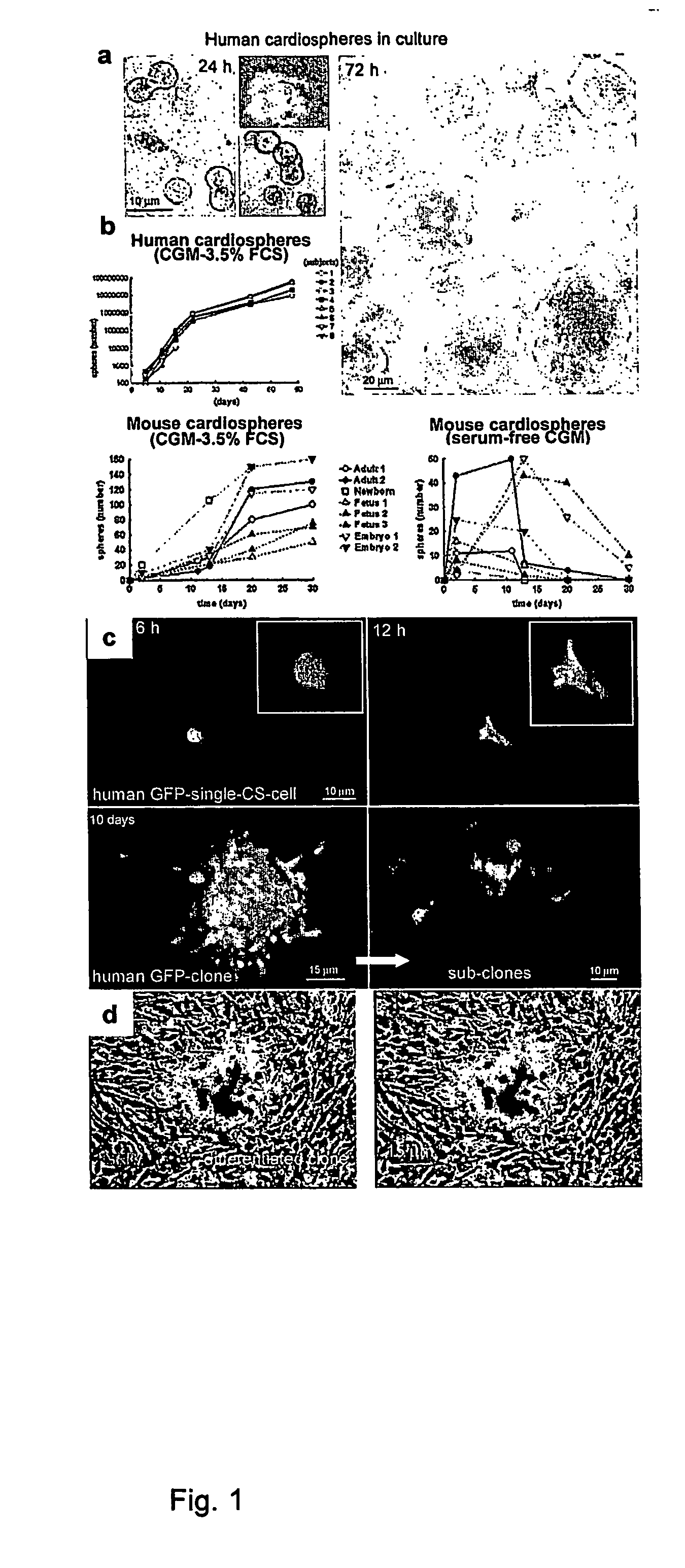
[0024] An object of the invention is a method to obtain stem cells able to repair damaged myocardiac tissue, comprising the following steps: a) take a biopsy specimen of cardiac tissue and keep it in an appropriate culture medium; b) treat the specimen under appropriate conditions with mild mechanical and / or chemical and / or enzymatic techniques to obtain tissue fragments sized large enough to allow the diffusion of nutrients present in the medium; c) leave the tissue fragments to adhere to appropriate solid supports and maintain them in a medium containing convenient serum and / or growth factors; d) allow the cells to grow, changing the medium partially or completely, until multicellular structures form that are either weakly adherent or do not adhere to the support; e) separate said multicellular structures from the rest of the culture; f) treat said multicellular structures by mild dissociation until most of the small phase-bright spherical cells detach but maintain their morphologic and functional characteristics; g) plate the cells on culture substrates treated with polylysine or other agents promoting the adhesion of the culture to the support in a medium containing at least the minimal essential constituents for the growth of mammalian cells; h) possibly repeat steps d) to g) at least once; i) select the cells that aggregate in phase-bright spheroid formations (cardiospheres); l) electively promote the formation of new cardiospheres by mild dissociation thereof and new formation; m) eventually cryopreserve the cardiospheres for use after thawing.
[0027] Experts in the field will understand that the CS derived with the procedure of the invention may be able to generate continuous cell lines following spontaneous transformation or transformation induced by chemical, physical or biologic agents.

Problems solved by technology

The limited ability of adult cardiomyocytes to undergo mitosis and to regenerate the myocardium after injury leads to a permanent deficiency in the number of functioning cells, with the development and progression of cardiac insufficiency.
However, the problem of having a source and an availability of SC remains (5-7).
While embryonic SC (undifferentiated cells from the embryo that can produce a wide range of specialized cells and that can be derived from the cell mass inside blastocytes which, in humans, form 4-5 days after fertilization of the ovum) have a marked capability to proliferate and differentiate, their potential immunogenicity, arrhythmogenicity, and ethical issues in particular, have limited their use.
Moreover, embryonic SC are pluripotent, consequently their use carries a potential risk of generating teratomas (as occurs in animal models).
Embryonic SC have the potential capability to generate these cardiomyocyte phenotypes in vitro but the yield is insufficient.
Furthermore, the in vivo proliferative capability of cardiomyocytes derived from embryonic SC appears to be limited by the growth of multinucleate cells.
The inability of skeletal myoblasts to transdifferentiate into cardiomyocytes and to couple with them could give rise to arrhythmias or other anomalies
But the functional and electrophysiologic properties of HSC- and MSC-derived cardiomyocytes are not well characterized, and the use of undifferentiated cells instead of cardiomyocytes could give rise to in vivo differentiation into fibroblasts rather than muscle cells or to the development of tumors.
Furthermore, there is no in vivo evidence from cardiomyopathic animals that supports the applicability of the method.
Adherent cells cloned by limiting dilution give rise to mesenchymal cells, including osteoblasts, chondrocytes, adipocytes and myocytes, although they cannot be clearly identified due to often inappropriate morphologic criteria and markers.
However, no data are given about the characteristics of the isolated SC.
Furthermore, the cells isolated with the previous method do not give rise to the formation of an essential component of the heart tissues, i.e. vessels and endothelium.

Method used


Image

  • Method for the isolation and expansion of cardiac stem cells from biopsy
  • Method for the isolation and expansion of cardiac stem cells from biopsy
  • Method for the isolation and expansion of cardiac stem cells from biopsy

Examples


Embodiment Construction

[0016] The method of the present invention employs heart biopsy tissue as starting material, hence an elective material that cannot be used in the method described in patent application WO 99 / 49015, since the material was insufficient. After fragmenting the biopsy specimen and possibly using dissociating agents (e.g. trypsin, EDTA and collagenase), the fragments are plated and added to a medium containing 10% FBS; 10-30 days later, fibroblast-like adherent cells grow from the explants over which small round, phase-bright cells migrate that tend to cluster but are either not or only weakly adherent. The cells are isolated by washing and mild dissociation (e.g. EDTA, trypsin-EDTA for 2-3 min). The cells are then plated on polylysine-treated cellular substrates in an appropriate medium unlike that used in the previous technique, in that it is horse-serum-free and contains other growth factors; after 2-3 days cell aggregates (cardiospheres) arise that tend to grow as floating formations...


PUM


Abstract

Method for the isolation, expansion and preservation of cardiac stem cells from human or animal tissue biopsy samples to be employed in cell transplantation and functional repair of the myocardium or other organs. Cells may also be used in gene therapy for treating genetic cardiomyopathies, for treating ischemic heart diseases and for setting in vitro models to study drugs.

Description

FIELD OF INVENTION [0001] The invention concerns a method for the isolation and expansion of cardiac stem cells derived from postnatal cardiac tissue biopsy. [0002] The invention deals with a method for the isolation, expansion and preservation of cardiac stem cells from human or animal tissue biopsy samples to be employed in cell transplantation and functional repair of the myocardium or other organs. [0003] The cells may also be used in gene therapy, for treating genetic cardiomyopathies by expressing the healthy gene in cells from biopsies of subjects with genetic defects, propagating the cells in vitro and then transplanting them in the patient; for treating ischemic heart diseases by inducing the release of angiogenetic growth factors by the transplanted cells; and for the setting of an in vitro models to study drugs. PRIOR ART [0004] Stem cells (SC) are able to replicate and to differentiate in response to appropriate signals, thus enabling the formation or regeneration of spe...

Claims


Application Information

Patent Timeline
IPC IPC(8): C12N5/08C12N5/06A61K35/12C12N5/077
CPCA61K35/12C12N5/0657C12N2533/32C12N2501/175C12N2501/11C12N2501/115C12N5/069A61P9/00A61P9/10
Inventor GIACOMELLO, ALESSANDROMESSINA, ELISABATTAGLIA, MASSIMOFRATI, GIACOMO
Owner UNIV DEGLI STUDI DI ROMA LA SAPIENZA
Learn More