Materials and methods for colorectal cancer screening, diagnosis and therapy
a colorectal cancer and material technology, applied in the field of materials and methods for colorectal cancer screening, diagnosis and therapy, can solve the problems of colon cancer treatment options of unpredictable and sometimes limited value, eye development defect,
- Summary
- Abstract
- Description
- Claims
- Application Information
AI Technical Summary
Problems solved by technology
Method used
Image
Examples
example 1
Methods and Materials
[0194] Methods and material used or referred to in subsequent examples are set forth directly below.
Antibodies
[0195] Monoclonal mouse anti-vimentin, β-catenin (Transduction Laboratories), Ki-67 (Pharmingen) and chromogranin A (Ab-3, NeoMarkers), monoclonal rat anti-BrdU (Harlan Seralab) and polyclonal rabbit anti-Prox-1 were obtained from the indicated commercial sources. The fluorochrome-conjugated secondary antibodies were obtained from Jackson Immunoresearch.
[0196] For production of Prox-1 antibodies cDNA encoding Prox-1 homeobox domain and prospero domain (amino acids 578-750 of human Prox-1, SEQ ID NO: 3) was subcloned into pGEX2t vector to produce GST-Prox-1 fusion construct. This construct was expressed in E. coli and the GST-Prox-1 fusion protein from E. coli was purified using glutathione Sepharose according to the manufacturer's instructions (Amersham, Piscataway, N.J.). Fusion protein was used to immunize rabbits according to a standard protocol....
example 2
Prox-1 mRNA is Elevated in Colorectal Tumors
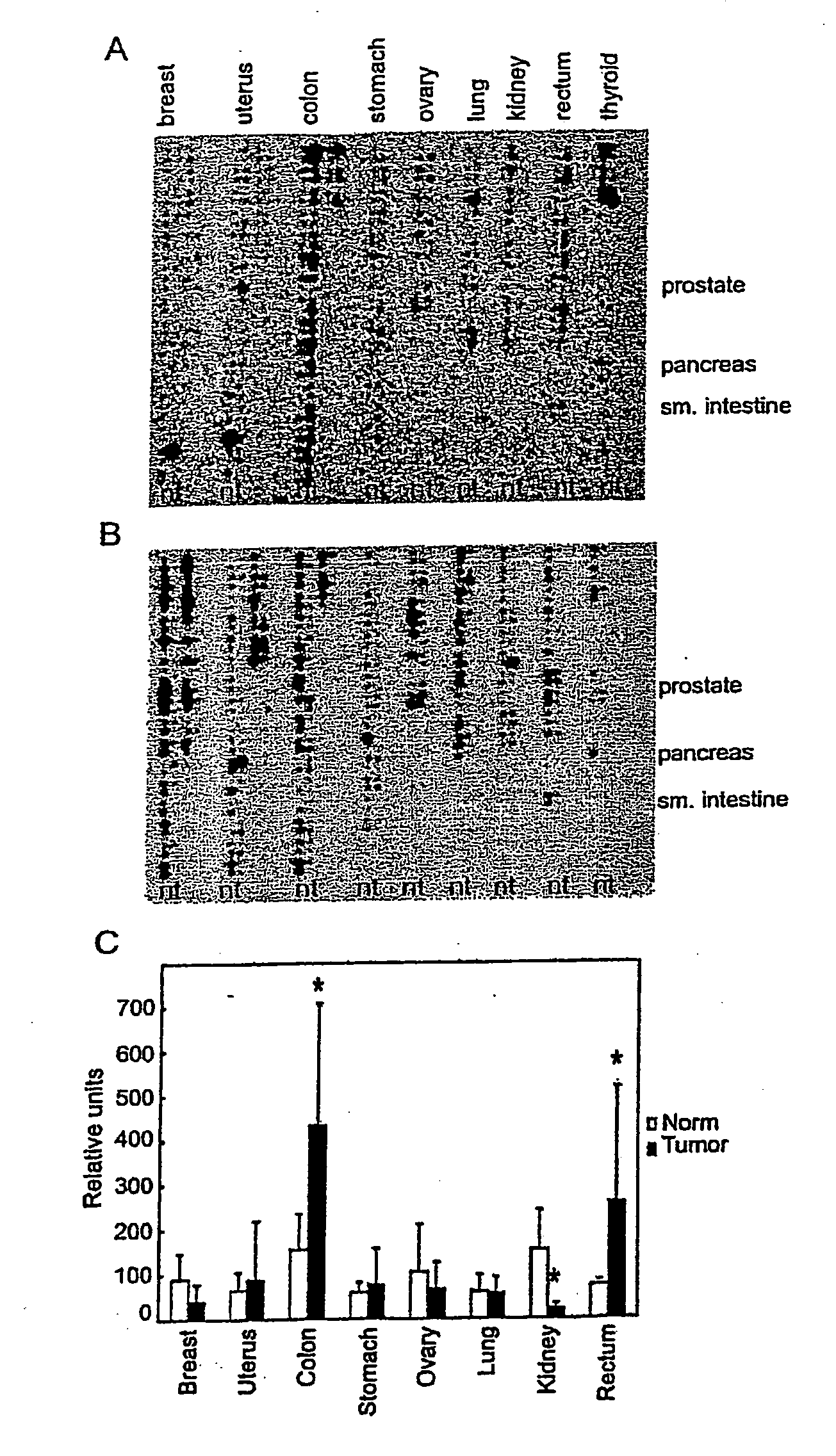
[0209] Experiments were conducted to assess the expression of Prox-1 mRNA in human cancers using a cancer gene profiling array filter, which contains cDNAs from about 250 human cancers and corresponding normal control tissues. Prox-1 mRNA was significantly increased in 35 out of 53 samples of colorectal cancers. In contrast, only rarely or not at all was any increase seen in samples from breast, uterine, lung, kidney, ovarian, or thyroid tumors (FIG. 1A, B, and C). Probes for Prox-1 (FIG. 1A) and the lymphatic endothelial marker LYVE-1 (FIG. 1B) were used.
[0210]FIG. 1C demonstrates quantification of dot blot in FIG. 1A, the asterisk indicating tumor samples in which Prox-1 expression is significantly different from that of the normal tissue (PCancer Res. 62: 1669-75 (2002)). Therefore, the filter to the probe for the lymphatic endothelial hyaluronan receptor LYVE-1 was hybridized. Unlike Prox-1, LYVE-1 levels were higher in the normal sa...
example 3
Prox-1 is Expressed in Round but not in Adherent Subclones of the SW480 Colon Adenocarcinoma Cell Line
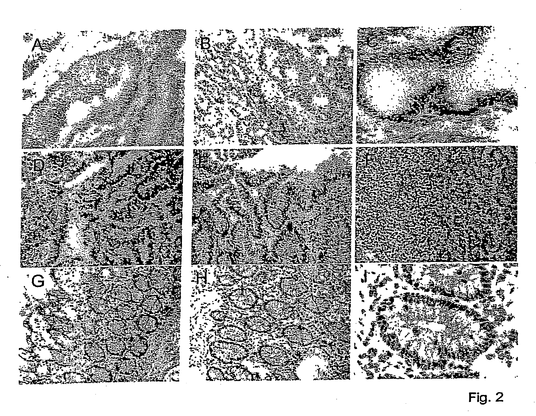
[0213] Additional studies were conducted to compare Prox-1 expression in various cells. No Prox-1 expression was seen in the majority of tumor cell lines studied. However, Prox-1 was mRNA was present in hepatocellular carcinoma cell line HepG2 and the colon carcinoma cell line SW480. BEC, blood endothelial cells, CAEC, coronary artery endothelial cells, and LEC, lymphatic endothelial cells, served as negative and positive controls. Immunofluorescent staining of Prox-1 revealed strong expression in all HepG2 cells, whereas only a subset of SW480 cells were Prox-1 positiveDouble immunofluorescent staining for Prox-1 and for β-catenin or for the F-actin marker phalloidin demonstrated that Prox-1 expression is restricted to weakly adherent round SW480 cells which did not display focal adhesions or actin stress fibers, and that Prox-1 was very weakly expressed the adherent cells. The ex...
PUM
| Property | Measurement | Unit |
|---|---|---|
| pH | aaaaa | aaaaa |
| pH | aaaaa | aaaaa |
| volumes | aaaaa | aaaaa |
Abstract
Description
Claims
Application Information
 - R&D
- Intellectual Property
- Life Sciences
- Materials
- Tech Scout
- Unparalleled Data Quality
- Higher Quality Content
- 60% Fewer Hallucinations
Browse by: Latest US Patents, China's latest patents, Technical Efficacy Thesaurus, Application Domain, Technology Topic, Popular Technical Reports.
© 2025 PatSnap. All rights reserved.Legal|Privacy policy|Modern Slavery Act Transparency Statement|Sitemap|About US| Contact US: help@patsnap.com


