Optical coherent tomographic (OCT) imaging apparatus and method using a fiber bundle

a fiber bundle and optical coherence tomography technology, applied in the field of optical coherence tomography, can solve the problems of insufficient resolution to delineate the microstructure of biological tissues at the level, restricted lateral resolution, and difficult to achieve small diameter endoscopic probes, etc., to achieve the effect of reducing back reflection

Inactive Publication Date: 2007-02-15
RGT UNIV OF CALIFORNIA
View PDF4 Cites 74 Cited by
  • Summary
  • Abstract
  • Description
  • Claims
  • Application Information

AI Technical Summary

Benefits of technology

[0009] A fiber-optic imaging bundle for use with an OCT interferometer and scanner is constructed, which is 3.2 mm in diameter, has various lengths, is composed of a large number (here, 50,419 individual fiber cores) of 12 μm fibers. Various lengths of fiber can be used, e.g. 12 inch fibers were used for hamster trachea image, and 6″ bundles for an infrared (IR card). Fiber bundles are polished at an angle (8 degrees) at each end to reduce back reflection. The OCT sample light beam is focused by an objective lens to a 10 μm beam spot on the proximal entrance plane of the fiber-optic bundle. The light beam spot is scanned laterally on the entrance plane of the fiber bundle. The light emerging from the fiber cores at the distal end of the fiber bundle is collimated by an achromatic doublet lens and refocused onto the sample by another achromatic doublet lens or GRIN lens. However, the objective lens may not be necessary for images.

Problems solved by technology

However, this resolution is insufficient to delineate the microstructure of biological tissues at the level required to detect many abnormalities, such as early cancers.
Since the mirror size and numerical aperture are limited by the endoscope size, the lateral resolution is subsequently restricted.
Three dimensional scanning can be accomplished by repeating the scans with an added directional motion component, but this may be technically challenging in small diameter endoscopic probes, particularly when precisely aligning components.
In confined spaces of endoscopic probes, the sweep distances obtainable using MEMS probes may also be quite limited.
The use of flexible fiberoptic bundles to deliver OCT directly to a tissue sample has not been previously achieved.

Method used


Image

  • Optical coherent tomographic (OCT) imaging apparatus and method using a fiber bundle
  • Optical coherent tomographic (OCT) imaging apparatus and method using a fiber bundle
  • Optical coherent tomographic (OCT) imaging apparatus and method using a fiber bundle

Examples


Embodiment Construction

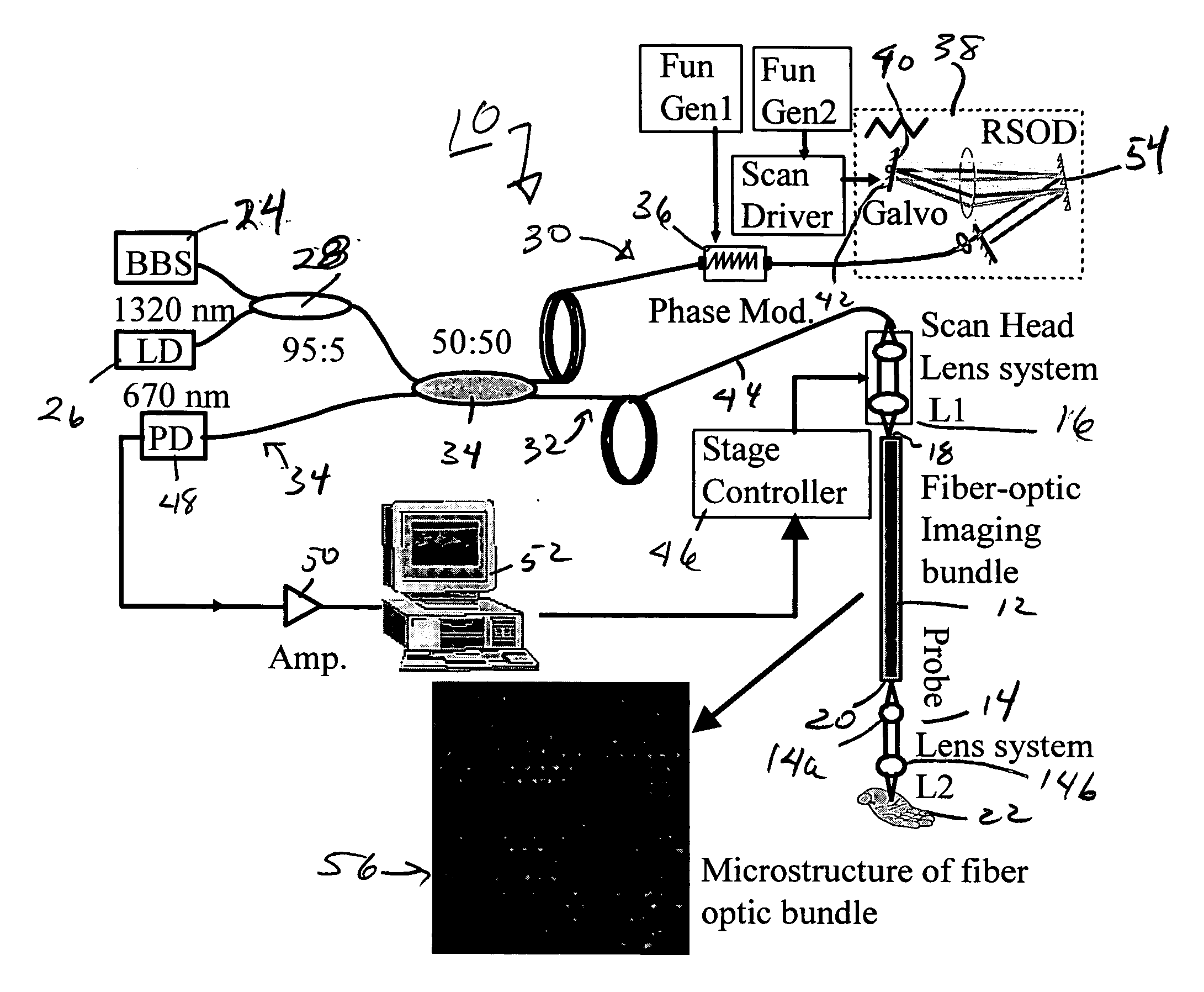
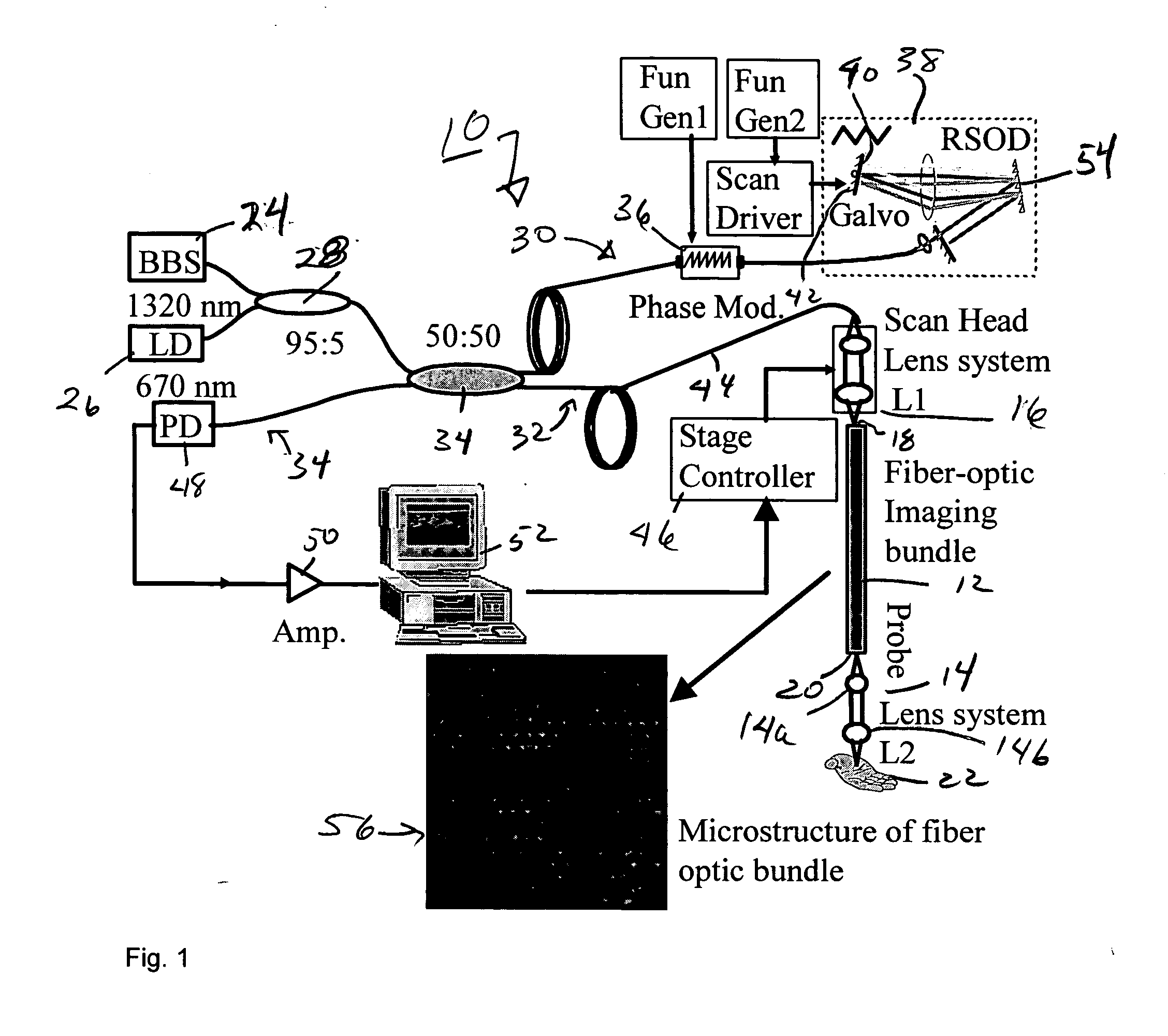
[0025] In the illustrated embodiment of FIG. 1, we present a new fiber bundle OCT imaging method and apparatus 10 with front view scanning which is comprised of a fused coherent fiber bundle 12 and an objective lens system 14. In this system 10, the scanning mechanism 16 is placed at the proximal fiber bundle entrance 18. The bundle 12, made up of several thousand cores, preserves the spatial relationship between the entrance 18 and the output 20 of the bundle 12. A cross section of the bundle 12 is shown in the enlarged inset 56 of FIG. 1 showing the dense honey-comb packing of the cores. Therefore, one or two directional scanning can be readily performed on the proximal bundle surface 18 to create 2 or 3 dimensional images. Because of this fiber bundle design, the scanning mechanism 16 can be placed proximally and no moving parts or driving current are needed within the endoscope (not shown) in which at least the distal portion of bundle 12 is included with lens system 12. This de...


PUM


Abstract

A fiber-optic bundle based optical coherence tomography (OCT) probe method is demonstrated in a multimode optical fiber bundle based OCT system. The system can achieve a lateral resolution of 12 μm and an axial resolution of 10 μm using a super-luminescent diode source. This imaging approach eliminates any moving parts in the probe and has a primary advantage for use in extremely compact and safe OCT endoscopes to image internal organs and great potential to be combined with confocal endoscopic microscopy.

Description

RELATED APPLICATIONS [0001] The present application is related to U.S. Provisional Patent Application, Ser. No. 60 / 672,473 filed on Apr. 18, 2005, which is incorporated herein by reference and to which priority is claimed pursuant to 35 USC 119.GOVERNMENT RIGHTS [0002] The invention was funded in part with a grant from the Department of Defense, Contract FA 9550-04-1-0101, Philip Morris: USA-32598, National Science Foundation (BES-86924) and the National Institutes of Health (EB-00293, NCI-91717, and RR-01192. The U.S. Government has certain rights to the invention.BACKGROUND OF THE INVENTION [0003] 1. Field of the Invention [0004] The invention relates to the field of optical coherence tomography and in particular to optical endoscopic scanning probes used with optical coherence tomographic interferometers. [0005] 2. Description of the Prior Art [0006] As a noninvasive imaging technique, optical coherence tomography (OCT) provides high resolution morphological, structural, and func...

Claims


Application Information

Patent Timeline
Patent Type & Authority Applications(United States)
IPC IPC(8): A61B6/00
CPCA61B5/0066A61B5/0073A61B5/0068
Inventor CHEN, ZHONGPINGXIE, TUQIANGMUKAI, DAVIDBRENNER, MATTHEW
Owner RGT UNIV OF CALIFORNIA
Why Eureka
Try Eureka