Protein cages for the delivery of medical imaging and therapeutic agents
a technology for applied in the field of protein cages for the delivery of medical imaging and therapeutic agents, can solve the problems of soluble material entrapment inside the cage, and achieve the effects of facilitating the aggregation and crystallization of ions, increasing the number of introduced materials present, and being highly mobil
- Summary
- Abstract
- Description
- Claims
- Application Information
AI Technical Summary
Benefits of technology
Problems solved by technology
Method used
Image
Examples
example 1
Modifications to Protein Cages for Enhanced Gd3+ Binding
[0136] We have taken advantage of our structural knowledge of the Ca2+ binding in wild type virions in an attempt to enhance binding of gadolinium (Gd3+) for eventual use as a possible MRI contrast agent. The Ca2+ binding sites in wild type virions results from the precise orientation of acidic residues contributed from adjacent coat protein subunits at the quasi three-fold axis (Speir, J. A., et al., 1995, Structure 3:63-78; and Zhao, X., 1998, Ph.D. Purdue University). There are 180 Ca2+ binding sites per virion. Ca2+ binding at these sites is thought to satisfy the charge repulsion created at pH 6.5 by the cluster of acidic residues, and to assist with creating shell curvature during virion assembly. Ca2+ is normally required for in vitro assembly of CCMV at >pH 6.5. We have demonstrated that Gd3+ can act as a substitute for Ca2+ in the pH-dependent assembly assay. We are attempting to enhance assembly-dependent Gd3+ bindin...
example 2
Electrostatic Modifications to Protein Cages
[0151] Entrapment and Growth of Anionic Metal Species
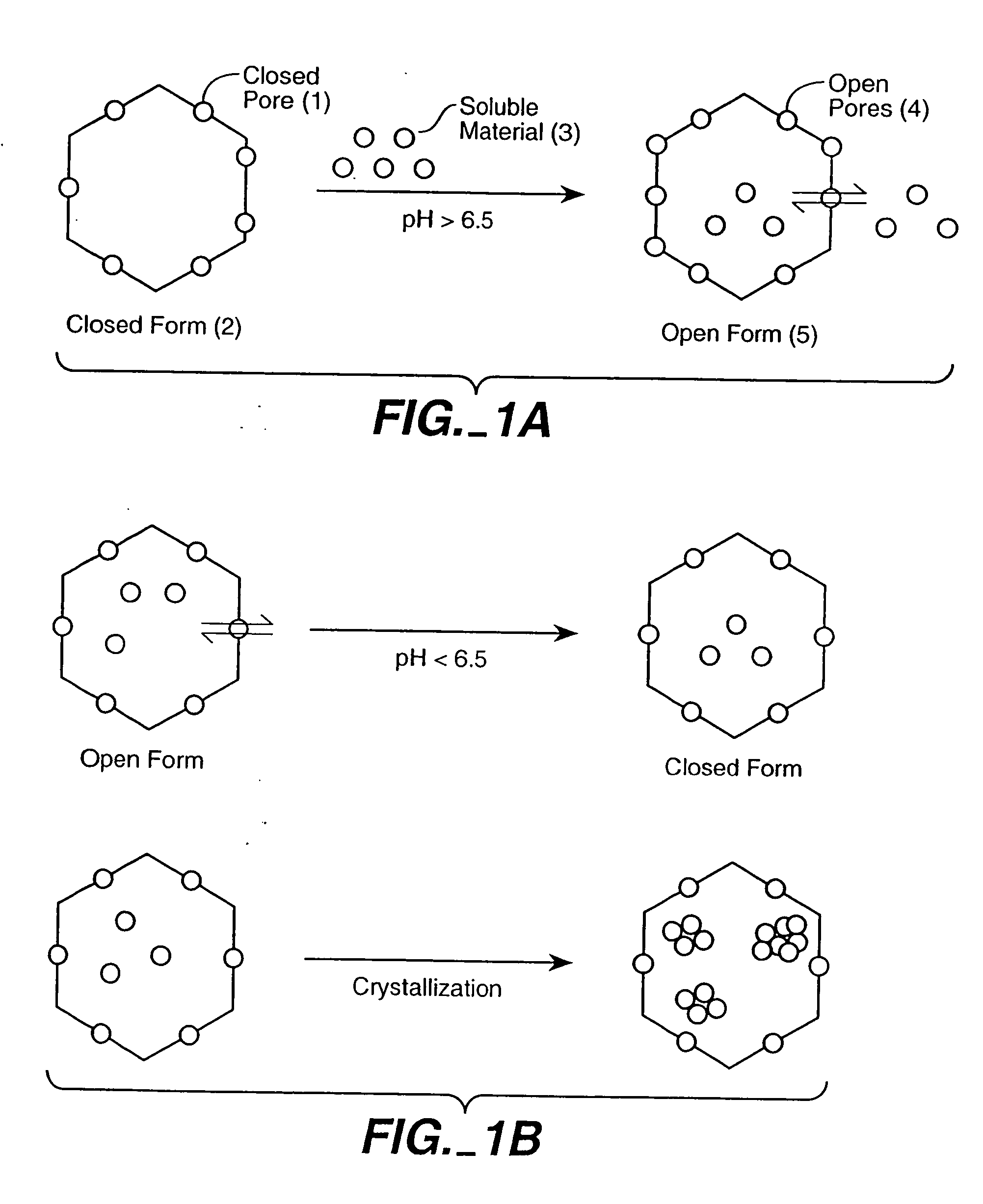
[0152] We have crystallized a range of polyoxometalate species in CCMV and the Norwalk Virus. This was accomplished by providing an interface for molecular aggregation, based on complementary electrostatic interactions between the protein and the anion metal species, which creates a locally high concentration at the protein interface. Briefly outlined, the empty virions were incubated with the precursor ions (WO42-, VO3-, MoO42-) at approximately neutral pH. Under these conditions the virus exists in its open (swollen) form and allows all ions access to the cavity. The pH of the virus solution was then lowered to approximately pH 5.0. This induced two important complementary effects i) The inorganic species underwent a pH dependent oligomerization to form large polyoxometalate species such as H2WO4210- (Douglas, T., and M. J. Young., 1998, Nature 393:152-155) which were readily crystal...
example 3
Bioengineering of New Chemical Switches for the Regulated Entrapment / Release of Materials
[0175] We have demonstrated that pH dependent expansion at the quasi three-fold axes is the result of deprotination of the acidic residues comprising the Ca2+ binding sites. The loss of protons at the elevated pH results in a close cluster of negative charges that must be accommodated either by the binding of Ca2+ or by the physical expansion (i.e. swelling) induced by electrostatic repulsion. We have taken advantage of CCMV's reversible swelling properties as a control mechanism to introduce and to release materials from the central cavity of the protein cage (see e.g. Examples 1 and 2). This reversible switching property of CCMV provides an exciting opportunity for development of elegant control mechanisms for entrapment and release of therapeutic agents.
[0176] pH Activated Chemical Switches
[0177] Gating in the wild-type virion results from electrostatic repulsion of carboxylate groups in t...
PUM
| Property | Measurement | Unit |
|---|---|---|
| size | aaaaa | aaaaa |
| size | aaaaa | aaaaa |
| temperature | aaaaa | aaaaa |
Abstract
Description
Claims
Application Information
 - R&D
- Intellectual Property
- Life Sciences
- Materials
- Tech Scout
- Unparalleled Data Quality
- Higher Quality Content
- 60% Fewer Hallucinations
Browse by: Latest US Patents, China's latest patents, Technical Efficacy Thesaurus, Application Domain, Technology Topic, Popular Technical Reports.
© 2025 PatSnap. All rights reserved.Legal|Privacy policy|Modern Slavery Act Transparency Statement|Sitemap|About US| Contact US: help@patsnap.com


