Ultrasound-based treatment methods for therapeutic treatment of skin and subcutaneous tissues

a technology of ultrasound waves and skin, applied in the field of ultrasound-based treatment methods for skin and subcutaneous tissues, tissue treatment methods using ultrasound waves, can solve the problems of hair follicle disruption, high cost, complicated skin treatment, etc., and achieve the effects of reducing the risk of infection

Inactive Publication Date: 2007-04-05
BOARD OF RGT THE UNIV OF TEXAS SYST
View PDF19 Cites 108 Cited by
  • Summary
  • Abstract
  • Description
  • Claims
  • Application Information

AI Technical Summary

Benefits of technology

[0021] In another aspect of the present invention, a method for rejuvenating the skin of a mammal is described, the method comprising irradiating the skin with ultrasonic radiation at a frequency suitable to penetrate to the subcutaneous tissue layer of the skin for a duration of time from about 1 millisecond to about 30 minutes, such that the ultrasonic radiation damages the subcutaneous tissue, and allows for the natural re-growth of the cellular structure.

Problems solved by technology

Regardless of the purpose, skin treatments are often complicated, expensive, and in some cases, largely ineffective.
While the ultrasound is used primarily for fat destruction, this technique remains an invasive technique as ultrasound probes are inserted through the skin in order to effect the destruction of tissue, and is not without complications.
The use of ultrasound and ultrasonic energy in methods of hair removal has also been reported, albeit with limited successes.
The resulting cavitation of the area surrounding the hair follicle causes the hair follicle to be disrupted.

Method used


Image

  • Ultrasound-based treatment methods for therapeutic treatment of skin and subcutaneous tissues
  • Ultrasound-based treatment methods for therapeutic treatment of skin and subcutaneous tissues
  • Ultrasound-based treatment methods for therapeutic treatment of skin and subcutaneous tissues

Examples


example 1

Cavitation Studies

[0088] A study on the cavitation threshold and activity in pure water and in water containing poly(lactic-co-glycolic acid) (PLGA) nanoparticles as well as in vivo in nude mice. FIGS. 4A and 4B illustrate the cavitation signals obtained at the same ultrasound pressure from pure water (4A) and from containing PLGA nanoparticles (4B), respectively. As is readily apparent from these figures, the cavitation signal obtained from water with PLGA nanoparticles was significantly greater. The signals were integrated using a procedure described in detail in the article by Larina, I. V., et al., [Technol Cancer Res. Treat., Vol. 4(2): pp. 217-226 (2005)] to measure cavitation activity at different pressure (cavitation activity is proportional to concentration of cavitation bubbles and strength of individual cavitation events). In further tests, and as illustrated in FIGS. 5A and 5B, cavitation activity was measured in both pure water (FIG. 5A) and in water containing PLGA na...

example 2

In Vivo Studies

[0090] In the in vivo experiments, Optison™ (Amersham, Mallinkrodt) or PLGA nanoparticles were injected into the tail vein of nude mice and the cavitation signals and activity were measured using the same approach as used in the experiments with water, described above. FIG. 6A shows cavitation activity measured from the subcutaneous tissue of a mouse injected with Optison™ prior to irradiation. As can be seen from the photomicrograph, cavitation activity can be detected with a threshold of about 48 bar.

[0091] However, the cavitation activity decreased rapidly due to degradation of Optison™ upon ultrasound irradiation. Injection of another mouse with PLGA nanoparticles also induced cavitation activity in the irradiated tumor (with almost same threshold), as shown in FIG. 6B. However, the PLGA nanoparticles produce stable cavitation for a longer time because they degrade at a much slower rate. These studies demonstrate that PLGA nanoparticles: (1) substantially lower ...

example 3

Ultrasound-Induced Tissue Damage Studies

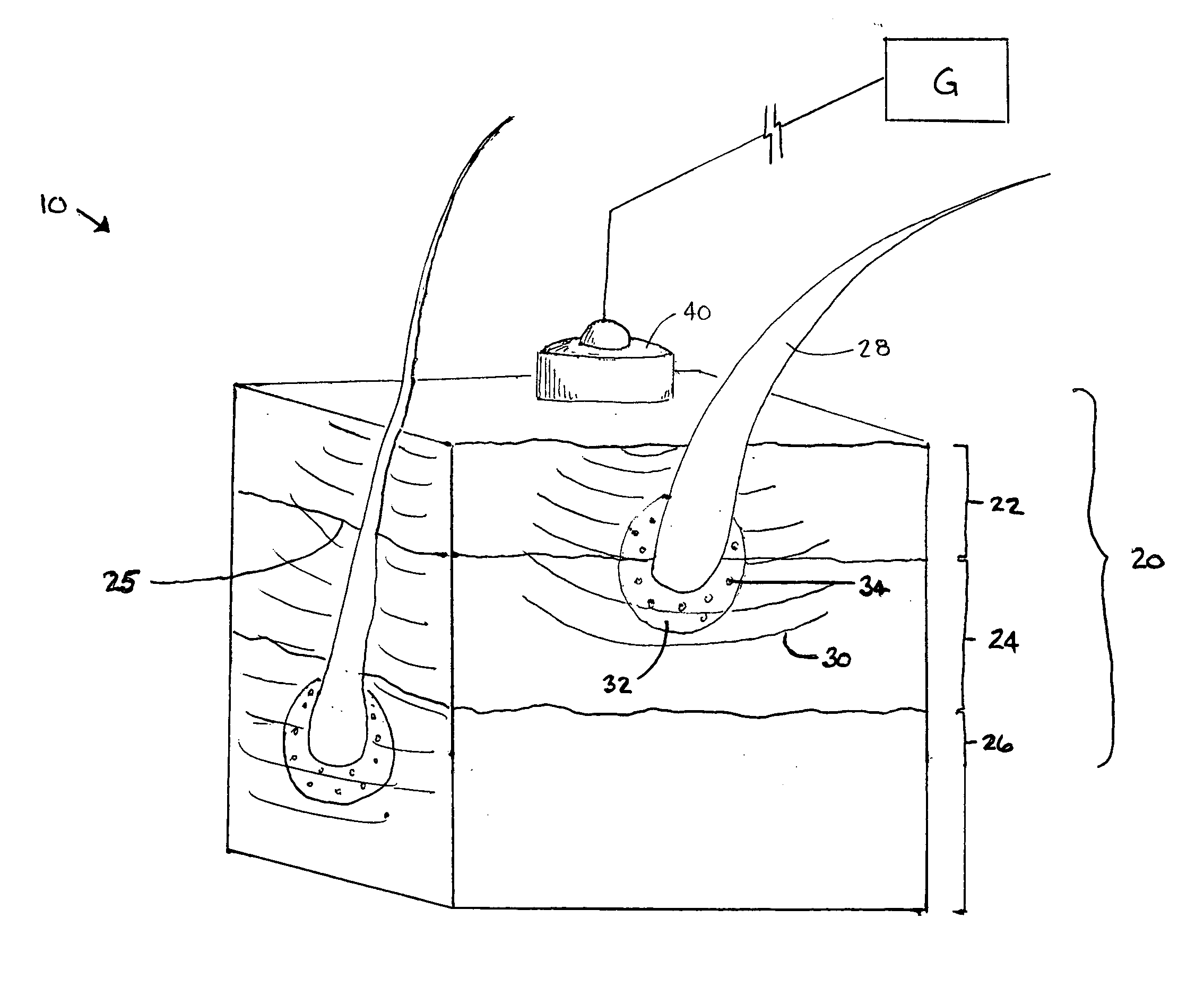
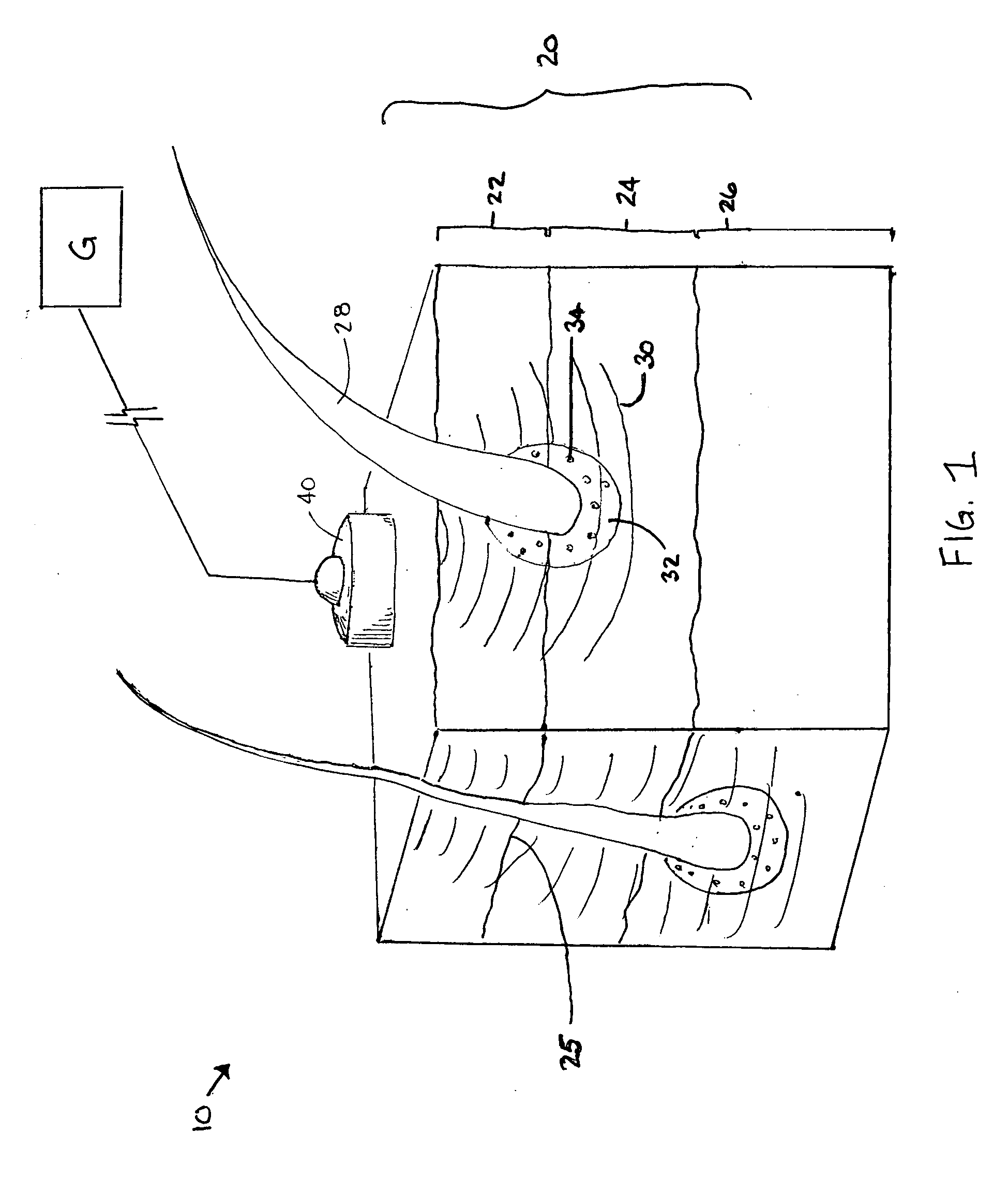
[0092] In addition to the benefits of can induce precise damage in multiple areas of tissue at a desired depth using such a system. FIG. 7A shows ultrasound-induced damage in multiple areas at different depth from the skin surface and lateral locations. The single areas of damage have millimeter or submillimeter size. FIG. 7B shows a damaged area with a submillimeter width and one-millimeter length. This size and depth of the damage areas is desirable for treatment of skin and / or subcutaneous tissues (e.g. skin rejuvenation) and cannot be achieved at depths greater than 0.5 mm with standard laser- (or in general, light-) based techniques / systems. The ultrasound beam can be moved laterally over the skin surface to produce desirable pattern of damaged areas at the desired, subcutaneous depths.


PUM


Abstract

The disclosure provides a method and apparatus for noninvasive and minimally-invasive treatment of skin and subcutaneous tissues with ultrasound with or without nano- or microparticles. The treatment includes, but is not limited to, hair removal, skin rejuvenation (wrinkle removal), scar removal, treatment of spider veins and varicose veins, removal of birthmarks, acne treatment, wound treatment, abnormal pigmentation and stretch mark removal, abnormal tissues in skin and subcutaneous layers, and tattoo removal. Skin and subcutaneous tissues which can be treated with the methods described include, but are not limited to, the dermis, epidermis, subcutaneous fat, connective tissue, muscle tissue, blood vessels, scar tissues, tendons, and cartilage tissues, and abnormal tissues in skin and subcutaneous layers. The disclosure is especially applicable to hair removal and skin rejuvenation.

Description

CROSS REFERENCE TO RELATED APPLICATIONS [0001] This application claims priority to Provisional Patent Application No. 60 / 722,492, filed Sep. 30, 2005, which is incorporated herein by reference in its entirety.STATEMENT REGARDING FEDERALLY SPONSORED RESEARCH OR DEVELOPMENT [0002] Not applicable. REFERENCE TO APPENDIX [0003] Not applicable. BACKGROUND OF THE INVENTION [0004] 1. Field of the Invention [0005] The invention relates to methods and apparatus for the non-invasive and / or minimally invasive treatment of the skin and subcutaneous layers of the skin for therapeutic applications and, more specifically, to tissue treatment methods using ultrasound waves. [0006] 2. Description of the Related Art [0007] Treatments of the skin are widely know and used, both for therapeutic and cosmetic reasons. Therapeutically, the uses include the treatment of cancerous cells, and the treatment of burn victims. Cosmetic applications are much more numerous, and include face-lifts and neck-lifts, tre...

Claims


Application Information

Patent Timeline
Patent Type & Authority Applications(United States)
IPC IPC(8): A61N5/00
CPCA61B2017/00747A61B2017/00769A61N7/00A61N2007/0008
Inventor ESENALIEV, RINAT O.
Owner BOARD OF RGT THE UNIV OF TEXAS SYST
Why Eureka
Try Eureka