Contrast agent for combined modality imaging and methods and systems thereof
a combined modality and contrast agent technology, applied in the field of diagnostic imaging, can solve the problems of limited exposing patients and doctors to radiation, and deep tissue in vivo optical imaging has relatively poor spatial resolution and anatomical registration, and achieves high molecular sensitivity
- Summary
- Abstract
- Description
- Claims
- Application Information
AI Technical Summary
Benefits of technology
Problems solved by technology
Method used
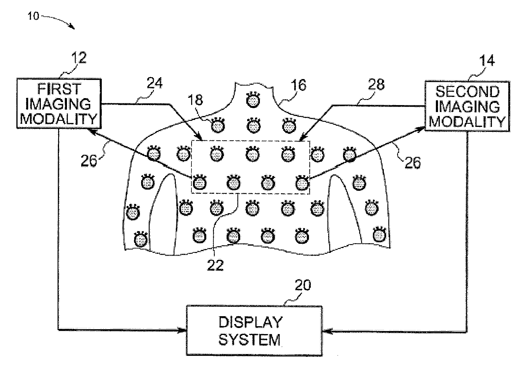
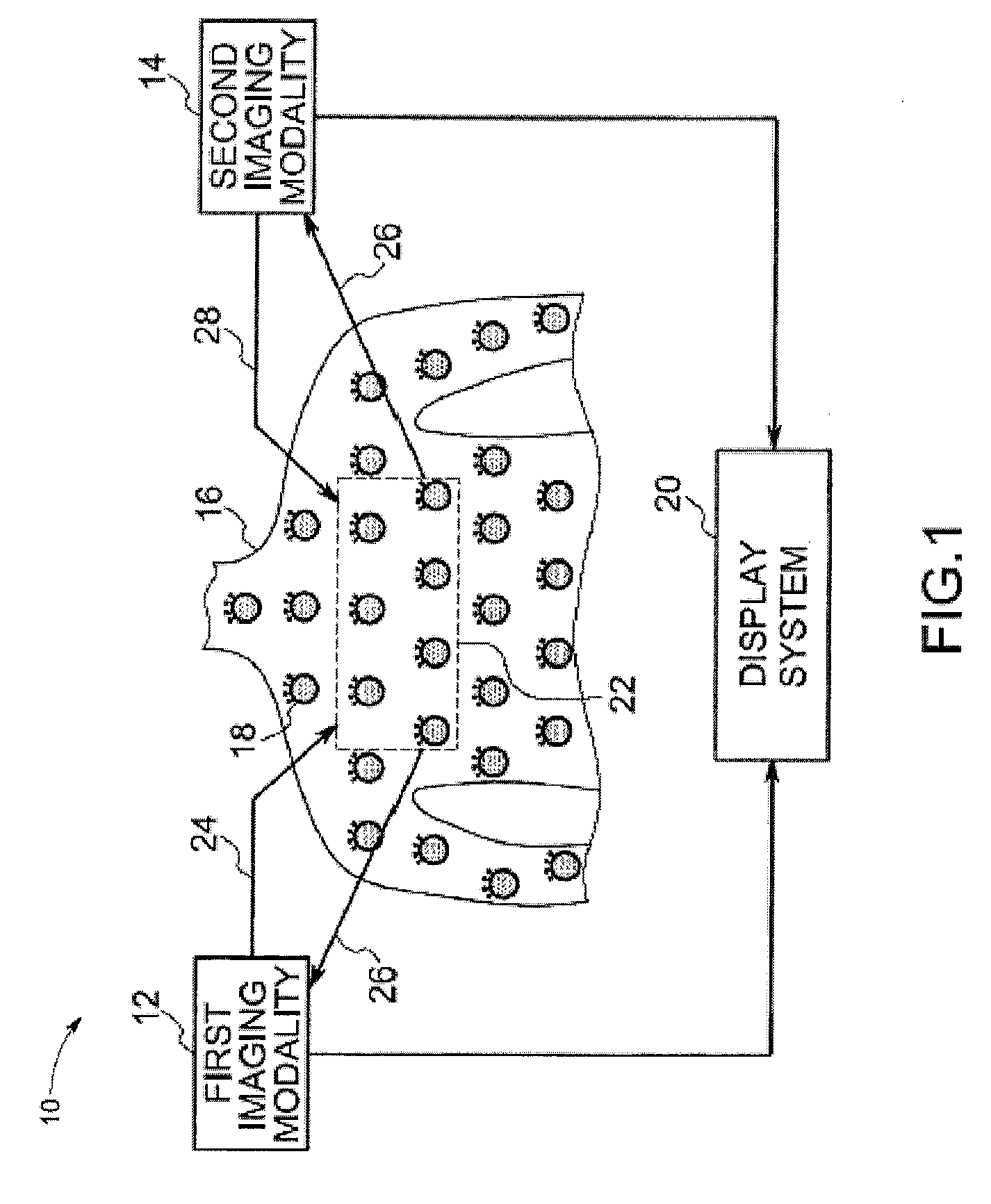
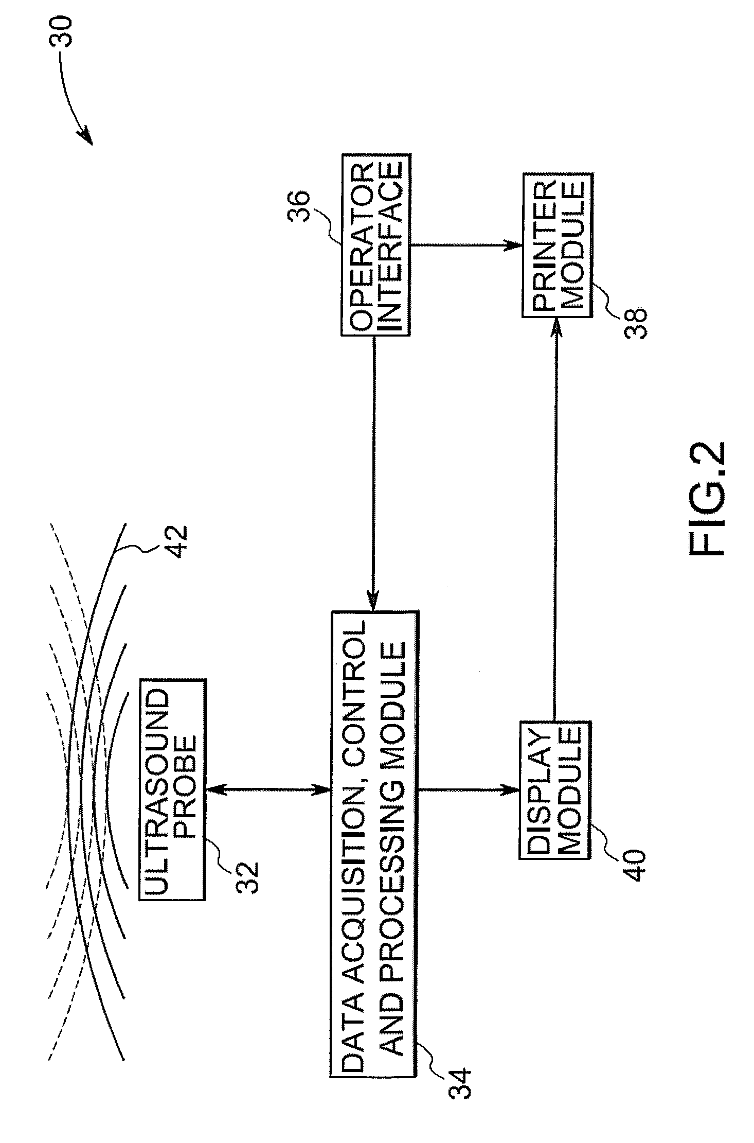
Image

Examples
example 1
Preparation of Non-fluorescent Microbubbles
[0106] Although non-fluorescent microbubbles are commercially available, we synthesized non-fluorescent microbubbles as follows. Two different scaffolds were selected for the preparation of non-fluorescent microbubbles: a surfactant-based system composed of Tween 80 and Span 60 (ST68), and the Human Serum Albumin (HSA) protein. The non-ionic surfactant based system, which has been previously studied, is stabilized by hydrophilic / hydrophobic interactions of the surfactant forming stable micellar-like type of systems. This system would provide flexibility to manipulate the density of dyes on the shell by the addition of different ratios of fluorescently labeled- versus non-labeled surfactants, assuming that these would arrange evenly around the shell due to the micellar type of system.
[0107] The commercially available microbubble Optison® is based on HSA, which includes multiple lysine (Lys) groups that may be used for covalent attachment o...
example 1a
Preparation of Surfactant-Based Microbubbles
[0108] The surfactant solution was prepared as follows: 1.48 g of Span 60 and 1.5 g of NaCl were ground together in a mortar with a pestle until homogeneous mixture was formed. Then, 10 mL of phosphate buffer solution (PBS) solution were added and mixture was mixed to slurry. The slurry was poured into a 50 mL beaker. 10 mL of PBS were added to the mortar followed by the addition of 1 mL of Tween 80. These two were mixed and then combined with the slurry. The mortar was rinsed with an additional 30 mL of PBS and added to the beaker.
[0109] For the preparation of ST68 microbubbles the parameters that were changed include, volume of the solution, sonication time, continuous vs. non-continuous sonication, sonication intensity, type of bath in which solution was immersed during sonication and depth of horn tip into solution. The sonicator used (Sonics and Materials, Inc. VCX 750 Model, CT, USA) was set to a frequency 20 KHz. The ultrasound co...
example 1b
Preparation of Human Serum Albumin-Based Microbubbles
[0111] The initial conditions explored for microbubbles formation were done using HSA solutions that were prepared using lyophilized HSA from Sigma (Cat #: A9511-25G). The parameters that were changed include the solvent used to dissolve the HSA (85 mM NaCl, PBS solution), volume of the solution, sonication time, continuous vs. non-continuous sonication, sonication intensity, size of the probe, type of bath in which solution was immersed during sonication and depth of horn tip in solution.
[0112] The initial conditions tried yielded two results. Either unstable large bubbles were formed, which would continuously grow in size after sonication until bursting back into the HSA solution, or the protein would denature and form a gel. The results are summarized below in Table 2 for summary of results.
TABLE 2Son.ContSon.ProbetimeNotintensityHorn tipsizeSolventVolume (mL)(min)cont(%)position(in.)BathResult85 mM103C40center⅛NBgelledNaCl...
PUM
 Abstract
Description
Claims
Application Information
 - R&D
- Intellectual Property
- Life Sciences
- Materials
- Tech Scout
- Unparalleled Data Quality
- Higher Quality Content
- 60% Fewer Hallucinations
Browse by: Latest US Patents, China's latest patents, Technical Efficacy Thesaurus, Application Domain, Technology Topic, Popular Technical Reports.
© 2025 PatSnap. All rights reserved.Legal|Privacy policy|Modern Slavery Act Transparency Statement|Sitemap|About US| Contact US: help@patsnap.com