Medical imaging modality

a medical imaging and modality technology, applied in the field of medical imaging modality, can solve the problems of affecting the accuracy of patient data, and unable to deliver good anatomical images, etc., and achieves the effects of reliable detection, precise localization of tissue anomalies, and good access to patients

Inactive Publication Date: 2007-05-03
SIEMENS AG
View PDF13 Cites 42 Cited by
  • Summary
  • Abstract
  • Description
  • Claims
  • Application Information

AI Technical Summary

Benefits of technology

[0013] The object of the invention is thus to specify a medical imaging modality of the type mentioned at the start of which the images make a reliable detection and precise localization of tissue anomalies, especially of malign tissue with tumor incidences possible and which offers good access to the patient so that alongside the image recording operative and / or minimally-invasive interventions in the patient can be undertaken and checked.

Problems solved by technology

PET does not deliver good anatomical images, so that the spatial localization and assignment of the focus of the disease source detected produces difficulties.
This reconciliation process for the image data sets, which is also referred to as a registration, as a rule has only a restricted accuracy and despite extensive (computer-aided) automation frequently requires time-consuming user interaction.
A significant disadvantage of this concept lies in the difficult access to the patient during examination caused by the two closed detector tubes being arranged adjacent one another.
This type of arrangement can not only increase the unease of the patient, it is also as a rule not possible to undertake interventions during the examination procedure, e.g. minimally-invasive or surgical interventions.
Although conventional angiography delivers high-resolution images of the vessel system, it is however not in a position to represent the surrounding soft tissue or any deposits on the wall of the vessel.
This means that under some circumstances valuable time for the initiation of the further therapy steps is lost, in which case the state of health of the patient may possibly drastically deteriorate.

Method used


Image

  • Medical imaging modality
  • Medical imaging modality
  • Medical imaging modality

Examples


Embodiment Construction

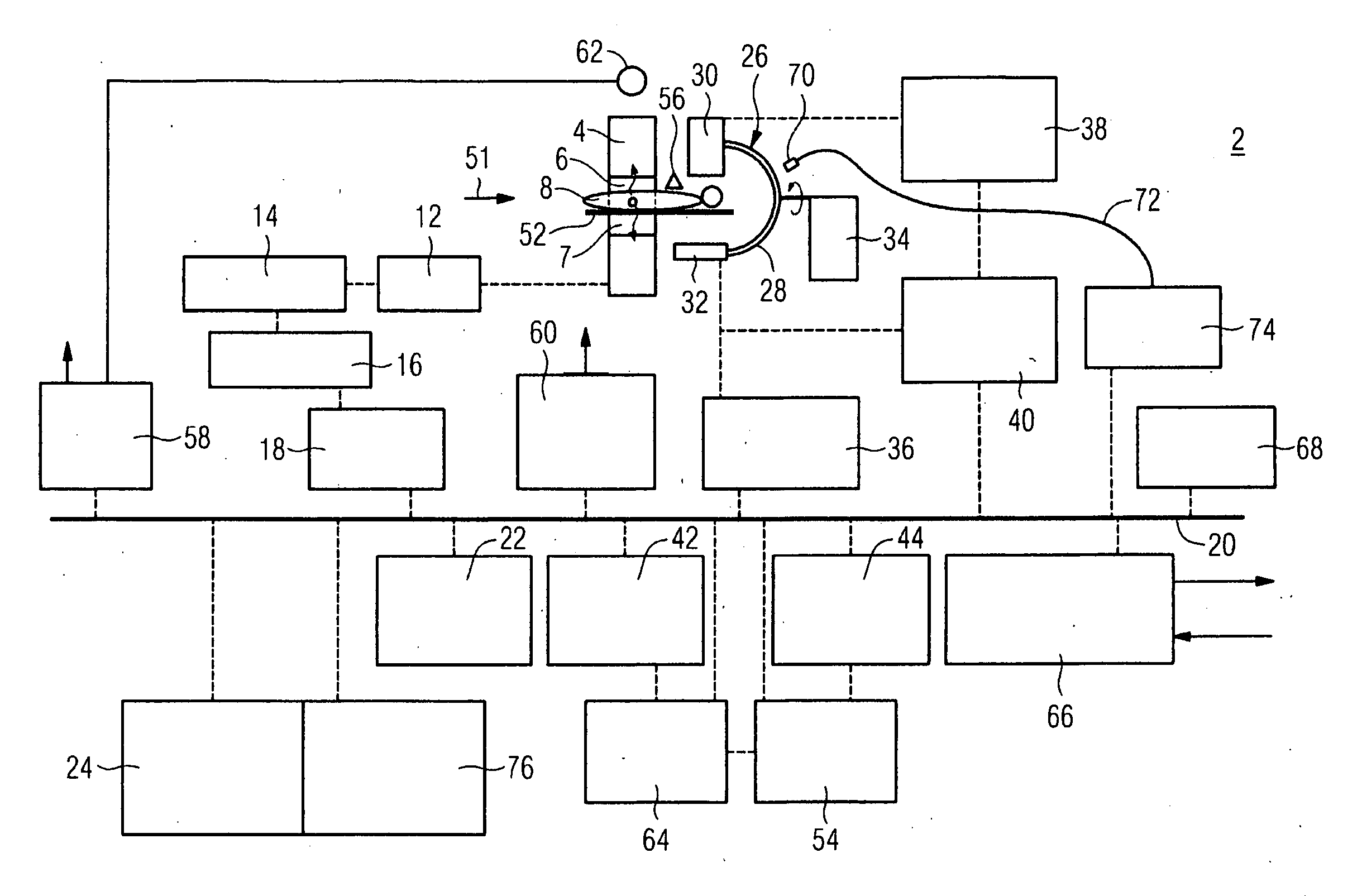
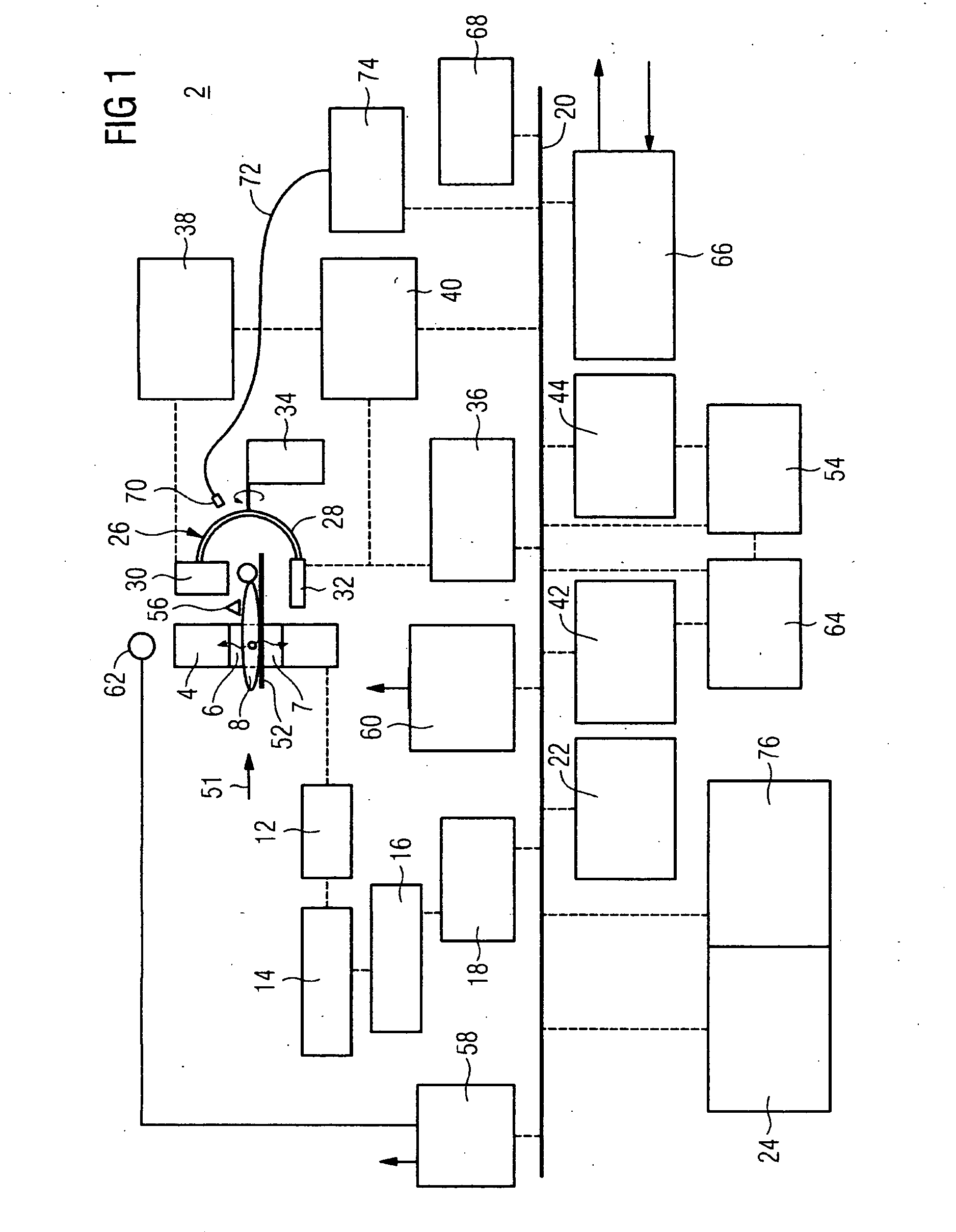
[0036] The medical examination and treatment device 2 shown in the schematic overview in FIG. 1, referred to in short below as a modality, comprises a PET unit based on the principle of Positron Emission Tomography (PET) The PET unit features a PET detector ring equipped with a plurality of scintillation or semiconductor detectors as well as with associated photo multipliers and pre-amplifiers to amplify the primary signals. The closed PET detector ring 4, shown in cross section here, is also referred to as a gantry. The detector elements of the PET detector ring 4 register—resolved in space and lime—in the cylindrical cavity 6 of the detector ring, energy-rich gamma quanta 7 emitted by a radiographic source. The radiographic source is in this case a human being, namely the patient 8 to be examined, into whom a slightly radioactive tracer is injected before the examination which accumulates in specific organs, especially in tumors, and which thus is distributed inhomogeneously in th...


PUM


Abstract

A medical imaging modality with a PET detector ring for Positron Emission Tomography connected on the data side to a PET image processing unit is to be structured in such a way that, on the basis of the images generated in the modality, a reliable detection and precise localization of metabolism anomalies, especially of malign tissue with tumor incidence is possible. The modality is further intended to offer good access to the patient, so that operative or minimally-invasive interventions on the patient can be undertaken and checked alongside the image recording. To this end there is provision, in accordance with the invention, for an ACT recording device for angiographic computer tomography connected on the data side to an ACT image processing unit to be arranged adjacent to the PET detector, whereby a common display unit for display of PET images and / or ACT images generated in the relevant image processing unit is assigned to the PET image processing unit and the ACT image processing unit.

Description

CROSS REFERENCE TO RELATED APPLICATIONS [0001] This application claims priority of German application No. 10 2005 048 853.6 filed Oct. 12, 2005, which is incorporated by reference herein in its entirety. FIELD OF THE INVENTION [0002] The invention relates to a medical imaging modality with a PET detector ring for Positron Emission Tomography connected on the data side to a PET image processing unit. BACKGROUND OF THE INVENTION [0003] Diseases of the vessels such as strokes, aneurysm and abdominal aorta aneurysms are among the most frequently occurring diseases in the world. With these diseases, but also especially with tumor diseases or similar, rapid and secure diagnosis and immediate initiation of therapy are of particular significance for the recovery process. [0004] The diagnosis of these types of diseases is supported by imaging methods. One imaging method which can supply a high level of diagnostic information is Positron Emission Tomography (PET). The method is based on the r...

Claims


Application Information

Patent Timeline
Patent Type & Authority Applications(United States)
IPC IPC(8): A61B5/05
CPCA61B6/032A61B6/481A61B6/504A61B6/037G01T1/1603G01T1/1611G01T1/2985A61B6/5235
Inventor MASCHKE, MICHAEL
Owner SIEMENS AG
Why Eureka
Try Eureka