Detection of somatic cells in milk

a somatic cell and milk technology, applied in the field of milk somatic cell detection, can solve the problems of milk in transport risk degradation and contamination, time and effort wasted in collecting and shipping samples, and the single most costly disease of the dairy industry is mastitis, so as to achieve the effects of low cost, high cost, and high efficiency

Inactive Publication Date: 2007-05-17
RGT UNIV OF CALIFORNIA
View PDF3 Cites 1 Cited by
  • Summary
  • Abstract
  • Description
  • Claims
  • Application Information

AI Technical Summary

Benefits of technology

[0008] Given the above-mentioned issues of high cost, great effort and wasted time related to milk collection and transportation, it is the object of the present invention to provide a method of determining SCC levels in a fast, efficient and low cost manner. Advantages of the present invention include simple and quick detection of quarters and / or cows with high SCCs, thereby improving milk quality.
[0009] In one aspect, the present invention provides methods of detecting a nucleic acid in a milk sample. The method includes contacting the milk sample with a metal ion chelator. The milk sample is also contacted with a detergent. After contacting the milk sample with a metal ion chelator and a detergent, the nucleic acid is detected. Detecting the nucleic acid in the milk sample is thereby accomplished.

Problems solved by technology

The single most costly disease of the dairy industry is mastitis, which is an inflammation of the mammary gland.
Time and effort is wasted to collect and ship samples and milk in transport risks degradation and contamination.

Method used


Image

  • Detection of somatic cells in milk
  • Detection of somatic cells in milk
  • Detection of somatic cells in milk

Examples


example 1

Assay Development

[0065] Three extraction procedures examples are provided. Method one included a fat removal step via centrifugation and the use of a commercial DNA extraction kit (DNeasy Tissue Kit, Cat. No. 69504, Qiagen, Valencia, Calif., USA). The DNA extraction method was performed as follows: Two milliliters of milk was centrifuged for 10 minutes at 5000 g. The fat and aqueous layer were removed. The pellet was resuspended in 200 μL of phosphate buffered saline. The steps for DNeasy Protocol for Cultured Animal Cells in the DNeasy Tissue Kit Handbook were then followed. In a cuvette, all of the final DNA solution (˜200 μL), 787 μL of 10 mM Tris, 1 mM EDTA, pH 7.5 (TE) and 13 μL of PicoGreen stock reagent were added. The cuvette was inverted to mix the solution and then inserted into the sensor and record the output voltage. Calibration with method one was the most sensitive to changes in SCC because the assay produced a purer form of DNA. The calibration equation based on met...

example 2

Sensor and Circuit Design

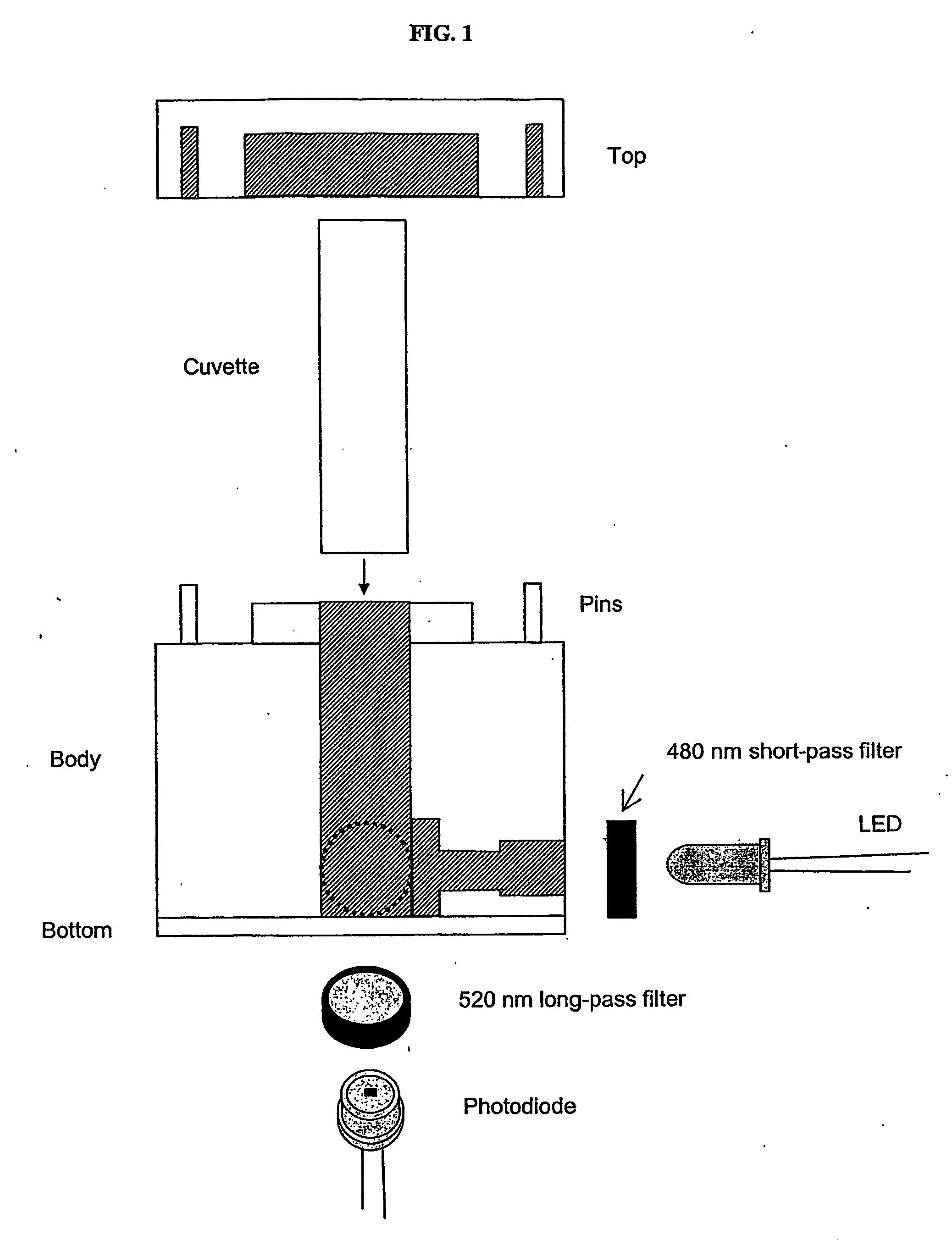
[0070] The sensor housing consisted of four pieces: the top, bottom, and two sides (FIG. 1). Side one contained a 470 nm light-emitting diode (LED) (BL-BBX3V4V-B02, American Bright, San Jose, Calif., USA) and a 480 nm short-pass edge filter (35-2039, Ealing Catalog, Inc., Rocklin, Calif., USA). Side two held a photodiode (BPW21R, Vishay, San Jose, Calif., USA) and a 520 nm long-pass edge filter (35-2153, Ealing Catalog, Inc., Rocklin, Calif., USA). The LED and photodiode were positioned perpendicular to each other. Sides one and two and the bottom piece were screwed together to form a block, which served as the main unit to hold a 1.5 mL sample cuvette (14-385-942, Fisherbrand, Fisher Scientific). The top piece completed the housing and was secured to the unit with pins.

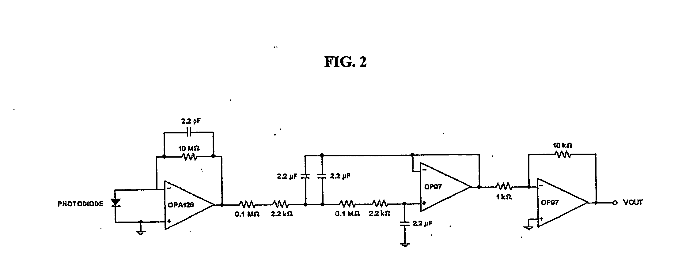
[0071] The circuit consisted of three stages: a photovoltaic amplifier, a low-pass filter, and a final gain stage using an inverting amplifier (FIG. 2). An optical filter that passed wavele...

example 3

Calibration

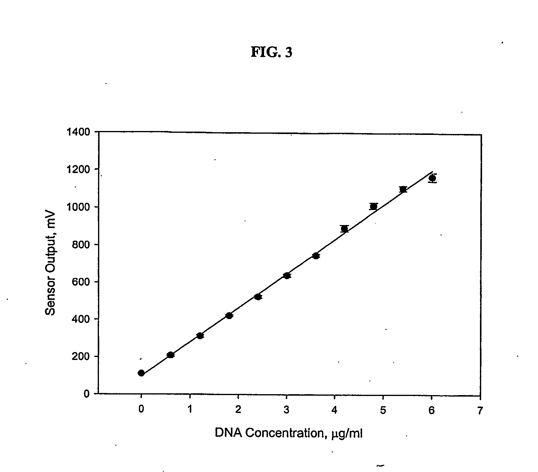
[0072] The sensor was calibrated with known concentrations of ctDNA in TE. Concentrations were prepared by diluting 500 ug / mL of ctDNA with TE to a final volume of 987 μL in a cuvette. The final solution was mixed with 13 μL of PicoGreen stock reagent by inversion. PicoGreen was given one minute to bind to ctDNA before the cuvette was inserted into the sensor for a reading. dsDNA was tested at concentrations between 0 to 6 μg / mL, which roughly corresponded to SCCs in the range of 0 to 1,000,000 cells / mL. Two replicates were completed at each concentration.

[0073] Preliminary calibrations were also completed with extracted DNA from raw milk with known SCCs using methods one, two, and three. Cows with low, medium and high levels of SCC were chosen based on monthly DHI reports. Based on the National Mastitis Council guidelines and federal and state regulations, SCCs less than or equal to 200,000 cells / mL were considered to be low, SCCs greater than 200,000, but less than or...


PUM

PropertyMeasurementUnit
wavelengthaaaaaaaaaa
cutoff frequencyaaaaaaaaaa
pHaaaaaaaaaa

Abstract

A simple, rapid and accurate method for detecting nucleic acids from milk are disclosed. The methods are useful in determining somatic cell count levels and diagnosing mastitis in cows.

Description

CROSS REFERENCE TO RELATED APPLICATIONS [0001] This application claims priority to U.S. Provisional Application Ser. No. 60 / 556,243, filed Mar. 24, 2004 and U.S. Provisional Application Ser. No. 60 / 490,126, filed Jul. 25, 2004, both of which are herein incorporated by reference in their entirety for all purposes.BACKGROUND OF THE INVENTION [0002] The single most costly disease of the dairy industry is mastitis, which is an inflammation of the mammary gland. Mastitis affects the dairy farmer financially through decreased milk yield, discarded milk, culling, drugs and veterinary expenses, and increased labor (DeGraves et al., Vet. Clinics N. Amer.: Food Animal Prac. 9:421, 1993; Blowey et al., Mastitis Control in Dairy Herds: An illustrated and practical guide 1995). The National Mastitis Council (1996) estimated the annual cost per cow to be $185, and the total annual cost of mastitis to be $1.8 billion. Blosser (J. Dairy Science 62:119, 1979) and Jasper et al. (21st Annual Mtg. Nat'...

Claims


Application Information

Patent Timeline
Patent Type & Authority Applications(United States)
IPC IPC(8): C12Q1/68A01J5/013A61BC07H21/02C07H21/04C12P19/34
CPCA01J5/0131A01J5/0135C12Q1/6806
Inventor DELWICHE, MICHEAL J.WU, JANICE
Owner RGT UNIV OF CALIFORNIA
Why Eureka
Try Eureka