
X-ray imaging arrangement

Inactive Publication Date: 2007-05-31
SECTRA MAMEA
View PDF13 Cites 14 Cited by
  • Summary
  • Abstract
  • Description
  • Claims
  • Application Information

AI Technical Summary

Benefits of technology

[0023] flexible to allow for dose minimization over a large range of object radii, and
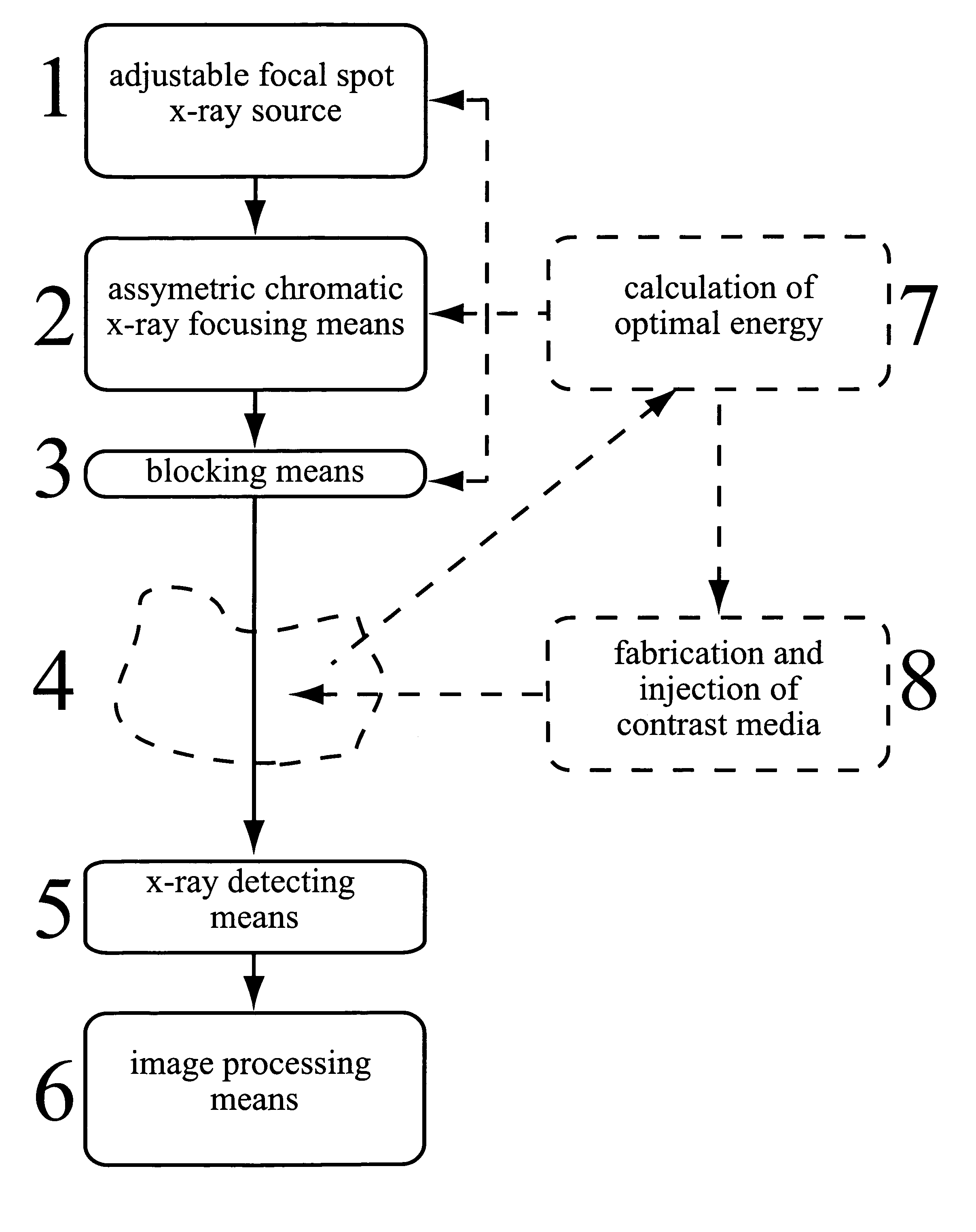
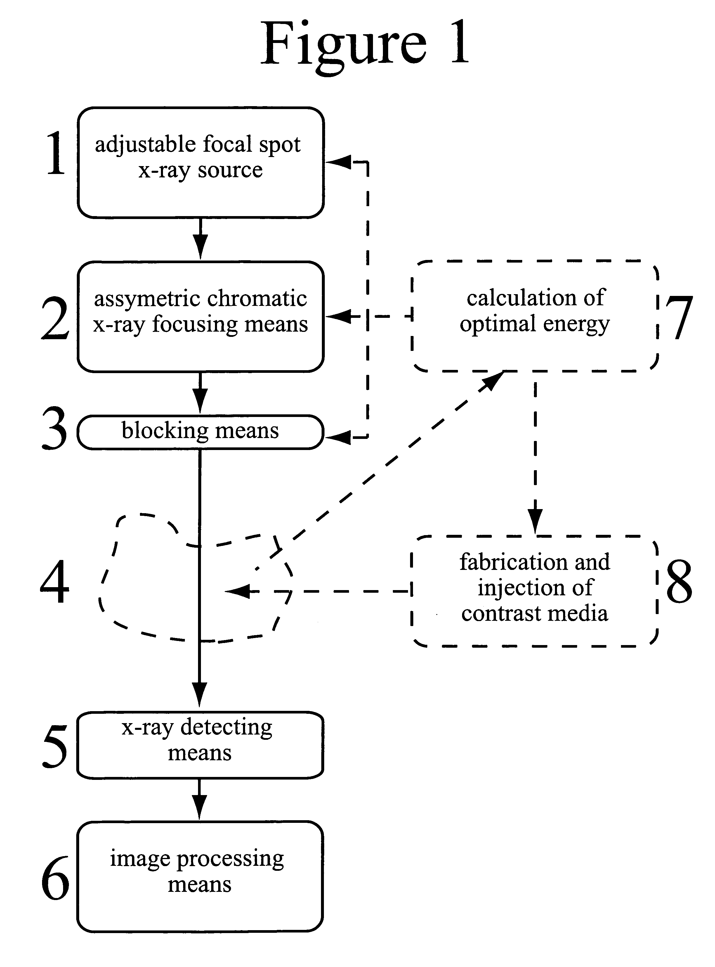
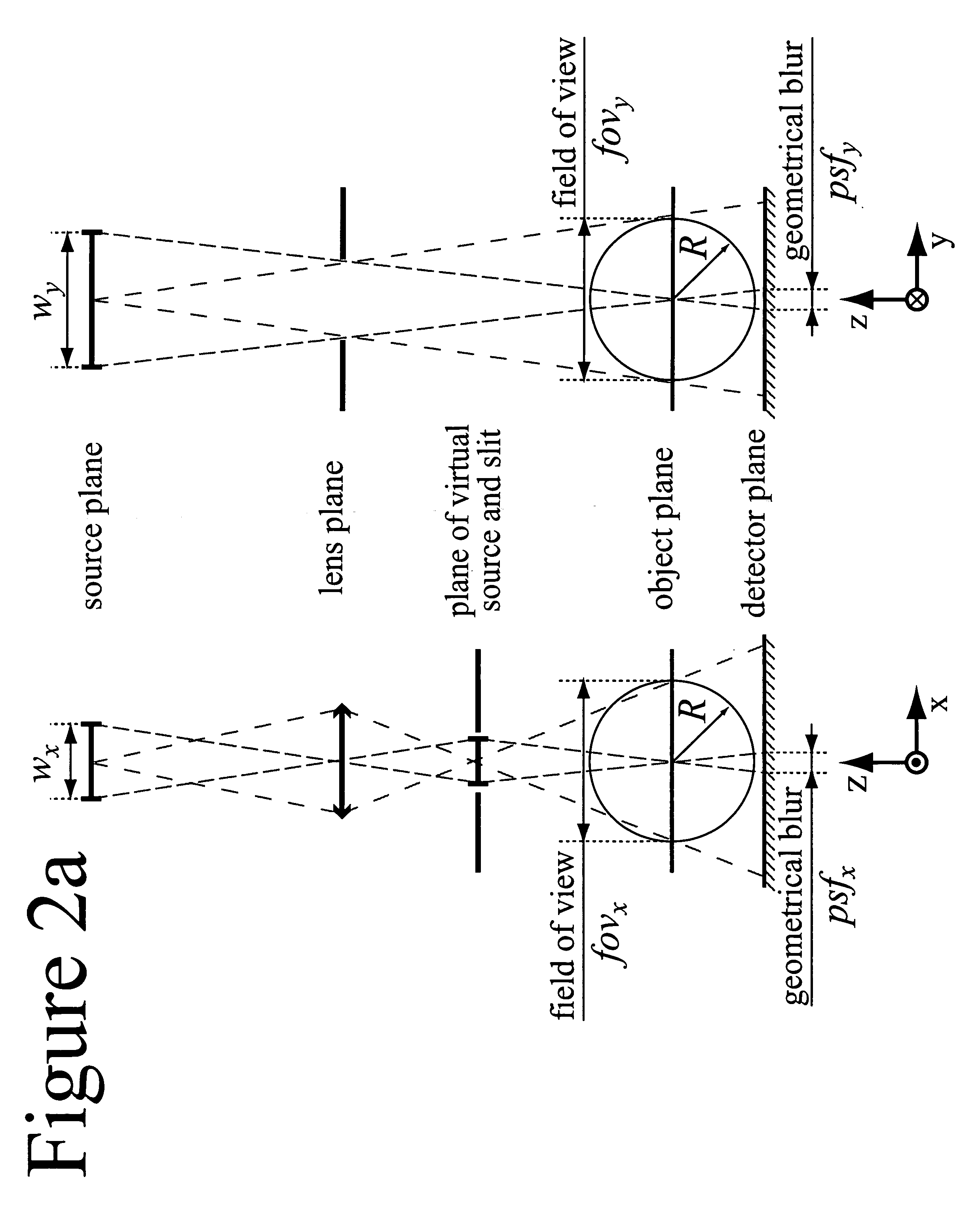
[0029] By choosing an appropriate asymmetric focal spot of the x-ray source it is possible to compensate for asymmetric focusing effects of the x-ray optics. If the optics comprises the mentioned arrangement of one-directional focusing x-ray lenses, the x-ray source focal spot is preferentially a line focus.
[0030] The system is intended to image an object, which is optionally injected with a contrast agent for target or tissue specific labeling, the contrast agent preferably containing a bulk material with an absorption edge situated between at least two peaks of characteristic radiation from the source. This allows for sensitively detecting the contrast agent inside the object using critical edge subtraction.
[0031] Optimization in terms of signal-to-noise ratio and dose to the object can optionally be obtained by selecting a combination of target material for the x-ray tube and contrast agent material, at a suitable energy according to the size of the object. Due to the flexibility of this system, such a combination can be found at many energies and a critical edge subtraction image can be obtained along with a structural image at optimum energy. This keeps the dose at a minimum at the same time as high sensitivity labeling allows for imaging of function and tissue character, as well as improvement of the contrast of body structures.
[0032] The detector can be energy discriminating to allow for images at all necessary energies to be acquired simultaneously. If the x-ray optics comprise the mentioned one-directional focusing lenses, it can be advantageous to use an electronic row detector to increase effectiveness and to reject scattered radiation.

Problems solved by technology

The important role of medical imaging for diagnosing various kinds of disease is unquestionable.
Two major disadvantages of all kinds of x-ray imaging are, however, a) the radiation dose put upon the subject (compared to for instance MRI), which increases with spatial resolution and signal to noise ratio, and b) the lack of information about function and tissue character (compared to PET, SPECT and f-MRI).
The drawback is that the x-ray attenuation of iodine is fairly weak, and large amounts of the substance have to be injected into the animal or human, being imaged.
This is a problem since iodinated contrast agents are not harmless.
Often however, heavy elements are foreign to the body and will be disposed of quickly, and most are toxic even in small amounts.
Metals for instance, due to the huge surface area to volume ratio if small particles are used, and the slightly hostile environment of the blood, might upon intravenous injection form ions which interfere with the body systems in various ways.
The above mentioned low sensitivity of x-ray imaging to iodinated contrast agents previously has made such attempts practically impossible because the high concentration needed for detection would be very hard to direct to a specific location in the subject and once there, the agent would in many cases perturb the very function it is supposed to measure.
This is a promising approach, however antibodies are large macromolecules (usually in the kDa range), and the concentration of epitopes in target tissue may be low, which means that the number of label molecules on each antibody, despite the anticipated increase in contrast of three times, needs to be very large.
The label complex might thus be very bulky and show limited extravasation from the vasculature system, as well as slow diffusion through the interstitial space.
The major obstacle in performing critical edge subtraction is to obtain acceptably narrow bandwidth tunable x-radiation—already at an energy difference in the order of a few keV the method becomes ineffective—and at the same time maintaining a high enough flux for reasonable exposure times. Therefore, it is widely believed that synchrotron sources are necessary to practice the method.
Synchrotrons are, however, rare and expensive establishments, hardly available for routine research, and the requirement of such a source clearly limits the usefulness of the method.
Nevertheless, because potential target materials are restricted by several factors including thermal properties, at a different energy, which might be suitable for a particular imaging situation, such a close match of the target material might not be possible for a given contrast agent.
The need for energy discrimination puts restrictions on the choice of detector.
However, not all materials are suitable to use in a detector.

Method used


Image

  • X-ray imaging arrangement
  • X-ray imaging arrangement
  • X-ray imaging arrangement

Examples


Embodiment Construction

[0043] Monochromatic x-ray imaging in general and computed tomography in particular has mainly two advantages compared to conventional polychromatic x-ray imaging [F.A. Dilmanian. Computed tomography with monochromatic x-rays. Am. J. Physiol. Imaging, 3:175-193, 1992]; the first of which is improving the image quality and by that the resolution, and the second one is enhancing the contrast per photon and accordingly minimizing the dose to the object.

[0044] Firstly, a polychromatic beam will be filtered by the object, and since the probability of x-ray absorption decreases with energy, the mean energy of the beam will be shifted towards higher energies by passing the object. This effect is called beam hardening and will cause image artifacts in tomographic imaging because the reconstruction algorithm assumes the signal to be independent of the ray path. Beam hardening can be compensated for by different methods, however only to a certain extent, and the only way to get rid of the ef...


PUM


Abstract

An x-ray system for narrow bandwidth imaging of in particular small objects is provided. X-radiation from an x-ray source (1) is focused by chromatic x-ray optics (2) on an x-ray energy dependent distance from the optics. Asymmetric focusing of the x-ray optics is compensated for by choosing an asymmetric focal spot of the source. The energy selective focusing makes possible blocking unwanted x-ray energies (3) from reaching an object (4). In that way optimization of the energy according to the size of the object can be done to minimize dose and maximize signal-to-noise ratio (7). Furthermore, a critical edge subtraction image can be obtained at the object dependent optimal energy if the object is injected with a contrast agent having an absorption edge close to the optimal energy (8). Radiation is registered (5) and processed (6) to combine structural and energy subtraction images.

Description

TECHNICAL FIELD OF THE INVENTION [0001] The present invention generally relates to imaging, especially X-ray imaging and arrangements and methods associated therewith. The invention also concerns medical decision support and improvements of existing diagnostic imaging methods, particularly for small objects. BACKGROUND OF THE INVENTION [0002] The important role of medical imaging for diagnosing various kinds of disease is unquestionable. Especially for many forms of cancer, the second most common cause of death in the western world today, early detection is vital for the chances of survival. Also for cardiovascular disease, the most common cause of death in the western world, imaging of particularly the vascular system plays an important role for diagnosis. [0003] In recent years, there has been a growing interest in the use of clinical imaging methods also within biomedical research for imaging animal models of disease and function. Traditional studies of animal models usually have...

Claims


Application Information

Patent Timeline
IPC IPC(8): G01N23/04
CPCA61B6/06A61B6/4035A61B6/4241A61B6/481A61B6/482A61B6/504A61B6/542A61B2503/40
Inventor CEDERSTROM, BJORNDANIELSSON, MATSFREDENBERG, ERIK
Owner SECTRA MAMEA
Learn More