Isolation and expansion of animal cells in cell cultures

a cell culture and cell technology, applied in the field of development biology, cell culture, tissue culture, etc., can solve the problems of requiring a prodigious effort and restricting the use of such cells for further manipulation or us

Inactive Publication Date: 2007-06-07
TISSUETECH INC
View PDF4 Cites 69 Cited by
  • Summary
  • Abstract
  • Description
  • Claims
  • Application Information

AI Technical Summary

Benefits of technology

[0030] When referring to a cell culture system “essentially free of” a substance (e.g., intact AM or feeder cells) is meant that that substance is not pr

Problems solved by technology

In practice, however, defining and refining the conditions that allow primary cell expansion without phenotypic changes (e.g., differentiation from an epithelial cell phenotype to a fibroblastic phenotype) ha

Method used


Image

  • Isolation and expansion of animal cells in cell cultures
  • Isolation and expansion of animal cells in cell cultures
  • Isolation and expansion of animal cells in cell cultures

Examples


example 1

Preservation and Expansion of Primate Keratocyte Phenotype by Downregulating TGF-β Signaling in a Low Calcium Serum-Free Medium

[0078] Because TGF-β signaling is conserved among a large variety of different cell types, the results set forth below and methods described herein can be adapted for other cell types with minor modifications.

Methods

[0079] Three rhesus monkeys (Macaca Mulatta), 4 years old, and rabbit and mouse cornea were obtained from an approved tissue-sharing program after euthanasia. An entire anterior corneoscleral segment was removed from the globe by cutting near the limbus with Wescott's scissors. A central cornea was obtained with an 8.0 mm Barron's trephine and immediately transferred to KSFM medium (cat# 17005-042, GIBCO Invitrogen corporation, Carlsbad, Calif.). After removing Descemet's membrane and the corneal endothelium, the corneal epithelium was removed by dispase digestion for 16 h at 4° C. and the remaining corneal stroma was incubated at 37° C. for 1...

example 2

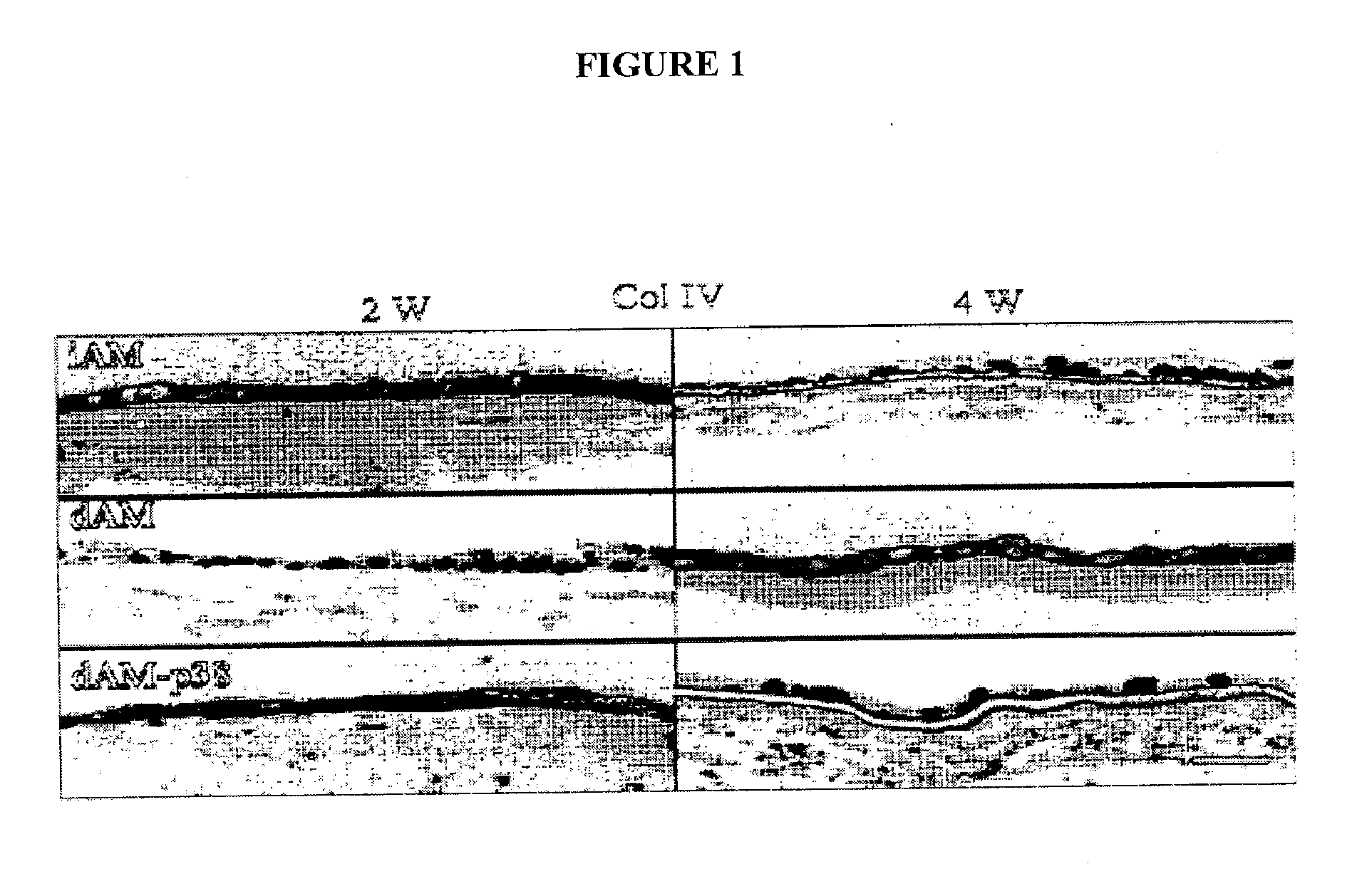
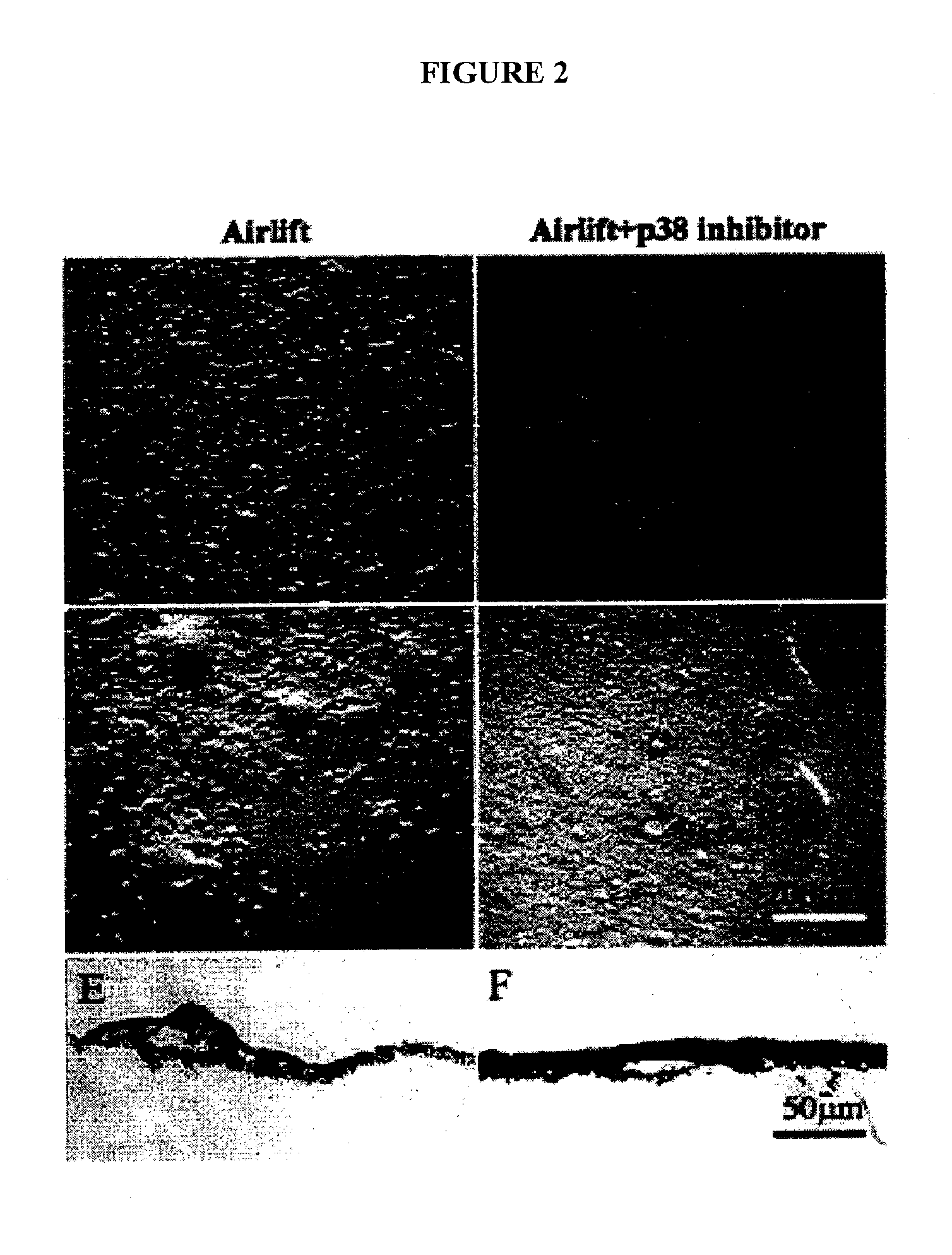
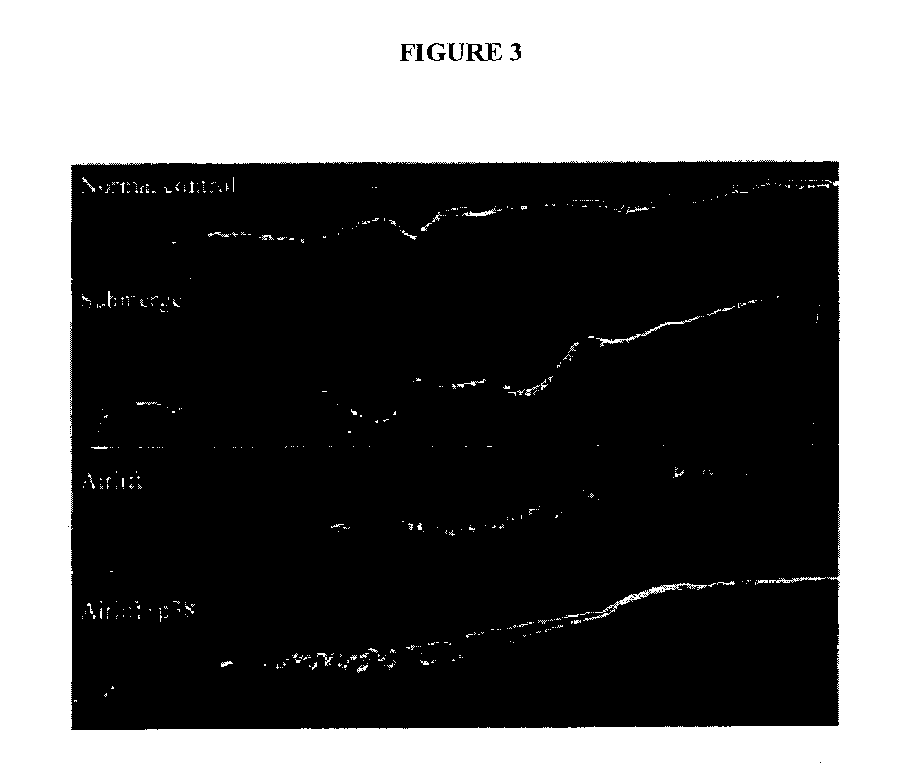
Keratocan Expression of Murine Keratocytes is Maintained on AM by Downregulating TGF-β Signaling

[0092] Keratocytes display a dendritic morphology and express keratocan. When cultured using conventional methods, however, keratocytes lose their dendritic morphology and cease expression of keratocan. As described below, keratocytes were expanded on AM and examined to determine if they maintained their characteristic phenotype, including the expression of keratocan.

Methods

[0093] Isolation and Culture of Keratocytes on Plastic or AM—Albino mice eyes were enucleated by forceps, washed profusely in PBS, and incubated in DMEM containing 20 mM HEPES, 15 mg / ml dispase II (Roche, Indianapolis, Ind.) and 100 mM sorbitol at 4° C. for 18 h (see Espana et al., Invest Ophthalmol Vis Sci. 44:4275-4281, 2003; Kawakita et al., Invest Ophthalmol Vis Sci. 45:3507-3512, 2004). The entire corneal epithelium loosened by this treatment was subsequently removed by vigorous shaking. Under a dissecting micr...

example 3

Clonal Initiation and Expansion of Murine Limbal Progenitor Cells in a Fibroblast-Free, Matrix-Free, and Serum-Free Niche

Methods

[0108] A flat mount preparation of freshly isolated intact human limbal epithelial sheet showed that p63 -positive (p63 being an epithelium-specific transcription factor) basal cells are grouped in clusters, indicating that progenitor cells are intermiixed with TACs in the limbal basal epithelium. Because TACs are known to have a negative paracrine influence on limbal epithelial progenitor cell renewal, it was hypothesized that elimination of TAC's paracrine influence by seeding at a low density and prolonging the culturing time beyond TAC's life span (about 3 weeks) would improve clonal initiation and expansion of limbal epithelial progenitor cells. Single cells dissociated from isolated mouse corneal / limbal sheets by trypsin / EDTA were seeded at a density of 40 cells / cm2 in a defined keratinocyte serum-free medium (KSFM) (Gibco-BRL, Carlsbad, Calif.) con...


PUM


Abstract

Described are methods for isolating/purifying and expanding animal stem cells and stem-cell-like cells. Isolation methods include conditions comprising preferentially digesting non-stem cells and non-stem-cell-like cells in a population and preferentially adhering stem cells and stem-cell-like cells in a population. Expansion methods include culturing such cells under conditions comprising modulation of TGF-β signaling, inhibition of cell signaling mediated by p38 MAP kinase using small molecular weight inhibitors, expansion of the cells on human amniotic epithelial cells as feeder layers, control of cell seeding density, control of levels of Ca2+ in the culture media, rapid adhesion on a substrate or by a combination of such conditions. More particularly, what is disclosed relates to methods and systems for expanding animal cells in ex vivo cell cultures, while preventing cellular differentiation, and selectively enriching stem cells. The embodiments also disclose a culture system for ex vivo expansion of limbal epithelial cells or mesenchymal cells, as well as surgical grafts made there from.

Description

CROSS-REFERENCE TO RELATED APPLICATION [0001] The present application is a continuation-in-part of U.S. patent application Ser. No. 11 / 476,376, filed on Jun. 28, 2006, which claims the benefit of U.S. provisional patent application Nos. 60 / 695,051 filed Jun. 29, 2005; 60 / 695,576, filed Jun. 30, 2005; and 60 / 703,188, filed Jul. 28, 2005; the present application is also a continuation-in-part of U.S. patent application Ser. No. 10 / 833,502 filed on Apr. 28, 2004, which claims the benefit of U.S. provisional patent application 60 / 473,007, filed May 22, 2003; and the present application claims the benefit of U.S. provisional patent application 60 / 801,491, filed May 17, 2006.STATEMENT AS TO FEDERALLY SPONSORED RESEARCH [0002] Certain aspects of this disclosure were made with United States government support under grant number RO1 EY06819 awarded by the National Institute of Health. The United States government may have certain rights in the invention. FIELD OF THE INVENTION [0003] The emb...

Claims


Application Information

Patent Timeline
IPC IPC(8): C12N5/06C12N5/08C12N5/071C12N5/073C12N5/074
CPCC12N5/0605C12N5/0629C12N5/063C12N2500/14C12N2500/25C12N2500/99C12N2501/15C12N2533/92C12N2500/90
Inventor TSENG, SCHEFFERHE, HUALI, WEICHEN, YING-TINGHAYASHIDA, YASUTAKA
Owner TISSUETECH INC
Why Eureka
Try Eureka